Transport in Animals
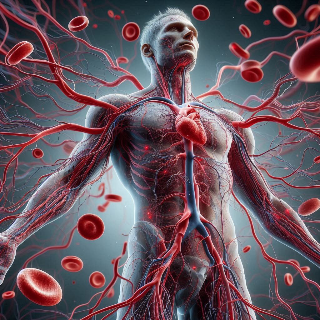
The Circulatory System
The circulatory system is a system of blood vessels with a pump and valves to ensure one way flow of blood. It is different in different organisms:
- Fish:
- Single circulation
- 2 chambered heart
- Blood passes through heart once in every circuit around the body
- Mammals:
- Double circulation
- 4 chambered heart
- Blood passes through heart twice in every circuit around the body
Double circulation has an advantage over single circulation:
- Blood travelling through capillaries in the lungs lose a lot of pressure, therefore it returns back to the heart to be pumped to the rest of the body with high pressure
- This helps provide cells with oxygen and nutrients faster and more frequently
The Heart
Blood is pumped away from the heart into arteries and returns back to the heart in veins
The walls of the atria are thinner than the walls of ventricles because ventricles need to pump blood out of the heart and so need to generate a higher pressure

The walls of the ventricles also differ:
- Left ventricle wall is thick as it needs to pump blood at a high pressure to the entire body
- Right ventricle wall is thinner than the left as it only needs to pump blood to the lungs, a nearby organ
The septum separates the 2 sides of the heart, and therefore prevents mixing of oxygenated and deoxygenated blood
Valves
- Valves prevent the backflow of blood
- They are pushed open when the atria contract
- They are pushed close the ventricles contract, so that blood doesn’t flow back into the atria
- The semilunar valves open when the ventricles contract so blood can exit the heart, and they close to prevent the backflow of blood into the ventricles
Pathway of Blood through the Heart
- Deoxygenated blood arrives at the RA via the vena cava
- It passes the tricuspid valve to reach the RV
- It passes through a semilunar valve to go to the lungs via the pulmonary artery
- The lungs purify the blood and sends it to the LA via the pulmonary vein
- It passes through the bicuspid valve to the LV and under high pressure through a semilunar valve to the rest of the body via the aorta
- The body uses up the oxygen and nutrients from the blood and the blood is deoxygenated once again

Exercise and Heart Rate
The activity of the heart may be monitored by an ECG, pulse rate and listening to sounds of valves closing.
During exercise the heart rate increases and will take some time to return back to normal after the end of a workout. This is because muscle cells have respired anaerobically and produced lactic acid. The heart:
- Must transport the lactic acid to the liver (site where it will be broken down)
- Has to transport enough oxygen to break down the lactic acid
- Increases its rate to remove waste products at a faster rate
Coronary Heart Disease (CHD)
The heart is an organ and it also requires a supply of blood for providing nutrients for respiration. The coronary arteries deliver blood to the walls of the heart.
If a coronary artery becomes partially or completely blocked by fatty deposits called plaques (mainly formed from cholesterol), the arteries are not as elastic as they should be and therefore cannot stretch to accommodate the blood which is being forced through them, leading to coronary heart disease:
- Partial blockage of the coronary arteries creates a restricted blood flow to the cardiac muscle cells and results in severe chest pains called angina
- Complete blockage means cells in that area of the heart will not be able to respire and can no longer contract, leading to a heart attack
This can be caused because of:
- Poor diet
- Stress
- Smoking
- Genetic predisposition
- Age
- Gender
Preventing CHD
- Quit smoking
- Exercise regularly – helps with weight loss and decreasing blood pressure and cholesterol levels
- Reduce fats in diet – reduces cholesterol and helps with weight loss
- Aspirin can be taken daily to reduce risk of blood clots forming in arteries
Treatment for CHD
- Angioplasty:
- A narrow catheter tube is passed into a narrow artery
- It is used to insert a metal stent at the site of blockage
- The stent opens up and forces the artery open
- Coronary Bypass:
- A piece of blood vessel is taken from the patient’s leg, arm, or chest and used to create a new passage for the flow of blood to the cardiac muscle, bypassing the blocked area
Blood
Blood is a tissue that consists of:
- Red Blood Cells (RBCs) – transports oxygen (haemoglobin carries it)
- Plasma – transports blood cells, ions, soluble nutrients, hormones and carbon dioxide
- Platelets – clotting
- White Blood Cells (WBCs) :
- Phagocytes: phagocytosis
- Lymphocytes: antibody production

Blood Clotting
Clotting is important in preventing blood loss and entry of pathogens. The process is:
- On exposure to air, platelets release enzymes
- These enzymes cause soluble fibrinogen to convert into insoluble fibrin
- Fibrin forms an insoluble mesh across the wound and traps RBCs
- This forms a clot
Transfer of Materials between Capillaries and Tissue Fluid
- Plasma (other than proteins) leak out of capillary walls to form the tissue fluid
- O2 and other required materials diffuse into the tissue fluid from capillaries
- O2 and other required materials diffuse into cells
- CO2 and other wastes diffuse out of cells into the tissue fluid, then the capillaries
- Some tissue fluid returns to the capillaries by diffusion
- Rest of it enters minute tubes called lymph vessels which returns plasma back to the bloodstream (at the vena cava)
Blood Vessels
Blood vessels include arteries, veins and capillaries:
| Arteries | Veins |
| Carry blood away from the heart | Carry blood towards the heart |
| Carry blood at high pressure | Carry blood at low pressure |
| Have thick muscular walls (to withstand pressure) | Have thin walls |
| Have a narrow lumen (to maintain high pressure) | Have a wide lumen |
| Speed of flow is fast | Speed of flow is slow |
| Contains no valves | Contains valves (prevents backflow of blood as it’s at low pressure) |
Capillaries:
- Carry blood at low pressure within tissues
- Carry oxygenated and deoxygenated blood
- Have walls which are 1 cell thick (to aid diffusion)
- Have leaky walls (so that plasma can easily leak out and form tissue fluid)
- Speed of flow is slow
- Highly branched (giving more surface area for diffusion)
Blood Vessels in the Human Body

Arterioles and Venules
- Arteries divide to form narrow arterioles that connect to capillaries
- Veins divide to form narrow venules that connect to capillaries
Shunt Vessels
- Sometimes the cardiovascular system need to redistribute the blood to specific areas of the body. This redirection of blood flow is caused by the use of a shunt vessel
- They can open and close to control the amount of blood flowing to a specific area
Lymphatic System
The lymphatic system is formed from a series of tubes which flow from tissues back to the heart/bloodstream. These tubes are known as lymph vessels.
A lymph node is present before the lymph vessels and it acts like a filter. Lymphocytes are present in these nodes, which remove harmful microbes that can infect the body
Diseases and Immunity
Diseases
Key terms:
- A pathogen is a disease-causing organism
- A transmissible disease is a disease in which the pathogen can be passed from one host to another
The pathogen for a transmissible disease may be transmitted:
- By direct contact – through blood or other body fluid
- Indirectly – from contaminated surfaces, food, animals or air
The human body has defences:
- Mechanical barriers – skin and hairs in the nose/trachea
- Chemical barriers – mucus, hydrochloric acid in the stomach and tears
- Cells – phagocytosis and antibody production by WBCs
All of these can be enhanced by vaccines.
Antigens and Antibodies
Antibodies lock on to antigens leading to direct destruction of pathogens, or marking of pathogens for phagocytes. Each antigen is specific, therefore an antibody which is specific to a particular antigen will only be able to lock on to it.
The antibodies attach to antigens and cause agglutination (clumping together), so that the pathogenic cell cannot move easily. Many of these pathogens are clumped up and phagocytes will destroy all of them at once.

Memory cells are there after the first time that this entire process takes place. These retain the ability to produce antibodies. Unfortunately, these cells don’t last forever. Some may last for short periods and some may be very long lasting.
Active Immunity
Key term:
- Active immunity is defence against a pathogen by antibody production in the body
Active immunity may be natural or artificial, and always produces memory cells:
- Natural: the pathogen will infect the body and antibodies are produced
- Artificial: vaccines
Process of Vaccination
- A chemical or weak cell is given to a person
- It provokes the immune system to make appropriate antibodies
- Memory cells are produced, giving long-term immunity
Passive Immunity
Passive immunity is a short-term defence against a pathogen by antibodies acquired by another individual. This doesn’t produce memory cells.
An example of this is that antibodies pass from the placenta of a mother to an unborn child and from a mother to an infant via breast-milk. This is important as it helps young people fight off infections until they are older and stronger and their immune system is more responsive.
Immune System Diseases
Some diseases are caused by the immune system targeting and destroying body cells. This includes:
- Type 1 diabetes – the body destroys its own insulin producing cells
- Transplant rejection – lymphocytes from the recipient may recognise antigens on the surface of the donor’s organ and will slowly destroy it
Controlling the Spread of Diseases
The spread of diseases can be controlled in several ways:
- Vaccines
- Hygienic food preparation
- Good personal hygiene
- Sewage treatment
- Waste disposal