
⚠️ Disclaimer: The information provided in this article is for educational purposes only and does not constitute medical advice. RevisionTown does not provide diagnosis, treatment, or medical recommendations. Always consult a qualified healthcare professional regarding any medical condition, symptoms, or concerns.
Read More – 🏥 Medical Disclaimer
Comprehensive Report on Retinal
1. Overview
What is retinal?
The term “retinal” primarily refers to the retina, which is a crucial part of the eye’s anatomy. The retina is a thin layer of light-sensitive tissue that lines the back of the eyeball. As the innermost of the three layers of the eye, it plays a vital role in the visual process by converting light that enters the eye into electrical signals that are transmitted to the brain via the optic nerve . The retina acts as a key bridge between the light that enters your eyes and the images you see, with special cells reacting to light and passing signals to your brain that allow you to perceive the world around you.
The retina consists of multiple layers of specialized cells organized in a complex structure. There are two main types of photoreceptor cells in the retina that are responsible for detecting light: rods and cones. Rods function primarily in dim light and provide monochromatic vision, while cones function in well-lit conditions and are responsible for color perception and high-acuity vision .
Affected body parts/organs
The retina is located at the back of the eyeball, behind the iris and lens. When light enters the eye, it is focused by the cornea and lens onto the retina. The retina has two main functional areas:
Macula: Located in the center of the retina, the macula contains a high concentration of cones that are essential for seeing colors and fine details. This central vision allows activities like reading, recognizing faces, and driving .
Peripheral retina: This surrounds the macula and allows for side vision when looking straight ahead. The rods in the peripheral retina also help with vision in low light conditions .
Prevalence and significance of retinal diseases
Retinal diseases represent a significant global health concern. They are a major cause of vision impairment and blindness worldwide. The prevalence of retinal disorders varies depending on the specific condition, but collectively they affect millions of people globally.
Key statistics on prevalence:
Age-related macular degeneration (AMD) is a leading cause of vision loss among people aged 50 and older, with the global burden expected to increase significantly due to aging populations. The number of people affected by AMD is projected to increase from 196 million in 2020 to 288 million by 2040 .
Diabetic retinopathy affects about 30% of all people with diabetes and is a leading cause of vision loss in working-age adults.
Retinitis pigmentosa, the most common inherited retinal disease, has an average disease prevalence of approximately 1 in 4,500 individuals .
Stargardt disease, another inherited retinal condition, affects approximately 1 in 17,000 people .
The significance of retinal diseases extends beyond their prevalence. Vision loss from retinal disorders can severely impact quality of life, independence, and ability to work. These conditions often lead to significant economic burden through direct healthcare costs and indirect costs such as lost productivity. Additionally, as populations age worldwide, the burden of age-related retinal diseases is expected to grow substantially in the coming decades.
2. History & Discoveries
When and how was retinal first identified?
The study of the retina dates back centuries, with gradual advancements in understanding its structure and function. Key milestones include:
In the 16th century, Adrianus van Tright published the first drawing of the retina .
Samuel Thomas Soemmerring discovered and illustrated the macula lutea (yellow spot) in the retina in the late 18th century .
Who discovered it?
While no single person can be credited with “discovering” the retina, several scientists made significant contributions to our understanding of this complex tissue:
Hermann von Helmholtz developed the ophthalmoscope in the 19th century, featuring “three essential elements: a source of illumination, a method of reflecting the light into the eye, and an optical means of correcting an out-of-focus image of the fundus” . This invention revolutionized the study of the retina by allowing clinicians to examine it in living patients.
In 1604, Johannes Kepler worked out the optics of the eye and determined that the retina must be where sight begins, though he “left it up to other scientists to reconcile the inverted retinal image with our perception of the world as upright” .
Santiago Ramón y Cajal published the first major characterization of retinal neurons in 1894 in “Retina der Wirbelthiere” (The Retina of Vertebrates) .
Major discoveries and breakthroughs
Several major breakthroughs have shaped our understanding of the retina and improved treatment options for retinal diseases:
In the 19th century, Heinrich Müller combined “careful psychophysical measurements and geometrical optics to find the location of the photosensitive layer of the retina in the living eye” . He correctly deduced that vision begins in the photoreceptors located at the back of the retina.
The discovery of the rod photopigment rhodopsin was a significant milestone in understanding how the retina functions .
In the mid-20th century, Arnall Patz, MD, identified the connection between high oxygen levels in incubators and blindness in premature infants. This discovery resulted in “an almost immediate 60% reduction in the number of blind children in the United States at that time” .
Jules Gonin made a critical observation that retinal breaks were the cause of retinal detachment and introduced “Ignipuncture,” the first surgical treatment for retinal detachment .
In the field of retinal imaging, JW Nordenson invented the modern fundus camera, which became commercially available through the Carl Zeiss Company in 1926 .
Charles Schepens introduced the first hands-free binocular indirect ophthalmoscope, significantly improving the examination of the retina .
Evolution of medical understanding over time
Our understanding of the retina has evolved dramatically over time:
Early understanding was limited to basic anatomical observations and theories about how vision works.
The development of microscopy and histological techniques allowed scientists to identify the cellular components of the retina.
Advancements in genetics have led to the identification of numerous genes associated with inherited retinal diseases, with causative mutations reported in 289 genes .
Modern imaging technologies, such as optical coherence tomography (OCT), have revolutionized the study of retinal structure and the diagnosis of retinal diseases.
In recent decades, there has been a shift from focusing solely on the treatment of symptoms to addressing the underlying causes of retinal diseases through gene therapy, stem cell treatments, and other novel approaches.
The approval of voretigene neparvovec-rzyl (Luxturna) by the FDA in 2017 marked a milestone as “the first gene therapy to treat a rare, inherited form of childhood blindness” .
The ARGUS II Retinal Prosthesis, developed by Mark S. Humayun, MD, PhD, received FDA approval as “the first ever to offer limited vision to patients with late-stage retinitis pigmentosa” .
This evolution continues today with ongoing research into genetic therapies, regenerative medicine, and artificial retina development.
3. Symptoms
Early symptoms vs. advanced-stage symptoms
Retinal diseases often progress from early, sometimes subtle symptoms to more severe manifestations in advanced stages. The specific symptoms depend on the particular retinal condition, but some common patterns include:
Early Symptoms:
- Decreased night vision or difficulty seeing in low light (often an early sign of retinitis pigmentosa)
- Slight blurring or distortion of central vision
- Appearance of small dark specks or transparent strings (floaters) in the vision
- Subtle changes in color perception
- Occasional flashes of light
Advanced-Stage Symptoms:
- Significant loss of central vision (common in macular degeneration)
- Severe vision distortion where straight lines appear bent or wavy
- Extensive peripheral vision loss leading to tunnel vision (typical of advanced retinitis pigmentosa)
- Development of blind spots in the visual field
- Curtain-like shadows over the field of vision (often indicating retinal detachment)
- Severe difficulty with night vision
- Significant color vision deficiencies
Common vs. rare symptoms
Common Symptoms:
- Blurred or reduced visual acuity
- Floaters (small dark shapes or threads in the visual field)
- Distorted vision (metamorphopsia)
- Loss of peripheral vision
- Difficulty adapting to dark environments
- Flashes of light
Rare Symptoms:
- Complete and sudden loss of vision (may occur with retinal detachment or severe vascular events)
- Abnormal color perceptions or visual hallucinations
- Pain (retinal diseases are typically painless, so pain may indicate complications or different eye conditions)
- Photopsia (perception of lights, sparks, or colors not actually present)
- Oscillopsia (a visual disturbance in which objects appear to oscillate)
How symptoms progress over time
The progression of symptoms in retinal diseases varies depending on the specific condition:
Age-related Macular Degeneration (AMD):
- Typically begins with mild blurring of central vision and difficulty recognizing faces
- May progress to distorted vision where straight lines appear wavy
- In advanced stages, can lead to a central blind spot that enlarges over time
- Usually affects both eyes, though one eye may be affected before the other
Diabetic Retinopathy:
- Often asymptomatic in early stages
- As it progresses, patients may experience floating spots, blurred vision, or dark areas in vision
- Advanced disease can cause severe vision loss or blindness if left untreated
- Progression often correlates with duration and control of diabetes
Retinitis Pigmentosa:
- Typically begins with night blindness in adolescence
- Progresses to loss of peripheral vision, creating a “tunnel vision” effect
- Central vision may be preserved until late stages of the disease
- Complete blindness may eventually occur, though the rate of progression varies widely
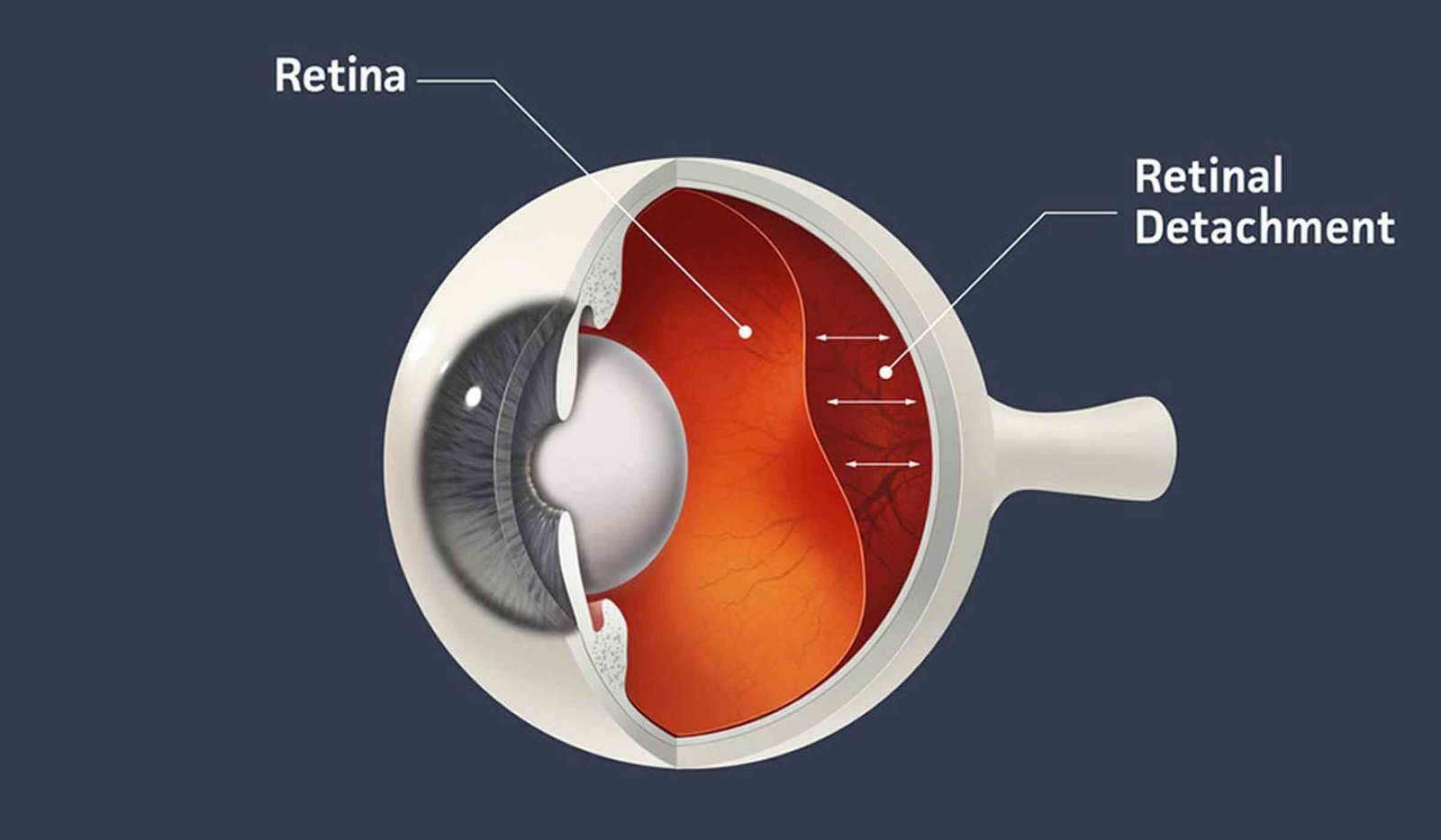
Retinal Detachment:
- Often begins with sudden onset of floaters and flashes of light
- Progresses to a curtain-like shadow over the visual field
- Without treatment, can rapidly lead to severe vision loss in the affected eye
- Symptoms typically develop over hours to days rather than weeks or months
Macular Hole:
- Initially causes blurring and distortion of central vision
- As the hole enlarges, a small blind spot may develop in central vision
- Without treatment, central vision in the affected eye may continue to deteriorate
Understanding the progression patterns of these symptoms is crucial for early diagnosis and intervention, which can often slow or halt the advancement of retinal diseases and preserve vision.
4. Causes
Biological and environmental causes
Retinal diseases arise from various biological and environmental factors:
Biological Causes:
- Age-related changes: Natural aging processes can lead to deterioration of retinal tissues, particularly in the macula, contributing to conditions like age-related macular degeneration
- Vascular abnormalities: Disruptions in blood supply to the retina can lead to ischemia and tissue damage
- Inflammatory processes: Immune responses can sometimes damage retinal tissues
- Metabolic dysfunction: Conditions like diabetes can disrupt normal metabolic processes in the retina
- Structural abnormalities: Physical changes in the eye’s structure can place stress on the retina
Environmental Causes:
- Exposure to ultraviolet (UV) light: Excessive exposure may contribute to retinal damage over time
- Toxins and chemicals: Certain substances can be toxic to retinal cells
- Smoking: Strongly associated with increased risk of age-related macular degeneration and other retinal disorders
- Diet: Nutritional deficiencies (particularly antioxidants) may increase susceptibility to retinal damage
- Oxygen levels: In premature infants, high oxygen levels in incubators can lead to retinopathy of prematurity
- Physical trauma: Injuries to the eye can damage the retina or lead to conditions like retinal detachment
Genetic and hereditary factors
Genetics plays a significant role in many retinal diseases:
- Retinitis pigmentosa and other inherited retinal dystrophies have clear genetic patterns, with mutations identified in multiple genes
- Stargardt disease, the most common form of juvenile macular degeneration, is typically inherited in an autosomal recessive pattern
- Some forms of age-related macular degeneration have genetic components, with several gene variants associated with increased risk
- Leber congenital amaurosis is an inherited retinal disorder caused by mutations in any of several genes
- Congenital retinoschisis is typically X-linked and caused by mutations in the RS1 gene
The genetic basis of inherited retinal diseases is complex, with causative mutations reported in 289 genes . These genetic factors determine not only whether a person develops a retinal disease but also how it progresses and responds to treatment.
Known triggers or exposure risks
Several triggers and exposures can initiate or exacerbate retinal diseases:
- Uncontrolled diabetes: Poor blood sugar control significantly increases the risk and progression of diabetic retinopathy
- Hypertension: Uncontrolled high blood pressure can damage retinal blood vessels
- Physical trauma: Direct injury to the eye or head can trigger retinal detachment, especially in predisposed individuals
- Intense light exposure: Looking directly at the sun or other extremely bright light sources can cause retinal damage
- Certain medications: Some drugs, such as hydroxychloroquine (Plaquenil), can cause retinal toxicity with long-term use
- Eye surgery: Previous eye operations can increase the risk of certain retinal complications
- Severe myopia: Being extremely nearsighted increases the risk of retinal detachment and other retinal problems
- Inflammation: Inflammatory conditions affecting the eye can damage retinal tissues
- Oxygen fluctuations: In premature infants, fluctuations in oxygen levels can trigger retinopathy of prematurity
Understanding these causes and triggers is essential for both preventive strategies and developing effective treatments for retinal diseases.
5. Risk Factors
Who is most at risk (age, gender, occupation, lifestyle, etc.)?
Several demographic, occupational, and lifestyle factors influence the risk of developing retinal diseases:
Age:
- Advanced age is a primary risk factor for many retinal conditions, particularly age-related macular degeneration
- The prevalence of AMD increases dramatically after age 50
- Diabetic retinopathy risk increases with the duration of diabetes
- Some inherited retinal diseases, like retinitis pigmentosa, often manifest symptoms in childhood or adolescence
Gender:
- Some retinal conditions show gender disparities, though these vary by specific disease
- Some studies suggest women may be at slightly higher risk for AMD
- Macular holes develop more often in women than men
- X-linked forms of retinitis pigmentosa primarily affect males
Occupation:
- Occupations with high exposure to sunlight or UV radiation (outdoor workers, welders)
- Jobs involving potential eye trauma (industrial workers, athletes in certain sports)
- Occupations with high levels of screen use may contribute to eye strain, though the direct link to retinal disease is less established
Lifestyle:
- Smoking significantly increases the risk of AMD and may worsen other retinal conditions
- Sedentary lifestyle contributes to conditions like diabetes and hypertension that affect retinal health
- Poor diet lacking in nutrients important for eye health (antioxidants, omega-3 fatty acids, lutein, zeaxanthin)
- Obesity increases risk for diabetic retinopathy and may contribute to AMD risk
Environmental, occupational, and genetic factors
Environmental Factors:
- Excessive sunlight exposure and UV radiation
- Air pollution exposure
- Altitude (high-altitude living may affect oxygen delivery to the retina)
- Exposure to certain chemicals or toxins
Occupational Factors:
- Jobs requiring prolonged close visual work without breaks
- Occupations with high risk of eye injury
- Exposure to industrial chemicals affecting eye health
- Working with lasers or bright lights without proper eye protection
Genetic Factors:
- Family history of retinal diseases
- Specific genetic mutations associated with inherited retinal dystrophies
- Genetic predisposition to related conditions like diabetes
- Genetic variants affecting inflammatory and immune responses
Impact of pre-existing conditions
Several pre-existing conditions significantly increase the risk of developing retinal diseases:
Diabetes:
- Major risk factor for diabetic retinopathy
- Risk increases with duration of diabetes and poor blood sugar control
- People with diabetes are also at higher risk for other retinal conditions
Hypertension:
- Can cause hypertensive retinopathy
- Increases risk of retinal vein occlusion
- May exacerbate other retinal conditions
Cardiovascular Disease:
- Conditions affecting circulation can impact retinal blood flow
- Atherosclerosis may increase risk of retinal vascular events
- Shared risk factors with several retinal diseases
Obesity:
- Associated with increased risk of diabetic retinopathy
- May contribute to AMD risk through inflammatory mechanisms
- Often coexists with other risk factors like hypertension and diabetes
Autoimmune Disorders:
- Conditions like lupus or rheumatoid arthritis may affect retinal health
- Treatment for these conditions sometimes includes medications with potential retinal toxicity
Previous Eye Conditions or Surgeries:
- History of retinal detachment increases risk for future detachments
- Previous eye injuries or surgeries
- Severe myopia (extreme nearsightedness)
Understanding these risk factors is crucial for identifying high-risk individuals who may benefit from more frequent screening and earlier intervention, potentially preventing or minimizing vision loss from retinal diseases.
6. Complications
What complications can arise from retinal diseases?
Retinal diseases can lead to various complications affecting both vision and quality of life:
Vision-Related Complications:
- Permanent vision loss or blindness
- Reduced visual acuity (inability to see fine details)
- Loss of central vision, affecting activities like reading and recognizing faces
- Loss of peripheral vision, affecting mobility and spatial awareness
- Distorted vision (metamorphopsia), where straight lines appear wavy
- Color vision deficiencies or changes
- Reduced contrast sensitivity
- Difficulty adapting to changes in lighting (particularly low light)
- Visual hallucinations (Charles Bonnet syndrome) in cases of significant vision loss
Secondary Eye Complications:
- Neovascular glaucoma (abnormal blood vessel growth leading to increased eye pressure)
- Vitreous hemorrhage (bleeding into the gel-like substance filling the eye)
- Retinal scarring
- Macular edema (swelling in the central retina)
- Retinal fibrosis (formation of fibrous tissue)
- Secondary retinal detachment
Long-term impact on organs and overall health
The impact of retinal diseases extends beyond the eyes to affect overall health and function:
Physical Health Impacts:
- Increased risk of falls and injuries due to visual impairment
- Reduced physical activity and mobility
- Difficulties with daily tasks like cooking, cleaning, and self-care
- Challenges with medication management, potentially affecting treatment of other conditions
- Sleep disturbances due to disruption of circadian rhythms (particularly in conditions affecting photoreceptors involved in light perception)
Mental Health Impacts:
- Depression and anxiety related to vision loss and reduced independence
- Social isolation
- Reduced participation in enjoyable activities
- Cognitive decline (vision loss is associated with faster cognitive deterioration in older adults)
- Psychological stress from managing a chronic condition
Impact on Other Systems:
- In some cases, retinal diseases are part of syndromic conditions that affect multiple body systems
- Retinal changes sometimes reflect systemic vascular health, indicating potential issues in other organs
- Management of conditions like diabetic retinopathy often requires control of systemic disease
Potential disability or fatality rates
Retinal diseases are rarely directly fatal but can lead to significant disability:
Disability Impact:
- Vision loss is a leading cause of disability worldwide
- Age-related macular degeneration is a leading cause of legal blindness in developed countries
- In developing countries, retinal diseases are becoming a more prominent cause of vision loss as other preventable causes are better controlled
- The global burden of retinal diseases measured in disability-adjusted life years (DALYs) is substantial and growing
Quality of Life Impact:
- Reduced independence
- Inability to drive
- Difficulties with reading and writing
- Challenges with facial recognition affecting social interactions
- Employment limitations or inability to work
- Increased need for assistance and supportive services
Mortality Considerations:
- While retinal diseases themselves are not typically life-threatening, there are associations between severe retinal diseases and increased mortality
- This association may be due to shared risk factors or underlying conditions rather than direct causation
- For example, severe diabetic retinopathy is associated with higher mortality rates, likely reflecting the severity of the underlying diabetes
The complications of retinal diseases highlight the importance of early detection, appropriate management, and supportive services to maintain quality of life for affected individuals.
7. Diagnosis & Testing
Common diagnostic procedures
Several procedures are commonly used to diagnose and evaluate retinal diseases:
Clinical Eye Examination:
- Dilated eye examination: The pupil is dilated with eye drops to allow the ophthalmologist to see the back of the eye, including the retina
- Visual acuity testing: Measures how well a person can see at various distances
- Visual field testing: Evaluates peripheral vision
- Amsler grid test: Checks for distortions or blank spots in central vision, particularly useful for detecting macular diseases
- Slit-lamp examination: Uses a special microscope to examine structures at the front of the eye and, with additional lenses, the retina
Specialized Retinal Examinations:
- Indirect ophthalmoscopy: Provides a wide-angle view of the retina
- Gonioscopy: Evaluates the angle where the iris meets the cornea, which can be important in some retinal conditions associated with glaucoma
Medical tests (e.g., blood tests, imaging, biopsies)
Various tests help diagnose retinal diseases and assess their severity:
Imaging Tests:
- Optical Coherence Tomography (OCT): Uses light waves to create detailed cross-sectional images of the retina, showing its layers and helping detect fluid, thinning, or other abnormalities
- Fluorescein Angiography: A dye is injected into the bloodstream and photographed as it flows through the retinal blood vessels, revealing leakage, blockage, or abnormal vessels
- Fundus Autofluorescence: A non-invasive imaging technique that can reveal changes in the retinal pigment epithelium
- Fundus Photography: Documents the appearance of the retina for baseline comparison and monitoring changes over time
- Ultrasonography: Particularly useful when the retina cannot be directly visualized due to opacity in the eye’s media
Laboratory Tests:
- Blood tests for diabetes and other systemic conditions that can affect the retina
- Genetic testing to identify mutations associated with inherited retinal diseases
Functional Tests:
- Electroretinography (ERG): Measures the electrical responses of the retina to light stimulation, helping evaluate the function of photoreceptors
- Microperimetry: Assesses retinal sensitivity at specific points
- Color vision testing: Can help identify certain photoreceptor abnormalities
- Contrast sensitivity testing: Evaluates the ability to distinguish between subtle differences in shading
Other Assessments:
- Intraocular pressure measurement: Important for conditions where retinal damage may be related to pressure within the eye
- Optical biometry: Measures the eye’s dimensions, which can be relevant for some retinal conditions
Early detection methods and their effectiveness
Early detection of retinal diseases is crucial for preserving vision, as treatment is often most effective when initiated before significant damage occurs:
Screening Methods:
- Regular comprehensive eye examinations with pupil dilation
- Diabetic retinopathy screening programs for people with diabetes
- Genetic screening for those with family history of inherited retinal diseases
- At-home monitoring with Amsler grids for macular degeneration
- Digital retinal photography screening in primary care or community settings
Effectiveness of Early Detection:
- For diabetic retinopathy, regular screening can reduce the risk of severe vision loss by more than 90%
- Early detection of wet AMD allows for prompt treatment that can preserve vision in many cases
- For retinal detachment, early recognition of warning signs (flashes, floaters, visual field defects) and prompt treatment can prevent permanent vision loss
- For inherited retinal diseases, early diagnosis through genetic testing can help with family planning, genetic counseling, and potential eligibility for gene therapy trials
Emerging Detection Technologies:
- Artificial intelligence algorithms that analyze retinal images to detect disease
- Home-based OCT devices for monitoring macular diseases
- Smartphone-based retinal imaging systems for wider accessibility
- Teleophthalmology approaches for remote screening and diagnosis
Challenges in Early Detection:
- Many retinal diseases are asymptomatic in early stages
- Access to specialized eye care can be limited in some regions
- Patient awareness of the importance of regular eye examinations
- Cost and insurance coverage for specialized testing
The integration of new technologies with traditional clinical assessment continues to improve the early detection of retinal diseases, offering better opportunities for intervention before irreversible vision loss occurs.
8. Treatment Options
Standard treatment protocols
Treatment approaches for retinal diseases vary depending on the specific condition, but several standard protocols have been established:
For Age-related Macular Degeneration (AMD):
- Dry AMD: Nutritional supplements containing vitamins C and E, zinc, copper, lutein, and zeaxanthin (AREDS/AREDS2 formula)
- Wet AMD: Anti-VEGF injections (bevacizumab, ranibizumab, aflibercept, brolucizumab) to inhibit abnormal blood vessel growth
- Lifestyle modifications including smoking cessation, dietary improvements, and protection from UV light
For Diabetic Retinopathy:
- Tight control of blood glucose, blood pressure, and lipid levels
- Laser photocoagulation for proliferative diabetic retinopathy
- Anti-VEGF injections for diabetic macular edema
- Vitrectomy for advanced cases with vitreous hemorrhage or tractional retinal detachment
For Retinal Detachment:
- Laser surgery or cryotherapy to seal retinal tears
- Pneumatic retinopexy (injection of a gas bubble into the eye)
- Scleral buckle surgery (placing a silicone band around the eye to push the wall of the eye against the detached retina)
- Vitrectomy to remove vitreous gel and repair complex detachments
For Retinal Vein Occlusion:
- Anti-VEGF injections for macular edema
- Focal laser treatment
- Corticosteroid injections in some cases
For Macular Hole:
- Vitrectomy surgery with gas bubble placement to close the hole
- Post-operative face-down positioning to help the hole close
For Epiretinal Membrane:
- Observation for mild cases
- Vitrectomy with membrane peeling for cases causing significant visual symptoms
Medications, surgeries, and therapies
A range of medications, surgical procedures, and therapeutic approaches are used to treat retinal diseases:
Medications:
- Anti-VEGF agents (bevacizumab/Avastin, ranibizumab/Lucentis, aflibercept/Eylea) for conditions involving abnormal blood vessel growth or leakage
- Corticosteroids (triamcinolone, dexamethasone implants) for inflammatory conditions and macular edema
- Nutritional supplements (AREDS2 formula) for dry AMD
- Neuroprotective agents (under investigation for various conditions)
- Immunosuppressive medications for autoimmune retinal disorders
Surgical Procedures:
- Vitrectomy: Removal of the vitreous gel to access the retina for various procedures
- Retinal laser surgery: Creates burns that seal retinal tears or destroys abnormal blood vessels
- Cryopexy: Uses intense cold to create scar tissue that seals retinal tears
- Pneumatic retinopexy: Injection of a gas bubble to push detached retina back into place
- Scleral buckling: Placement of a silicone band around the eye to support detached areas
- Membrane peeling: Removal of abnormal tissue from the retinal surface
- Retinal prosthesis implantation (e.g., ARGUS II) for certain cases of profound vision loss
Therapeutic Approaches:
- Photodynamic therapy: Combines light-sensitive medication with laser treatment
- Laser photocoagulation: Uses focused light to seal leaking blood vessels or retinal tears
- Low vision rehabilitation: Helps patients maximize remaining vision
- Transpupillary thermotherapy: Uses infrared radiation to treat certain tumors
Emerging treatments and clinical trials
The field of retinal disease treatment is rapidly evolving, with numerous promising approaches in development:
Gene Therapy:
- Voretigene neparvovec (Luxturna): First FDA-approved gene therapy for an inherited retinal disease (RPE65 mutation-associated retinal dystrophy)
- Multiple gene therapies for other forms of inherited retinal diseases are in clinical trials, targeting conditions like X-linked retinitis pigmentosa, choroideremia, achromatopsia, and others
- Gene editing approaches using CRISPR-Cas9 technology are being investigated for retinal diseases
Stem Cell Therapy:
- Transplantation of retinal pigment epithelial (RPE) cells derived from human embryonic stem cells or induced pluripotent stem cells (iPSCs)
- Clinical trials using bone marrow-derived stem cells for retinitis pigmentosa have shown some promising results
- Retinal progenitor cell transplantation for various retinal degenerative conditions
Novel Drug Delivery Systems:
- Sustained-release implants to reduce the frequency of intravitreal injections
- Port delivery systems for continuous drug release
- Suprachoroidal delivery approaches for targeted therapy
Neuroprotection Strategies:
- Drugs aimed at preserving photoreceptors regardless of the underlying genetic cause
- Neurotrophic factors to promote retinal cell survival
Retinal Prostheses and Visual Assistive Devices:
- Advanced versions of retinal implants beyond the ARGUS II
- Optogenetic approaches to make non-photoreceptor retinal cells light-sensitive
- Visual prostheses that bypass the retina entirely and stimulate the visual cortex
Combination Therapies:
- Approaches that combine different treatment modalities, such as gene therapy with neuroprotective agents
- Targeting multiple molecular pathways simultaneously
Regenerative Medicine Approaches:
- Creation of retinal organoids (miniature retinas grown in laboratories) for disease modeling and potential transplantation
- 3D bioprinting of retinal tissues
Clinical trials for these emerging treatments continue to advance, offering hope for improved visual outcomes for patients with previously untreatable retinal conditions. These innovative approaches represent a paradigm shift from managing symptoms to addressing the underlying causes of retinal diseases and potentially restoring vision.
9. Prevention & Precautionary Measures
How can retinal diseases be prevented?
While not all retinal diseases can be prevented, several strategies can reduce risk or slow progression:
For Age-related Macular Degeneration:
- Smoking cessation – smoking significantly increases AMD risk
- Maintaining a healthy diet rich in leafy green vegetables, fish, and antioxidants
- Regular exercise and weight management
- Blood pressure control
- UV protection with sunglasses
- AREDS/AREDS2 nutritional supplements for those at intermediate or high risk
For Diabetic Retinopathy:
- Tight glycemic control – maintaining blood glucose levels within target range
- Blood pressure management
- Regular diabetic retinopathy screening examinations
- Prompt treatment of early retinopathy changes
- Cholesterol management
- Healthy lifestyle (diet, exercise, weight management)
For Retinal Detachment:
- Prompt treatment of retinal tears or holes
- Eye protection during high-risk activities (contact sports, industrial work)
- Regular eye examinations for those with high myopia or family history
- Being aware of warning symptoms (flashes, floaters, visual field changes)
For General Retinal Health:
- Regular comprehensive eye examinations
- Protection from excessive UV exposure
- Eye safety in hazardous environments
- Healthy lifestyle choices (nutrition, exercise, avoiding smoking)
Lifestyle changes and environmental precautions
Several lifestyle and environmental modifications can help protect retinal health:
Dietary Recommendations:
- Consume foods rich in antioxidants (berries, dark leafy greens)
- Include omega-3 fatty acids (found in fish like salmon and sardines)
- Adequate intake of nutrients important for eye health (lutein, zeaxanthin, vitamins A, C, E, zinc)
- Limit refined carbohydrates and unhealthy fats
- Maintain adequate hydration
Physical Activity:
- Regular exercise helps maintain healthy blood flow to the retina
- Activity supports overall cardiovascular health, which benefits retinal circulation
- Weight management reduces risk of conditions that affect retinal health
Eye Protection:
- Wear UV-blocking sunglasses outdoors
- Use protective eyewear during sports or hazardous activities
- Take regular breaks during prolonged screen use (20-20-20 rule: every 20 minutes, look at something 20 feet away for 20 seconds)
- Proper lighting for reading and close work
- Protect retinas from extremely bright light sources
Environmental Considerations:
- Maintain good indoor air quality
- Avoid smoke exposure (both direct smoking and secondhand smoke)
- Reduce exposure to air pollution when possible
Health Management:
- Control of chronic conditions like diabetes and hypertension
- Adherence to medication regimens
- Stress management (chronic stress can affect blood pressure and overall eye health)
- Adequate sleep to allow eye recovery and reduce strain
Vaccines (if applicable) or preventive screenings
While there are no vaccines specifically for retinal diseases, preventive screenings are crucial for early detection and intervention:
Recommended Screening Protocols:
For the General Population:
- Comprehensive dilated eye examination by age 40
- Follow-up examinations every 2-4 years for adults with no risk factors
- Annual or biennial examinations for adults over 60
For People with Diabetes:
- Type 1 diabetes: First screening within 5 years of diagnosis, then annually
- Type 2 diabetes: Screening at diagnosis, then annually
- More frequent monitoring if retinopathy is present
For People with Risk Factors:
- Family history of retinal disease: Earlier and more frequent screenings
- High myopia: Regular dilated examinations to check for retinal tears
- History of retinal problems: Follow-up schedule as recommended by ophthalmologist
- Users of medications with potential retinal toxicity: Baseline examination and periodic monitoring
Emerging Screening Approaches:
- Telemedicine-based retinal screening programs
- AI-assisted retinal image analysis for early detection
- Home monitoring technologies (such as apps for Amsler grid testing)
- Community-based screening initiatives for underserved populations
While complete prevention is not possible for many retinal conditions, especially those with strong genetic components, these preventive strategies can significantly reduce risk, delay onset, or slow progression of retinal diseases. Early detection through appropriate screening remains one of the most effective approaches for preserving vision.
10. Global & Regional Statistics
Incidence and prevalence rates globally
Retinal diseases represent a significant global health burden, with varying prevalence across different conditions:
Age-related Macular Degeneration (AMD):
- Global prevalence estimated at 196 million people in 2020, projected to increase to 288 million by 2040
- Early AMD affects approximately 9-25% of people over 65 years
- Late AMD affects approximately 0.5-2% of people over 65 years globally
- Higher prevalence in populations of European ancestry compared to Asian or African populations
Diabetic Retinopathy:
- Affects approximately 30% of all people with diabetes
- With the global diabetes population expected to reach 57 million by 2025, diabetic retinopathy prevalence is also projected to rise significantly
- Prevalence ranges from 10% in India to 43% in Indonesia within the Asia-Pacific region
Retinitis Pigmentosa:
- Average global prevalence of approximately 1 in 4,500 individuals
- Affects approximately 1.5 million people worldwide
- Incidence is relatively consistent across different regions but may be higher in populations with consanguineous marriages
Other Inherited Retinal Diseases:
- Stargardt disease: Approximately 1 in 17,000 people
- Usher syndrome: Approximately 1 in 25,000 people
- Leber congenital amaurosis: Approximately 1 in 42,000 people
- All inherited retinal diseases collectively: Approximately 1 in 3,450 people
Retinal Vein Occlusion:
- Branch retinal vein occlusion (BRVO) affects approximately 2.9% of older adults
- Central retinal vein occlusion (CRVO) affects approximately 1.6%
- Second most common retinal vascular disease globally
Retinal Detachment:
- Annual incidence of approximately 5 in 100,000 people
- Higher incidence (approximately 20 in 100,000) in middle-aged and elderly populations
Mortality and survival rates
While retinal diseases themselves are rarely directly fatal, they contribute to disability and can be associated with conditions that affect mortality:
Mortality Considerations:
- Patients with diabetic retinopathy, particularly proliferative diabetic retinopathy, show higher overall mortality rates compared to diabetic patients without retinopathy
- This likely reflects the severity of the underlying diabetes and vascular disease rather than a direct effect of the retinopathy
- AMD is associated with a modest increase in mortality risk in some studies, possibly related to shared risk factors
Survival Impact:
- The disability associated with vision loss can indirectly affect survival through increased risk of falls, medication errors, and reduced quality of life
- Retinal diseases that are manifestations of systemic conditions (e.g., retinal vasculitis in autoimmune diseases) may reflect disease activity that impacts survival
Quality of Life and Functional Survival:
- Vision loss from retinal diseases significantly impacts quality-adjusted life years
- The global burden measured in disability-adjusted life years (DALYs) shows that retinal diseases contribute substantially to years lived with disability
Country-wise comparison and trends
Retinal disease prevalence shows significant regional variations and trends:
Developed Countries:
- Age-related macular degeneration is the leading cause of blindness
- High access to anti-VEGF treatments has reduced the incidence of legal blindness from wet AMD
- Diabetic retinopathy screening programs have improved early detection and treatment
- Inherited retinal diseases represent a growing focus with advances in genetic diagnosis and experimental therapies
Developing Countries:
- Shift in causes of blindness from infectious diseases and cataract toward retinal diseases
- Rapidly increasing prevalence of diabetic retinopathy due to growing diabetes epidemic
- Limited access to advanced imaging and treatments in many regions
- Retinopathy of prematurity increasing in some areas as neonatal care improves but screening and treatment remain inadequate
Regional Variations:
- European countries show the highest prevalence of AMD
- Asian countries are experiencing rapidly rising rates of myopia-related retinal complications
- African countries still face significant challenges with infectious causes of retinal disease alongside growing non-communicable retinal conditions
- Latin American countries show increasing rates of retinopathy of prematurity
Economic Impact:
- The global retinal disorder treatment market was estimated at USD 12.57 billion in 2022 and is expected to expand at a compound annual growth rate of 9.3% from 2023 to 2030
- Macular degeneration treatments dominated the market with a share of 43.63% in 2022
- Indirect economic costs through productivity loss and caregiving requirements are substantial but less well quantified
These statistics highlight the growing global impact of retinal diseases and the need for improved prevention, detection, and treatment strategies worldwide, particularly as populations age and lifestyle-related risk factors become more prevalent.
11. Recent Research & Future Prospects
Latest advancements in treatment and research
Recent years have seen remarkable advances in the understanding and treatment of retinal diseases:
Gene Therapy Breakthroughs:
- FDA approval of voretigene neparvovec (Luxturna) in 2017 as the first gene therapy for an inherited retinal disease caused by RPE65 mutations
- Expansion of gene therapy approaches to target other genetic forms of retinal dystrophies, with multiple clinical trials showing promising results
- Development of more efficient viral vectors for gene delivery to retinal cells
- Advancements in the precision of gene editing technologies like CRISPR-Cas9 for potential application in retinal diseases
Stem Cell Therapy Progress:
- Clinical trials demonstrating safety and potential efficacy of various stem cell approaches for retinal diseases
- Development of methods to generate retinal organoids from induced pluripotent stem cells (iPSCs) for disease modeling and potential therapeutic use
- Early-stage clinical trial showing CD34+ stem cells are safe and potentially effective for retinitis pigmentosa
- Refinement of techniques for delivering stem cells to different retinal layers
Novel Drug Developments:
- Introduction of new anti-VEGF agents with longer duration of action, reducing treatment burden
- FDA approval of the first drugs to slow geographic atrophy progression in advanced dry AMD
- Development of combination therapies targeting multiple disease pathways simultaneously
- Advancements in sustained-release drug delivery systems
Imaging and Diagnostic Innovations:
- Higher resolution optical coherence tomography (OCT) and OCT angiography
- Artificial intelligence algorithms for automated detection and classification of retinal diseases
- Development of more sensitive functional testing methods to detect early disease
- Portable and smartphone-based retinal imaging systems to increase accessibility
Ongoing studies and future medical possibilities
Numerous exciting research directions promise to transform retinal disease management in the coming years:
Expanding Gene Therapy Applications:
- Development of gene therapy approaches for more common retinal diseases like dry AMD
- Optimization of gene delivery methods, including non-viral vectors
- Exploration of optogenetic approaches to make remaining retinal cells light-sensitive
- Advancements in genetic editing to correct mutations rather than just supplying functioning genes
Regenerative Medicine Frontiers:
- Creation of complete retinal tissue from stem cells for transplantation
- Stimulation of endogenous repair mechanisms in the retina
- Development of 3D bioprinting techniques for retinal tissue engineering
- Combination of cell replacement with supportive scaffolds and growth factors
Next-Generation Retinal Prosthetics:
- Development of higher-resolution electronic retinal implants
- Creation of photovoltaic devices that don’t require external power sources
- Integration of artificial intelligence with visual prosthetics to enhance image processing
- Brain-computer interfaces that bypass damaged retinal tissue entirely
Drug Discovery and Delivery Innovations:
- Identification of neuroprotective compounds to preserve retinal cells regardless of underlying cause
- Development of nanotechnology-based drug delivery systems
- Exploration of systemic treatments that cross the blood-retina barrier
- Novel anti-inflammatory and anti-fibrotic agents for retinal diseases
Precision Medicine Approaches:
- Pharmacogenomic studies to predict individual response to retinal disease treatments
- Development of personalized treatment algorithms based on genetic, imaging, and functional biomarkers
- Patient-specific disease modeling using iPSC-derived retinal organoids
- Targeted therapies based on individual molecular disease mechanisms
Potential cures or innovative therapies under development
While complete cures remain elusive for many retinal conditions, several innovative approaches show promise for dramatically improving outcomes:
Curative Approaches for Genetic Diseases:
- Refinement of gene replacement therapies to achieve more complete and lasting restoration of vision
- CRISPR-based gene editing to correct pathogenic mutations permanently
- Development of universal gene therapy approaches that could benefit multiple genetic forms of retinal disease
- Advancement of cellular reprogramming techniques to regenerate lost retinal cells
Restorative Technologies:
- Integration of advanced materials science with biology to create bio-hybrid retinal implants
- Development of injectable hydrogels that support retinal cell survival and function
- Engineered proteins that can replace or supplement dysfunctional molecular components in retinal cells
- Synthetic biology approaches to create novel visual cycle pathways
Preventive Strategies:
- Development of drugs that can prevent the onset of age-related retinal degeneration
- Genetic counseling and preimplantation genetic diagnosis to prevent transmission of inherited retinal diseases
- Lifestyle interventions based on improved understanding of environmental risk factors
- Early intervention protocols based on predictive biomarkers
Transformative Treatment Paradigms:
- Combination therapies that address multiple aspects of disease pathology simultaneously
- Development of one-time treatments that provide lifelong benefits
- Non-invasive therapies that can be self-administered
- Telemedicine platforms integrated with home monitoring technologies for continuous disease management
These innovative approaches represent a shift from managing vision loss to preserving and potentially restoring vision, offering hope to millions of people affected by retinal diseases worldwide. While challenges remain in translating these advances to widely available clinical applications, the pace of progress suggests a future with dramatically improved outcomes for retinal disease patients.
12. Interesting Facts & Lesser-Known Insights
Uncommon knowledge about retinal
Several fascinating aspects of the retina are not widely known outside specialized medical circles:
The retina is actually part of the central nervous system and is an extension of the brain, making it the only part of the brain that can be directly observed non-invasively .
The human retina consumes oxygen more rapidly than any other tissue in the body, making it exceptionally metabolically active .
Unlike most mammals, some birds have two foveas (areas of high visual acuity) in each eye, allowing them to have sharp vision in different directions simultaneously .
The cephalopod retina (in octopuses, squids) is structurally different from the vertebrate retina – it is “non-inverted,” with photoreceptors facing toward incoming light rather than away, suggesting independent evolutionary development .
The bandwidth of the human retina has been calculated to be approximately 8.75 megabits per second, while a guinea pig’s retinal transfer rate is 875 kilobits per second .
The retina contains intrinsically photosensitive retinal ganglion cells that are not primarily involved in vision but regulate circadian rhythms and pupillary light reflexes.
Myths and misconceptions vs. medical facts
Several myths and misconceptions about retinal health persist, but medical facts provide important clarification:
Myth: Reading in dim light damages the retina. Fact: While reading in poor light may cause eye strain and discomfort, it does not cause permanent damage to the retina.
Myth: Sitting too close to the TV or computer screens damages the retina. Fact: While prolonged screen use can cause digital eye strain, there is no evidence that it causes permanent retinal damage in healthy eyes.
Myth: Retinal diseases always lead to blindness. Fact: Many retinal conditions, if detected early and properly managed, may never progress to severe vision loss or blindness.
Myth: Nothing can be done for macular degeneration. Fact: While there is no cure for AMD, treatments can slow or halt progression of wet AMD, and nutritional supplements can reduce the risk of progression in certain forms of dry AMD.
Myth: All retinal diseases are inherited. Fact: While some retinal diseases have a strong genetic component, many others are influenced by age, environment, and other health conditions.
Myth: Eye exercises can prevent or treat retinal diseases. Fact: While eye exercises may help with certain vision problems like convergence insufficiency, they do not prevent or treat retinal diseases.
Myth: Vision lost to retinal disease can never be restored. Fact: While historically this was largely true, advances in gene therapy, stem cell research, and retinal prosthetics are beginning to challenge this assumption for certain conditions.
Impact on specific populations or professions
Retinal diseases can have unique impacts on certain populations and professions:
Professional Drivers:
- Even mild retinal diseases affecting visual fields or night vision can end careers
- Special licensing concerns with certain retinal conditions
- Increased screening recommendations for commercial drivers
Visual Artists:
- AMD and other macular diseases may dramatically affect perception of details and colors, fundamentally changing artistic expression
- Some artists with retinal diseases develop distinctive styles that reflect their changing vision
- Adaptation through modified techniques or tools can allow continued artistic practice
Night Shift Workers:
- Disruption of circadian rhythms may potentially impact retinal health
- Increased challenge for those with retinal conditions affecting night vision
- May benefit from specialized lighting accommodations
Children with Inherited Retinal Diseases:
- Educational accommodations needed as vision changes
- Psychological support particularly important for adapting to progressive vision loss
- Early intervention with low vision services can improve outcomes
- Specialized career counseling may be beneficial
Elderly Population:
- Age-related retinal diseases contribute significantly to loss of independence
- Increased risk of falls and related injuries
- Social isolation due to mobility and recognition challenges
- Compounding effect when combined with other age-related conditions
Individuals in Remote or Underserved Areas:
- Limited access to specialized retina care
- Delays in diagnosis and treatment can lead to worse outcomes
- Emerging telemedicine and portable diagnostic technologies may help bridge this gap
- Community-based screening programs becoming increasingly important
The impact of retinal diseases extends far beyond vision loss alone, affecting psychological well-being, career opportunities, independence, and quality of life. Understanding these broader impacts is essential for providing comprehensive care and support to affected individuals.
This report provides a comprehensive overview of retinal structure, function, diseases, and treatments. While significant advances have been made in understanding and treating retinal conditions, ongoing research continues to expand our knowledge and therapeutic options. Early detection, appropriate treatment, and supportive care remain essential components of managing retinal diseases and preserving vision.