Cellular structure and function
- Cells as the basic structural feature of life on Earth, including the distinction between prokaryotic and eukaryotic cells
- Surface area to volume ratio as an important factor in the limitations of cell size
- The need for internal compartments (organelles) with specific cellular functions
- The structure and specialisation of plant and animal cell organelles for distinct functions, including chloroplasts and mitochondria
- The structure and function of the plasma membrane in the passage of water, hydrophilic and hydrophobic substances via osmosis, facilitated diffusion and active transport
Cell Theory
Key Knowledge:
|
All living things are capable of performing seven key functions (things that cannot perform all seven functions are considered non-living):
- Metabolism, Reproduction, Sensitivity, Homeostasis, Excretion, Nutrition, Growth / Movement (simple mnemonic = MR SHENG)
In essence, living things undergo complex chemical reactions (metabolism) in order to maintain a stable living condition (homeostasis)
- These chemical reactions require inputs (gained via nutrition) and produce wasteful outputs (removed via excretion)
- To maintain stable conditions, living things must be able to detect changes (sensitivity) and respond accordingly (growth / movement)
- Continued survival also requires living things to be able to repair and replicate themselves (reproduction)
Cell Theory
The fundamental unit that is capable of performing all of the functions of life is the cell – and according to the cell theory:
- The cell is the smallest unit of life (nothing smaller than a cell is considered to be living – e.g. viruses are not alive)
- All living things are composed of cells (or cellular products) – i.e. living organisms may be unicellular or multicellular
- Cells arise from pre-existing cells – life cannot spontaneously generate (as shown by Pasteur’s biogenesis experiment)
Types of cells
Any living thing that performs all the functions of life and can operate as an independent entity is called an organism
- Organisms may be composed of one of two cell types (simpler prokaryotic cells or more complex eukaryotic cells)

All organisms can be classified into different groups (taxonomic ranks) according to certain shared characteristics
- At the highest level of classification are domains, followed by kingdoms (then phyla, class, order, family, genus and species)
Prokaryotes can be classified into two distinct domains:
- Bacteria – A diverse domain that includes all traditional bacterial species (including all pathogenic forms)
- Archaea – Includes most extremophiles (prokaryotes that are found in adverse environments – like high temperatures)
Eukaryotes belong to the domain Eukarya and may be classified into four distinct kingdoms:
- Protists – Include various unicellular and multicellular organisms that lack specialised tissue
- Fungi – Have cell walls made of chitin and obtain nutrition via heterotrophic absorption (i.e. they are decomposers)
- Plants – Have cell walls made of cellulose and synthesise organic nutrients via photosynthesis (i.e. they are producers)
- Animals – Lack a cell wall and obtain nutrition via heterotrophic ingestion (i.e. they are consumers)

Prokaryotic Cells
Prokaryotic cells are the most basic types of cells and contain four key cellular components:
- They are enclosed by a plasma membrane, which separates the internal contents from the external environment
- They contain an internal fluid (called the cytosol) in which various metabolic reactions and biological processes can occur
- The genetic material is composed of a single circular chromosome (called the genophore), located in a region called the nucleoid
- There are ribosomes (70S in size) that function to translate the genetic instructions into cellular activity (by making proteins)
Additionally, prokaryotic cells may contain certain additional cellular components:
- They may also contain autonomous circular DNA molecules called plasmids, which can be transferred via bacterial conjugation
- Hair-like extensions of the plasma membrane (called pili) may mediate surface attachments or facilitate plasmid exchange
- Longer projections called flagella contain microtubules and motor proteins that enable prokaryotic movement (via a whip-like motion)
- All bacterial cells contain a rigid outer cell wall made of peptidoglycan to help maintain the overall shape and structure of the cell
- Some bacteria may possess an additional outer covering called the slime capsule (glycocalyx) to help prevent desiccation

Eukaryotic Cells
Eukaryotic cells have a more complex structure as they contain membrane-bound compartments that perform specific roles:
- Eukaryotic cells have a double-membrane nucleus that stores the genetic material as chromatin (uncondensed linear DNA)
- Within the nucleus is a region called the nucleolus, which is the site of ribosome assembly (ribosomes will be 80S in size)
- The mitochondrion is the site of aerobic cellular respiration and is responsible for the production of ATP (energy source)
- Lysosomes function to break down cellular components, whereas peroxisomes function to break down toxic metabolites
- Centrosomes produce microtubule spindle fibres and are involved in the process of cell division (e.g. mitosis or meiosis)
- The Golgi complex is a series of membrane stacks and vesicles that act to sort, store, modify and export cellular materials
- An intracellular membranous network called the endoplasmic reticulum (ER) transports materials between organelles
- The rough ER is embedded with ribosomes and transports proteins within the cell, while smooth ER transports lipids
Plant cells possess a number of additional cellular components that are no present within animal cells:
- They contain a rigid cell wall made of cellulose to provide mechanical support to the cell and prevent excess water uptake
- They have a large, central vacuole that helps to maintain hydrostatic pressure within the cell and regulate internal pH
- The leaf tissue will contain chloroplasts which are responsible for the process of photosynthesis (not present in root cells)
Fungal cells have a cell wall made of chitin, while protista vary greatly in organisation and do not have distinctive features

Endosymbiosis
The origin of eukaryotic cells can be explained by endosymbiotic theory (they evolved from symbiotic prokaryote interactions)
- Eukaryotic cells are believed to have evolved from the engulfment of a prokaryote by another prokaryote (via phagocytosis)
- The engulfed prokaryotic cell remained undigested as it contributed new functionality to the engulfing cell (e.g. photosynthesis)
- Over generations, the engulfed cell lost some of its independent utility and became a supplemental support structure (organelle)

Mitochondria and chloroplasts are both eukaryotic organelles suggested to have arisen via endosymbiosis
- Mitochondria were prokaryotes that could undertake aerobic respiration, while chloroplast were photosynthesising cyanobacteria
- Evidence that supports the extracellular origins of these organelles can be seen by the presence of certain prokaryotic features

Organelles
Key Knowledge:
|
Organelles are specialised sub-structures within a cell that serve a specific function (i.e. organelle = ‘little organ’)
Prokaryotic cells do not typically possess any membrane-bound organelles, whereas eukaryotic cells possess several
Universal Organelles (prokaryotes and eukaryotes)
 | Ribosomes |
|---|---|
| Structure: Two subunits made of RNA and protein; larger in eukaryotes (80S) than prokaryotes (70S) Function: Provides internal structure and mediates intracellular transport (less developed in prokaryotes) | |
 | Cytoskeleton |
| Structure: A filamentous scaffolding within the cytoplasm (fluid portion of the cytoplasm is the cytosol) Function: Provides internal structure and mediates intracellular transport (less developed in prokaryotes) | |
 | Plasma Membrane |
| Structure: Phospholipid bilayer embedded with proteins (not an organelle per se , but a vital structure) Function: Semi-permeable and selective barrier surrounding the cell |
Eukaryotic Organelles (animal and plant cells)
 | Nucleus |
|---|---|
| Structure: Double membrane structure with pores; contains an inner region called a nucleolus Function: Stores genetic material (DNA) as chromatin; nucleolus is site of ribosome assembly | |
 | Endoplasmic Reticulum |
| Structure: A membrane network that may be bare (smooth ER) or studded with ribosomes (rough ER) Function: Transports materials between organelles (smooth ER = lipids ; rough ER = proteins) | |
 | Golgi Apparatus |
| Structure: An assembly of vesicles and folded membranes located near the cell membrane Function: Involved in the sorting, storing, modification and export of secretory products | |
 | Mitochondrion |
| Structure: Double membrane structure, inner membrane highly folded into internal cristae Function: Site of aerobic respiration (ATP production) | |
 | Peroxisome |
| Structure: Membranous sac containing a variety of catabolic enzymes Function: Catalyses breakdown of toxic substances (e.g. H2O2) and other metabolites | |
 | Centrosome |
| Structure: Microtubule organising centre (contains paired centrioles in animal cells but not plant cells) Function: Radiating microtubules form spindle fibres and contribute to cell division (mitosis / meiosis) |
Plant Organelles (not found in animal cells)
 | Chloroplast |
|---|---|
| Structure: Double membrane structure with internal stacks of membranous discs (thylakoids) Function: Site of photosynthesis – manufactured organic molecules are stored in various plastids | |
 | Cell Wall |
| Structure: External outer covering made of cellulose (not an organelle per se , but a vital structure) Function: Provides support and mechanical strength; prevents excess water uptake | |
 | Vacuole (large and central) |
| Structure: Fluid-filled internal cavity surrounded by a membrane (tonoplast) Function: Maintains hydrostatic pressure (animal cells may have small, temporary vacuoles) |
Animal Organelles (not found in plant cells – although this is debatable)
 | Lysosome |
|---|---|
| Structure: Membranous sacs filled with hydrolytic enzymes Function: Breakdown / hydrolysis of macromolecules (plant cells may have a comparable structure) |
Membranes
Key Knowledge:
|
The plasma membrane separates the internal components of a cell from the external environment via two key properties:
- It is semi-permeable – some material cannot cross the membrane without assistance
- It is selective – the cell can regulate the passage of these materials according to need
Membrane Structure
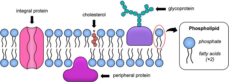
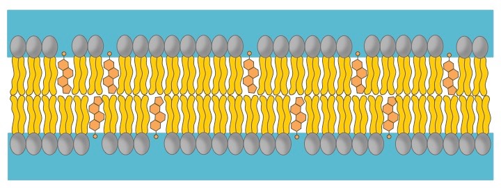
Plasma membranes consist of three principal components – a phospholipid bilayer, proteins and cholesterol (in animal cells only)
The organisation of these components is represented by the fluid-mosaic model, which reflects the fact that membranes are:
- Fluid – the phospholipid bilayer is viscous (due to weak hydrophobic associations), meaning membrane components can move position
- Mosaic – the phospholipid bilayer is embedded with proteins (and potentially cholesterol), resulting in a mosaic of components

1. Phospholipid Bilayer

Structure of Phospholipids
|
Arrangement in Membranes
|
Properties of the Phospholipid Bilayer
|
2. Membrane Proteins
Phospholipid bilayers are embedded with proteins, which may be either permanently or temporarily attached to the membrane
- Integral proteins are permanently attached to the membrane and are typically transmembrane (they span across the bilayer)
- Peripheral proteins are temporarily attached by non-covalent interactions and associate with one surface of the membrane
Membrane proteins can serve a variety of key functions, including:
- Transport: Membrane proteins can facilitate the passive or active movement of molecules that cannot freely cross the bilayer
- Metabolic Activity: Enzymes can be bound to the membrane to localise activity and receptors can detect signalling molecules
- Connections: Membrane proteins can join cells together or act as an attachment point for extracellular or intracellular components

3. Cholesterol
Cholesterol is a component of animal cell membranes, where it functions to maintain integrity and mechanical stability
- It is absent in plant cells, as these plasma membranes are surrounded and supported by a rigid cell wall made of cellulose
- Cholesterol is an amphipathic molecule (like phospholipids), meaning it has both hydrophilic and hydrophobic regions
Cholesterol interacts with the fatty acid tails of phospholipids to moderate the properties of the membrane:
- Cholesterol functions to immobilise the outer surface of the membrane, reducing fluidity
- It makes the membrane less permeable to very small water-soluble molecules that would otherwise freely cross
- It functions to separate phospholipid tails and so prevent crystallisation of the membrane
- It helps secure peripheral proteins by forming high density lipid rafts capable of anchoring the protein

Membrane Transport
Movement of materials across a membrane will depend on both the size and solubility of the material in question
- Small, lipophilic molecules can freely pass across the phospholipid bilayer (e.g. oxygen, carbon dioxide, water, steroids)
- Larger molecules (e.g. glucose) or polar / charged molecules (e.g. ions) will require membrane proteins in order to cross
Passive transport involves the movement of material along a concentration gradient (high concentration ⇒ low concentration)
- Because materials are moving down a concentration gradient, it does not require the expenditure of energy (ATP hydrolysis)
- There are three main types of passive transport mechanisms: simple diffusion, facilitated diffusion or osmosis
Active transport involves the movement of materials against a concentration gradient (low concentration ⇒ high concentration)
- Because materials are moving against the gradient, it requires the expenditure of energy (e.g. ATP hydrolysis)
Bulk transport (cytosis) involves materials entering or leaving the cell via vesicles (the membrane breaks and reforms)
- In this form of transport, materials do not cross the membrane directly and can move along or against the gradient
- This is an active process (requires ATP) but it is not active transport (as materials are circumventing the membrane)
1. Simple Diffusion
Simple diffusion involves the net movement of molecules from a region of high concentration to a region of low concentration
- This directional movement along a gradient is passive and will continue until molecules become evenly dispersed (equilibrium)
- Small and non-polar (lipophilic) molecules will be able to freely diffuse across cell membranes (e.g. O2, CO2, glycerol)
The rate of diffusion can be influenced by temperature (affects kinetic energy), molecular size and the size of the concentration gradient
2. Facilitated Diffusion
Facilitated diffusion is the passive movement of molecules across the cell membrane via the aid of a membrane protein
- It is utilised by molecules that are unable to freely cross the phospholipid bilayer (e.g. large, polar molecules and ions)
This process is mediated by two distinct types of transport proteins – channel proteins and carrier proteins
- Channel proteins have a hydrophilic pore and may be gated to regulate the passage of ions in response to certain stimuli
- Carrier proteins undergo a conformational change to translocate solutes and have a comparably slower rate of transport

3. Osmosis
Osmosis is the net movement of water molecules across a semi-permeable membrane from a region of low solute concentration to a region of high solute concentration (until equilibrium is reached)
- Water is considered the universal solvent – it will associate with, and dissolve, polar or charged molecules (solutes)
- Because solutes cannot cross a cell membrane unaided, water will move to equalise the two solutions
- At a higher solute concentration there are less free water molecules in solution as water is associated with the solute
- Osmosis is essentially the diffusion of free water molecules and hence occurs from regions of low solute concentration

Solutions may be loosely categorised as hypertonic, hypotonic or isotonic according to their osmotic effect
- Solutions with a relatively higher solute concentration are categorised as hypertonic (and will gain water)
- Solutions with a relatively lower solute concentration are categorised as hypotonic (and will lose water)
- Solutions that have the same solute concentration are categorised as isotonic (there will be no net water flow)
4. Active Transport
Active transport is the active movement of molecules across the cell membrane via the aid of a protein pump
- Active transport is essentially the reverse of facilitated diffusion (it moves materials against a concentration gradient)
- The pump binds a solute and then undergoes a conformational change to translocate solutes across the membrane
- The conformational change is mediated by the hydrolysis of ATP (to ADP + Pi) and is hence energy dependent

5. Bulk (Vesicular) Transport
The fluidity of membranes allows materials to be taken in or released by cells without crossing the phospholipid bilayer
- The membrane is principally held together by weak hydrophobic associations between the fatty acid tails of the phospholipids
- These weak interactions can be spontaneously broken and reformed in a process that is energy dependent (via ATP hydrolysis)
- Membrane segments can be excised to form internal vesicles, while new segments are added when membrane and vesicle fuse
Endocytosis
Endocytosis involves large substances (or bulk amounts of smaller substances) entering the cell without crossing the membrane
- The membrane forms a flask-like depression (i.e. invagination) which envelopes the extracellular material to be internalised
- The invagination is then sealed off to form an intracellular vesicle containing the material

There are two main types of endocytosis:
- Phagocytosis – The process by which solid substances are ingested (usually to be transported to the lysosome)
- Pinocytosis – The process by which liquids / dissolved substances are ingested (allows faster entry than via protein channels)
Exocytosis
Exocytosis involves substances exiting the cell without crossing the membrane (these substances must be packaged in vesicles)
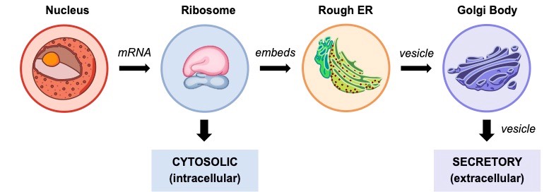
- Polypeptides synthesised within the rough ER will be packaged into a vesicle and transported to the Golgi apparatus
- The Golgi complex will potentially modify the protein (e.g. glycosylation) and then export it out of the cell via a vesicle (exocytosis)
- Proteins can either be secreted by the Golgi immediately (constitutive secretion) or stored for a delayed release (regulatory secretion)
- Proteins synthesised via this pathway can also be shuttled to other organelles besides the Golgi (e.g. mitochondria, lysosome, etc.)

SA:Vol Ratio
Key Knowledge:
|
Cells need to produce chemical energy (via metabolism) to survive and this requires the exchange of materials with the environment
- The rate of metabolism of a cell is a function of its mass / volume (larger cells need more energy to sustain essential functions)
- The rate of material exchange is a function of its surface area (large membrane surface equates to more material movement)
Surface Area : Volume Ratio
The relationship between metabolic energy requirements and capacity for material exchange is represented by the SA:Vol ratio
- Cells need a high SA:Vol ratio, as it means there is sufficient capacity to exchange the materials needed for vital metabolic processes
- As a cell grows, the volume (units3) will increase faster than the surface area (units2), leading to a lower SA:Vol ratio
- Hence growing cells tend to divide and remain small in order to maintain a sufficiently high SA:Vol ratio necessary for survival
Cells and tissues that are specialised for gas or material exchanges will increase their surface area to optimise material transfer
- Intestinal tissue of the digestive tract may form a ruffled structure (villi) to increase the surface area of the inner lining
- Alveoli within the lungs have membranous extensions called microvilli, which function to increase the total membrane surface
Organelles will also increase their surface area to volume ratio in order to improve the efficacy of their specialised functions
- The inner mitochondrial membrane is highly folded into cristae in order to maximise ATP production via aerobic respiration
- Chloroplasts contain membrane discs (thylakoids) arranged into stacks (grana) to increase the light dependent reactions

Cell Size
Cells and their components are measured according to the metric system (all measurements relative to the standard of 1 metre)
- Most cells and organelles are typically measured in micrometres (μm), which represent one millionth (10–6) of a metre
- Smaller components such as cell membranes, viruses and DNA molecules may be measured in nanometres (10–9)

Microscopes
Microscopes are scientific instruments that are used to visualise objects that are too small to see with the naked eye
- There are two main types of microscope: optical (light) microscopes and electron microscopes
Light Microscopes
- Use lenses to bend light and magnify images by a factor of roughly 100-fold
- Can be used to view living specimens in natural colour
- Chemical dyes and fluorescent labelling may be applied to resolve specific structures
Electron Microscopes
- Use electromagnets to focus electrons resulting in significantly greater magnifications and resolutions
- Can be used to view dead specimens in monochrome (although false colour rendering may be applied)
- Transmission electron microscopes (TEM) pass electrons through specimen to generate a cross-section
- Scanning electron microscopes (SEM) scatter electrons over a surface to differentiate depth and map in 3D

Magnification
To calculate the linear magnification of a drawing or image, the following equation should be used:
- Magnification = Image size (with ruler) ÷ Actual size (according to scale bar)
To calculate the actual size of a magnified specimen, the equation is simply rearranged:
- Actual Size = Image size (with ruler) ÷ Magnification

