
⚠️ Disclaimer: The information provided in this article is for educational purposes only and does not constitute medical advice. RevisionTown does not provide diagnosis, treatment, or medical recommendations. Always consult a qualified healthcare professional regarding any medical condition, symptoms, or concerns.
Read More – 🏥 Medical Disclaimer
What are white spots on skin?
White spots on skin (hypopigmentation) refer to areas of skin that appear lighter than the surrounding normal skin tone due to a reduction or absence of melanin, the pigment responsible for giving skin its color. These spots can vary greatly in size, shape, pattern, and distribution depending on their underlying cause.
White spots can manifest as small, confetti-like macules, larger patches, or even extensive areas of depigmentation. They may appear suddenly or develop gradually, remain stable or change over time, and can be temporary or permanent depending on the specific condition causing them.
Affected body parts/organs
White spots can appear on virtually any part of the body, though certain conditions have predilections for specific areas:
- Face and neck: Common locations for vitiligo, pityriasis alba, and post-inflammatory hypopigmentation
- Trunk (chest and back): Frequently affected by tinea versicolor and idiopathic guttate hypomelanosis
- Upper arms and shoulders: Common sites for idiopathic guttate hypomelanosis and tinea versicolor
- Hands and feet: Often affected by vitiligo, particularly around fingers and wrists
- Legs: Common location for idiopathic guttate hypomelanosis, especially on sun-exposed areas
- Scalp and hair: Some conditions like vitiligo can affect hair-bearing areas, causing white patches of hair (poliosis)
- Mucous membranes: Vitiligo can sometimes affect mucous membranes of the mouth and genitals
Beyond the skin itself, some conditions causing white spots may be associated with abnormalities in melanocytes (pigment-producing cells), immune system dysfunction, or other systemic effects depending on the specific disorder.
Prevalence and significance
White spots on skin represent a diverse group of conditions with varying prevalence rates:
Vitiligo: Affects approximately 0.5-2% of the global population, with an estimated worldwide prevalence of about 1%. A recent systematic review reported highest prevalence in Central Europe and South Asia (0.52% of the general population in both regions). In the United States, prevalence is estimated between 0.5-1%.
Tinea versicolor: Affects about 1% of people in temperate climates but up to 40% in tropical regions with hot, humid environments.
Pityriasis alba: Affects approximately 5% of children in the United States, predominantly occurring in children and adolescents between ages 3-16.
Idiopathic guttate hypomelanosis: Extremely common in adults over 40, affecting nearly 50-80% of individuals over 40 years old, and up to 90% of those aged 81-90. It’s particularly common in fair-skinned individuals with significant sun exposure history.
Post-inflammatory hypopigmentation: Prevalence varies widely as it can occur after many inflammatory skin conditions.
The significance of white spots extends beyond their physical appearance:
Psychological impact: White spots, especially on visible areas like the face and hands, can significantly affect quality of life, self-esteem, and mental health. Conditions like vitiligo are associated with higher rates of depression, anxiety, and social isolation.
Diagnostic significance: Some white spots may be indicators of underlying autoimmune disorders, infections, or other systemic conditions requiring investigation and management.
Social implications: In some cultures and societies, visible skin discoloration can lead to stigmatization, discrimination, and social ostracism.
Economic burden: Treatment of persistent white spots can be costly and long-term, creating financial strain for affected individuals.
The significance varies depending on the specific condition, its extent, visibility, and the individual’s skin tone (white spots are often more noticeable on darker skin).
2. History & Discoveries
First identification of white spots on skin
The recognition of white spots on skin dates back to ancient times, with different conditions being described throughout history:
Vitiligo: One of the earliest recorded descriptions of vitiligo appears in the Ebers Papyrus from 1500 BCE in ancient Egypt, where it was described as a condition characterized by white spots spreading across the skin. The term “vitiligo” itself is believed to have been coined by the Roman physician Celsus in the 1st century CE, derived from the Latin word “vitium” meaning “blemish” or “defect.”
Tinea versicolor: This fungal condition was first properly described by the Swedish botanist and physician Erik Acharius in 1809, though skin discoloration caused by fungal infections had been noted by various cultures for centuries.
Pityriasis alba: The first detailed medical descriptions emerged in the early 20th century, with the term “pityriasis alba” (meaning “white scaly condition”) becoming established in dermatological literature in the 1950s.
Idiopathic guttate hypomelanosis: This condition was formally described in the medical literature in the 1960s, although it had likely been observed clinically for much longer.
Key discoverers and researchers
Several key figures have contributed significantly to our understanding of conditions causing white spots on skin:
Robert Willan (1757-1812): The English physician is considered by many to be the founder of modern dermatology. He developed the first systematic classification of skin diseases, including several that cause white spots.
Thomas Bateman (1778-1821): Building on Willan’s work, Bateman further refined the classification of skin diseases and provided detailed descriptions of hypopigmentation disorders.
Walter F. Lever (1909-1992): Made significant contributions to the histopathological understanding of various skin disorders, including those causing hypopigmentation.
Aaron B. Lerner (1920-2007): Pioneered research on melanin and melanocytes, contributing greatly to the understanding of pigmentation disorders. He isolated and characterized melanocyte-stimulating hormone (MSH).
Jean-Claude Bystryn (1932-2010): Made significant contributions to understanding the autoimmune basis of vitiligo.
John E. Harris: Contemporary researcher who has advanced the immunological understanding of vitiligo and developed new treatment approaches.
Major discoveries and breakthroughs
The understanding of white spots on skin has evolved significantly over time, with several key milestones:
1950s-1960s: Recognition of vitiligo as an autoimmune condition, with researchers identifying autoantibodies against melanocytes.
1960s-1970s: Identification of Malassezia (Pityrosporum) yeasts as the cause of tinea versicolor, which was previously thought to be a form of tinea (ringworm).
1970s-1980s: Development of phototherapy, particularly narrow-band UVB and PUVA (psoralen plus UVA) as treatments for vitiligo and other hypopigmentation disorders.
1980s-1990s: Improved understanding of the association between vitiligo and other autoimmune conditions, leading to more comprehensive evaluation and management approaches.
1990s-2000s: Identification of genetic factors involved in various hypopigmentation disorders, including the discovery of multiple genes associated with vitiligo susceptibility.
2000s-2010s: Development of targeted immunomodulatory therapies, including topical calcineurin inhibitors for treating various forms of hypopigmentation.
2010s-Present: Breakthrough understanding of the JAK-STAT pathway in vitiligo, leading to the development of JAK inhibitors as promising treatments for vitiligo and potentially other hypopigmentation disorders.
Evolution of medical understanding
The medical understanding of white spots on skin has evolved from purely descriptive to mechanistic:
Descriptive Era (Pre-1900s): White spots were primarily categorized based on their clinical appearance and pattern of distribution. Little was known about their causes.
Microscopic Era (Early 1900s): The development of skin biopsies and histopathological examination allowed physicians to differentiate between conditions based on cellular changes.
Biochemical Era (1940s-1970s): Understanding of melanin biochemistry and the role of melanocytes in skin pigmentation led to more precise categorization of hypopigmentation disorders.
Immunological Era (1970s-1990s): Recognition of the role of the immune system in conditions like vitiligo transformed understanding from simply a pigmentary disorder to an autoimmune condition.
Genetic Era (1990s-2010s): Identification of genetic factors and inheritance patterns in various hypopigmentation disorders added another dimension to understanding and classification.
Molecular Era (2010s-Present): Detailed understanding of signaling pathways and molecular mechanisms has led to targeted therapies and a more nuanced approach to treatment.
This evolution has shifted the approach from symptom management to addressing underlying pathological mechanisms, leading to more effective treatments and potentially curative approaches for certain conditions in the future.
3. Symptoms
The manifestation of white spots on skin varies considerably depending on the underlying condition. Understanding these variations is crucial for accurate diagnosis and appropriate management.
Early symptoms vs. advanced-stage symptoms
Early Symptoms:
Vitiligo:
- Initially appears as small, well-defined pale or white macules
- Often begins on sun-exposed areas or around body openings (eyes, nose, mouth)
- May first appear after physical trauma (Koebner phenomenon)
Tinea Versicolor:
- Begins as small, faint, scaly spots that may be lighter or darker than surrounding skin
- Minimal itching or discomfort
- Usually appears on upper back, chest, and shoulders
Pityriasis Alba:
- Initially presents as slightly red, scaly patches, particularly on the face
- Gradual fading to pale or white patches
- Mild scaling and occasional itching
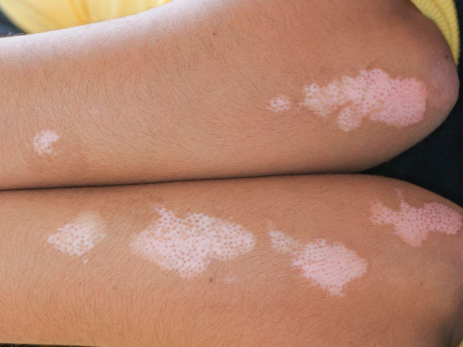
Idiopathic Guttate Hypomelanosis:
- Small (2-5 mm), discrete round white spots
- Appears on sun-exposed areas, especially lower legs and forearms
- Asymptomatic with no scaling or inflammation
Advanced-Stage Symptoms:
Vitiligo:
- Larger patches that may coalesce to cover extensive areas
- Potential progression to generalized or universal vitiligo (affecting most of the body)
- Possible involvement of mucous membranes and hair (poliosis)
- Development of trichrome (three-color) appearance with white center, lighter middle zone, and normal skin
Tinea Versicolor:
- Confluent patches covering large areas of trunk
- More noticeable color contrast, especially after sun exposure
- Possible spread to upper arms, neck, and rarely face
Pityriasis Alba:
- Multiple hypopigmented patches with more defined borders
- Reduction in scaling but persistence of hypopigmentation
- More noticeable contrast during summer months
Idiopathic Guttate Hypomelanosis:
- Increased number of spots with age
- Larger spots (up to 10 mm)
- Development of a porcelain-white appearance
Common vs. rare symptoms
Common Symptoms across most conditions:
- White or lighter patches compared to surrounding skin
- Asymptomatic presentation (no pain or significant discomfort)
- Increased visibility after sun exposure (as surrounding skin tans)
- Smooth texture (except in tinea versicolor, which is often scaly)
Rare or Condition-Specific Symptoms:
Vitiligo:
- Premature graying of hair in affected areas
- Inflammation or redness at the border of vitiligo patches
- Hyperpigmented border (more common in darker skin types)
- Trichrome (three-color) appearance
- Koebner phenomenon (development of new lesions at sites of skin trauma)
Tinea Versicolor:
- Mild itching or irritation, especially when sweating
- Yellow, tan, or pink discoloration (rather than white)
- “Spaghetti and meatballs” appearance under microscopic examination
Pityriasis Alba:
- Initial redness and inflammation before hypopigmentation develops
- Seasonal fluctuation (more noticeable in summer, less in winter)
- Association with atopic features (dry skin, asthma, hay fever)
Idiopathic Guttate Hypomelanosis:
- Slight depression or atrophy of affected spots
- Exclusivity to sun-exposed areas
- Resistance to repigmentation treatments
How symptoms progress over time
The natural history and progression of white spots varies by condition:
Vitiligo:
- Pattern: May follow segmental (limited to one body segment) or non-segmental (bilateral, symmetric) patterns
- Progression: Can be rapid, with new spots appearing over weeks to months, or slow, evolving over years
- Phases: Often has active phases (new spots developing) alternating with stable phases
- Long-term: May eventually stabilize or continue to progress throughout life
- Repigmentation: Spontaneous repigmentation occurs in 10-20% of cases, often appearing as small islands of pigment within white patches
Tinea Versicolor:
- Seasonal pattern: Often worsens in summer months (heat and humidity)
- Recurrence: High rate of recurrence, even after successful treatment
- Progression: Typically expands slowly but can rapidly cover large areas in humid conditions
- Post-inflammatory: Even after treatment, color normalization may take months
- Long-term: Chronic condition with tendency to recur without maintenance therapy
Pityriasis Alba:
- Duration: Individual lesions typically last 6 months to 2 years
- Resolution: Usually resolves spontaneously, especially after puberty
- Seasonal variation: More noticeable in summer (with tanning) and drier in winter
- Recurrence: New patches may develop as others resolve
- Long-term: Complete resolution by adulthood in most cases
Idiopathic Guttate Hypomelanosis:
- Age-related progression: Number of spots increases with age
- Cumulative pattern: Related to cumulative sun exposure over lifetime
- Persistence: Once present, spots typically remain permanently
- Expansion: Individual spots typically remain stable in size
- Long-term: Gradual increase in number but not necessarily in size of individual lesions
Understanding this progression helps in setting appropriate expectations for patients and planning long-term management strategies.
4. Causes
Biological causes
The biological mechanisms leading to white spots on skin generally involve disruption of melanin production or distribution. However, the specific pathways differ across conditions:
Vitiligo:
- Autoimmune destruction: The primary mechanism involves immune system targeting and destroying melanocytes
- Oxidative stress: Increased oxidative stress damages melanocytes, triggering an autoimmune response
- Melanocyte detachment: Defective adhesion mechanisms may cause melanocytes to detach from the basal layer and undergo apoptosis
- Neural factors: Neuropeptides and neurotransmitters may have toxic effects on melanocytes in segmental vitiligo
Tinea Versicolor:
- Fungal overgrowth: Caused by overgrowth of lipophilic Malassezia yeasts (formerly called Pityrosporum)
- Dicarboxylic acids: The yeast produces dicarboxylic acids (e.g., azelaic acid) that inhibit tyrosinase, the key enzyme in melanin production
- Pityriacitrin: A metabolite produced by Malassezia that acts as a UV filter, preventing normal tanning
Pityriasis Alba:
- Post-inflammatory: Mild eczematous inflammation leads to disruption of melanin transfer
- Irregular melanization: Irregular distribution of melanosomes in keratinocytes
- Mild dermatitis: Subtle inflammatory process affecting melanin production
Idiopathic Guttate Hypomelanosis:
- Melanocyte degeneration: UV-induced damage to melanocytes
- Accelerated aging: Considered a manifestation of photoaging and intrinsic aging
- Decreased melanin transfer: Reduced transfer of melanosomes from melanocytes to keratinocytes
- Melanocyte dysfunction: Functional impairment rather than complete absence of melanocytes
Post-inflammatory Hypopigmentation:
- Inflammatory damage: Inflammation disrupts melanin production
- Cytokine effects: Inflammatory cytokines alter melanocyte function
- Melanocyte inhibition: Temporary suppression of melanocyte activity
Environmental causes
Environmental factors play a significant role in triggering or exacerbating white spots on skin:
Ultraviolet (UV) Radiation:
- Vitiligo: UV exposure can trigger new lesions in susceptible individuals
- Idiopathic Guttate Hypomelanosis: Directly caused by cumulative sun damage
- Tinea Versicolor: UV exposure makes spots more visible as surrounding skin tans
Climate:
- Tinea Versicolor: Thrives in hot, humid environments, explaining its higher prevalence in tropical regions
- Pityriasis Alba: Dry climate exacerbates the condition, making scaling more apparent
Physical Trauma:
- Vitiligo: Demonstrates Koebner phenomenon, where new lesions develop at sites of skin trauma
- Post-inflammatory Hypopigmentation: Can develop after burns, abrasions, or other skin injuries
Chemical Exposure:
- Chemical leukoderma: Contact with certain chemicals (phenols, catechols) can cause depigmentation
- Occupational exposure: Industrial chemicals in rubber, adhesives, detergents, and disinfectants
Microbiome Disruption:
- Tinea Versicolor: Changes in skin microbiome allow overgrowth of normally commensal Malassezia yeasts
- Altered skin flora: Use of broad-spectrum antibiotics or immunosuppressants may disrupt normal skin flora
Genetic and hereditary factors
Genetic factors contribute significantly to susceptibility for developing white spots:
Vitiligo:
- Family history: 20-30% of patients have a family member with vitiligo
- Multiple genes involved: Over 50 genetic loci associated with increased risk
- HLA associations: Particularly HLA-A2, HLA-DR4, HLA-DR7
- Key genes: PTPN22, NLRP1, XBP1, and genes involved in melanocyte function and immune regulation
Pityriasis Alba:
- Association with atopy: Genetic predisposition to atopic dermatitis increases risk
- Filaggrin mutations: Defects in skin barrier function genes may contribute
Idiopathic Guttate Hypomelanosis:
- Genetic predisposition: More common in certain families
- HLA associations: Some evidence for HLA-DQ3 association
Tinea Versicolor:
- Host susceptibility factors: Genetic variations affecting immune response to commensal yeasts
- Less clear genetic pattern: Primarily driven by environmental factors
Other Genetic Hypopigmentation Disorders:
- Piebaldism: Autosomal dominant condition due to KIT gene mutations
- Tuberous sclerosis: TSC1 and TSC2 gene mutations causing ash-leaf spots
- Albinism: Various genetic defects in melanin synthesis pathways
Known triggers or exposure risks
Specific triggers that can precipitate or worsen white spots include:
Vitiligo Triggers:
- Physical trauma: Cuts, scrapes, burns, or surgical wounds (Koebner phenomenon)
- Sunburn: Severe sunburn can trigger new lesions
- Emotional stress: Major life stressors often precede onset or exacerbations
- Pregnancy and hormonal changes: Can trigger onset or progression
- Autoimmune disease onset: Development of other autoimmune conditions may coincide with vitiligo onset
Tinea Versicolor Triggers:
- Excessive sweating: Athletics, occupational heat exposure
- Immunosuppression: Corticosteroid use, HIV infection, organ transplantation
- Hormonal changes: Pregnancy, oral contraceptive use
- Malnutrition: Particularly deficiencies in zinc and other micronutrients
- Occlusive clothing or products: Non-breathable fabrics, heavy moisturizers
Pityriasis Alba Triggers:
- Excessive dryness: Low humidity environments, frequent bathing
- UV exposure: Sunbathing without protection
- Harsh soaps/detergents: Irritant products disrupting skin barrier
- Seasonal changes: Particularly winter-to-summer transitions
Idiopathic Guttate Hypomelanosis Risks:
- Cumulative sun exposure: Outdoor occupations, recreational sun exposure
- Tanning bed use: Artificial UV exposure
- Aging: Natural aging process increases susceptibility
- Fair skin: Fitzpatrick skin types I and II at higher risk
Understanding these causes and triggers helps in developing preventive strategies and targeted treatments for individuals with white spots on skin.
5. Risk Factors
Demographic risk factors
Several demographic factors influence the risk of developing white spots on skin:
Age:
- Vitiligo: Most commonly begins between ages 10-30, with about 50% of cases developing before age 20
- Pityriasis Alba: Primarily affects children and adolescents (ages 3-16)
- Idiopathic Guttate Hypomelanosis: Increases with age, affecting <50% of people aged 31-40, but 80-90% of those over 70
- Tinea Versicolor: Most common in adolescents and young adults (15-35 years)
Sex/Gender:
- Vitiligo: Affects males and females equally
- Idiopathic Guttate Hypomelanosis: More common in women, likely due to increased cosmetic awareness and clothing patterns exposing more skin
- Tinea Versicolor: No significant gender predilection
- Pityriasis Alba: Affects boys and girls equally
Skin Type/Ethnicity:
- Vitiligo: Occurs in all races but is more visibly apparent in darker skin tones
- Idiopathic Guttate Hypomelanosis: More common in fair-skinned individuals (Fitzpatrick types I-III)
- Tinea Versicolor: More noticeable in darker skin or tanned skin, where it often appears as hypopigmented (lighter) patches
- Pityriasis Alba: More noticeable in darker skin tones
Geographic Location:
- Vitiligo: Highest reported prevalence in India (up to 8.8% in some regions), Central Europe, and South Asia
- Tinea Versicolor: Up to 40% prevalence in tropical regions vs. 1% in temperate climates
- Idiopathic Guttate Hypomelanosis: Higher prevalence in regions with high UV exposure
Lifestyle and occupational risk factors
Various lifestyle and occupational factors can increase the risk of developing white spots:
Sun Exposure:
- Idiopathic Guttate Hypomelanosis: Direct correlation with cumulative lifetime sun exposure
- Vitiligo: Sunburn can trigger new lesions through Koebner phenomenon
- Pityriasis Alba: Becomes more noticeable after sun exposure as surrounding skin tans
Occupation:
- Outdoor workers: Agricultural workers, construction workers, lifeguards have higher risk of idiopathic guttate hypomelanosis
- Chemical industry workers: Exposure to phenols and other depigmenting agents increases risk of chemical leukoderma
- Healthcare workers: Frequent hand washing and sanitizer use may compromise skin barrier, increasing risk of irritant responses
Clothing Choices:
- Occlusive clothing: Tight, non-breathable fabrics create warm, humid environments favorable for tinea versicolor
- Sun-protective clothing: Reduced risk of UV-induced conditions like idiopathic guttate hypomelanosis
- Friction from clothing: Can trigger Koebner phenomenon in vitiligo-prone individuals
Hygiene Practices:
- Excessive washing: Can compromise skin barrier, predisposing to pityriasis alba
- Infrequent bathing: May contribute to overgrowth of Malassezia in tinea versicolor
- Use of harsh soaps: Can exacerbate skin inflammation leading to post-inflammatory hypopigmentation
Diet and Nutrition:
- Vitamin D deficiency: Associated with increased risk and severity of vitiligo
- Antioxidant-poor diet: May exacerbate oxidative stress involved in vitiligo pathogenesis
- Zinc deficiency: May play a role in susceptibility to tinea versicolor
Genetic and environmental interactions
The development of white spots often involves complex interactions between genetic predisposition and environmental triggers:
Vitiligo Gene-Environment Interactions:
- Individuals with genetic susceptibility (HLA types, PTPN22 variants) may develop vitiligo only after environmental triggers (stress, skin trauma)
- Oxidative stress from environmental pollutants can trigger disease in genetically predisposed individuals
Skin Barrier Dysfunction:
- Genetic predisposition to atopic dermatitis (filaggrin mutations) combined with environmental irritants increases risk of pityriasis alba
- Natural skin barrier effectiveness modifies susceptibility to Malassezia overgrowth in tinea versicolor
UV Sensitivity:
- Genetic variations in melanocortin-1 receptor (MC1R) that determine fair skin combine with sun exposure to increase risk of idiopathic guttate hypomelanosis
- Polymorphisms in DNA repair genes modify susceptibility to UV-induced melanocyte damage
Immune Response Variation:
- Genetic factors affecting immune regulation interact with infectious or environmental triggers in vitiligo
- HLA types influence how the immune system responds to Malassezia yeasts in tinea versicolor
Impact of pre-existing conditions
Several pre-existing medical conditions significantly modify the risk of developing white spots:
Autoimmune Disorders:
- Thyroid disease: 15-25% of vitiligo patients have thyroid abnormalities
- Type 1 diabetes: Associated with increased vitiligo risk
- Alopecia areata: Co-occurs with vitiligo more frequently than expected by chance
- Rheumatoid arthritis: Associated with increased risk of vitiligo
- Lupus erythematosus: May cause hypopigmented lesions and increase vitiligo risk
Skin Conditions:
- Atopic dermatitis: Strong association with pityriasis alba
- Psoriasis: Can lead to post-inflammatory hypopigmentation
- Seborrheic dermatitis: May predispose to tinea versicolor due to altered skin environment
Endocrine Disorders:
- Hyperthyroidism: Associated with increased vitiligo risk
- Addison’s disease: Increased association with vitiligo
- Polycystic ovary syndrome: Hormonal alterations may influence melanocyte function
Immunodeficiency States:
- HIV/AIDS: Increased risk of extensive tinea versicolor
- Immunosuppressive therapy: Predisposes to fungal infections including tinea versicolor
- Congenital immunodeficiencies: May alter normal immune surveillance of skin microbiome
Metabolic Disorders:
- Diabetes mellitus: Associated with microvascular changes that may affect melanocyte nutrition
- Malnutrition: Affects skin barrier function and immune response
- Obesity: Creates skin fold environments favorable for tinea versicolor
Understanding these risk factors is crucial for developing targeted prevention strategies and identifying individuals who may benefit from early intervention or more aggressive management approaches.
6. Complications
Physical complications
While many white spots on skin are primarily cosmetic concerns, several physical complications can develop:
Vitiligo Complications:
- Sunburn susceptibility: Depigmented areas lack melanin protection and burn easily
- Ocular abnormalities: Uveitis, iritis, and other eye inflammation
- Hearing abnormalities: Associated with Vogt-Koyanagi-Harada syndrome (a rare variant)
- Premature graying: Acceleration of hair graying process
- Secondary infections: Increased susceptibility in damaged skin barrier
Tinea Versicolor Complications:
- Persistent discoloration: Post-inflammatory hyper or hypopigmentation lasting months after treatment
- Folliculitis: Secondary inflammation of hair follicles
- Spread to unusual locations: In immunocompromised hosts, can spread to face and limbs
- Recurrent infections: High recurrence rate leading to chronic management challenges
Idiopathic Guttate Hypomelanosis Complications:
- Increased photodamage: Reduced melanin protection accelerates photoaging
- Skin cancer risk: Theoretical increased risk in chronically sun-damaged skin
- Cosmetic progression: Increasing number of spots over time
Pityriasis Alba Complications:
- Secondary infection: Scratching dry, itchy patches can lead to impetigo or other infections
- Lichenification: Chronic rubbing can lead to thickened skin
- Persistent hypopigmentation: In some cases, color changes may persist into adulthood
Psychological and social impact
The psychological and social burden of white spots can be substantial and often underestimated:
Psychological Effects:
- Body image concerns: Negative self-perception, particularly with visible lesions
- Depression and anxiety: Higher rates compared to the general population
- Social anxiety disorder: Fear of judgment or rejection based on appearance
- Decreased quality of life: Comparable to other major skin diseases like psoriasis
- Sleep disturbances: Associated with stress from chronic visible condition
Social Consequences:
- Stigmatization: Particularly in cultures with limited understanding of these conditions
- Discrimination: Employment and relationship discrimination reported, especially with extensive vitiligo
- Social withdrawal: Avoidance of social activities, especially those involving exposure of affected skin
- Educational/occupational impact: School absenteeism or career limitations due to appearance concerns
- Relationship difficulties: Challenges in intimate relationships and partner acceptance
Cultural Factors:
- Cultural stigma: In some cultures, conditions like vitiligo are incorrectly believed to be contagious or associated with spiritual impurity
- Marriage prospects: In certain societies, visible skin conditions can significantly impact marriage arrangements
- Varied impact by region: More severe psychosocial consequences reported in cultures emphasizing flawless skin
Long-term health implications
The long-term health consequences of white spots extend beyond the skin:
Vitiligo Long-term Implications:
- Autoimmune comorbidities: Increased lifetime risk of developing other autoimmune conditions
- Vitamin D deficiency: Reduced vitamin D synthesis in depigmented skin, especially in those avoiding sun
- Premature skin aging: Increased photoaging in unprotected depigmented areas
- Ocular complications: Higher risk of retinal abnormalities and uveitis
- Neurological associations: Potential links to certain neurological disorders
Systemic Associations:
- Metabolic syndrome: Some evidence for association with vitiligo
- Cardiovascular risk: Potential increased risk related to chronic inflammation
- Altered immune surveillance: Theoretical concerns about skin cancer detection in hypopigmented skin
Treatment-Related Consequences:
- Medication side effects: From long-term use of immunomodulators
- Phototherapy risks: Cumulative effects of UV exposure in treatment
- Steroid-associated complications: From prolonged use of topical or systemic corticosteroids
Rare but serious complications
While uncommon, several serious complications are worth noting:
Malignant Transformation:
- Squamous cell carcinoma: Rare development in chronic, scarring white spots
- Melanoma detection challenges: Delayed detection of melanoma arising in hypopigmented skin
Severe Autoimmune Syndromes:
- Autoimmune polyglandular syndrome: Multiple endocrine gland failure associated with vitiligo
- Vogt-Koyanagi-Harada syndrome: A serious multisystem disorder with vitiligo, uveitis, hearing loss, and neurological symptoms
Extensive Disease Complications:
- Universal vitiligo: Complete or near-complete depigmentation presenting unique management challenges
- Severe photosensitivity: In extensive vitiligo, leading to inability to participate in outdoor activities
- Heat and cold intolerance: Altered thermoregulation in extensively affected skin
Treatment Complications:
- PUVA-associated skin cancer: Long-term risk with psoralen plus UVA therapy
- Immunosuppression-related infections: With systemic immunomodulatory treatments
- Paradoxical worsening: Development of Koebner phenomenon from treatments like surgery or laser
Understanding these potential complications is essential for comprehensive management and appropriate patient counseling about long-term prognosis and monitoring needs.
7. Diagnosis & Testing
Clinical examination and history
The diagnostic process for white spots on skin begins with a thorough clinical examination and detailed medical history:
Key Elements of Clinical Examination:
- Distribution pattern: Symmetric (vitiligo) vs. asymmetric (segmental vitiligo) vs. localized (tinea versicolor)
- Morphology: Shape, size, border characteristics (well-defined in vitiligo, ill-defined in pityriasis alba)
- Surface texture: Smooth (vitiligo) vs. scaly (tinea versicolor, pityriasis alba)
- Color: Pure white (vitiligo) vs. off-white or light brown (tinea versicolor, idiopathic guttate hypomelanosis)
- Hair involvement: White hair in affected areas suggests vitiligo
- Mucous membrane involvement: Present in some cases of vitiligo
Critical History Elements:
- Onset and progression: Sudden vs. gradual, spreading vs. stable
- Associated symptoms: Itching, burning, sensitivity to sun
- Triggering factors: Stress, trauma, sunburn, illness
- Family history: Vitiligo, autoimmune disorders, atopic conditions
- Previous treatments: Response to treatments, relapses
- Occupational exposures: Chemicals, excessive sun exposure
- Personal history: Autoimmune conditions, atopic disorders, recent infections
Special Examination Techniques:
- Wood’s lamp examination: Enhancement of hypopigmented areas under UV light
- Dermoscopy: Magnified view of skin showing characteristic patterns
- Diascopy: Pressing glass against lesions to assess for vascular changes
Laboratory testing
Various laboratory tests can help confirm diagnosis and identify associated conditions:
Blood Tests:
- Complete blood count: To evaluate for associated hematologic abnormalities
- Thyroid function tests: TSH, T3, T4 for vitiligo patients (15-25% have thyroid disorders)
- Autoantibody panels: ANA, anti-thyroid antibodies, anti-parietal cell antibodies
- Vitamin D levels: Often decreased in vitiligo
- Blood glucose: Diabetes association with vitiligo
Microbiological Tests:
- KOH preparation: For tinea versicolor, showing characteristic “spaghetti and meatballs” appearance of yeast and hyphae
- Fungal culture: Rarely needed but can confirm Malassezia species in atypical presentations
- Wood’s lamp examination: Pale yellow-green fluorescence in some tinea versicolor cases
Immunological Tests:
- Melanocyte antibodies: Anti-melanocyte antibodies in vitiligo
- Cytokine profiles: Research tool for evaluating immune activity
- HLA typing: For research purposes, certain HLA types associated with increased vitiligo risk
Biopsy and histopathology
Skin biopsy can provide definitive diagnosis in challenging cases:
Vitiligo Histopathology:
- Complete absence of melanocytes in established lesions
- Lymphocytic infiltrate at the dermal-epidermal junction in active lesions
- Normal skin architecture with absent melanin
Tinea Versicolor Histopathology:
- Hyphae and yeast cells in stratum corneum
- Minimal inflammation
- Decreased melanin in hypopigmented variants
Pityriasis Alba Histopathology:
- Mild spongiosis (intercellular edema)
- Subtle inflammation
- Decreased melanin content without complete loss of melanocytes
Idiopathic Guttate Hypomelanosis Histopathology:
- Flattened epidermis with basket-weave hyperkeratosis
- Decreased melanin in basal layer
- Reduced but not absent melanocytes
- Solar elastosis in dermis
Special Stains and Techniques:
- Fontana-Masson stain: Highlights melanin
- MART-1 or HMB-45 immunostaining: Identifies melanocytes
- PAS stain: Highlights fungal elements in tinea versicolor
Advanced diagnostic methods
Several advanced techniques aid in difficult diagnoses or research settings:
Imaging Techniques:
- Reflectance confocal microscopy: Non-invasive imaging showing cellular details
- High-definition optical coherence tomography: Visualizes skin structure without biopsy
- In vivo multiphoton microscopy: Advanced research tool for melanin distribution
Spectrophotometric Analysis:
- Mexameter: Measures melanin index objectively
- Chromameter: Provides quantitative color assessment
- Digital image analysis: Computer-assisted evaluation of extent and severity
Molecular and Genetic Testing:
- PCR for fungal DNA: Identifies specific Malassezia species
- Genetic testing: For familial cases of vitiligo or associated syndromes
- Immunofluorescence studies: For autoimmune mechanisms in vitiligo
Emerging Technologies:
- AI-assisted diagnosis: Machine learning algorithms to differentiate white spot etiologies
- Mobile app-based monitoring: Digital tracking of progression
- 3D photography: For objective assessment of extent and response to treatment
Differential diagnosis
Accurate diagnosis requires consideration of several conditions that can mimic white spots:
Vitiligo Differential Diagnosis:
- Piebaldism: Congenital, stable white patches with typical distribution (forehead, ventral trunk)
- Tuberous sclerosis: Ash-leaf spots with other systemic findings
- Nevus depigmentosus: Present from birth, doesn’t evolve
- Chemical leukoderma: History of chemical exposure, often with distinct patterns
- Post-inflammatory hypopigmentation: History of preceding inflammation or injury
Tinea Versicolor Differential Diagnosis:
- Pityriasis alba: Typically affects face in children, less scaly
- Pityriasis rosea: Herald patch and Christmas tree distribution
- Seborrheic dermatitis: More erythematous, affects seborrheic areas
- Secondary syphilis: Other systemic symptoms, positive serology
- Mycosis fungoides (hypopigmented variant): Progressive, may have atypical lymphocytes on biopsy
Pityriasis Alba Differential Diagnosis:
- Atopic dermatitis: More erythematous, more itchy
- Tinea faciei: Expanding rings, positive fungal elements
- Post-inflammatory hypopigmentation: History of preceding inflammation
- Early vitiligo: Complete depigmentation, Wood’s lamp enhancement
- Nevus anemicus: Vascular anomaly, not a true pigmentary disorder
Idiopathic Guttate Hypomelanosis Differential Diagnosis:
- Guttate vitiligo: Complete depigmentation, may affect non-sun-exposed areas
- Post-inflammatory hypopigmentation: History of prior inflammation
- Lichen sclerosus et atrophicus: Atrophic, often involves genital area
- White sunspots: Larger, less defined, history of intense sunburn
- Hypopigmented mycosis fungoides: Progressive, may have suspicious histopathology
Thorough diagnostic evaluation allows for appropriate management tailored to the specific condition causing white spots on skin.
8. Treatment Options
Conservative management
Many cases of white spots on skin can be managed with conservative approaches:
General Measures for All Types:
- Sun protection: Broad-spectrum sunscreen (SPF 30+) for all hypopigmented areas
- Moisturization: Regular use of emollients to maintain skin barrier function
- Gentle skin care: Avoiding harsh soaps and excessive scrubbing
- Camouflage: Cosmetic coverage with specialized makeup or self-tanners
Vitiligo Conservative Management:
- Observation: For stable, limited disease in less visible areas
- Sun protection: Critical to prevent sunburn and contrast with normal skin
- Cosmetic camouflage: Medical makeup (Dermablend, Covermark)
- Self-tanners: Dihydroxyacetone-based products for temporary color
Tinea Versicolor Conservative Approaches:
- Selenium sulfide shampoo: Applied to affected areas as body wash (leave on 10 minutes)
- Over-the-counter antifungals: Clotrimazole, miconazole, terbinafine creams
- Zinc pyrithione soaps: For mild cases and maintenance
- Environmental modifications: Wearing breathable clothing, avoiding excessive heat and humidity
Pityriasis Alba Management:
- Moisturizers: Regular application to combat dryness
- Mild soap substitutes: Non-soap cleansers to prevent irritation
- Trigger avoidance: Reducing environmental irritants
- Time: Reassurance about natural resolution over time
Idiopathic Guttate Hypomelanosis Approaches:
- Sun protection: Preventing new lesions through UV protection
- Moisturizers: Improving overall skin appearance
- Acceptance: Understanding benign nature of condition
Pharmacological treatments
Various medications are used to treat white spots, depending on the underlying cause:
Vitiligo Pharmacotherapy:
- Topical corticosteroids: First-line for limited disease (triamcinolone, clobetasol)
- Topical calcineurin inhibitors: Tacrolimus and pimecrolimus, effective for face and skin folds
- Topical JAK inhibitors: Ruxolitinib cream (FDA-approved in 2021 for vitiligo)
- Vitamin D analogs: Calcipotriene, often combined with corticosteroids
- Oral JAK inhibitors: Systemic treatments for extensive disease (off-label)
- Systemic corticosteroids: Short courses for rapidly progressing disease
Tinea Versicolor Medications:
- Topical antifungals: Ketoconazole, econazole, ciclopirox
- Oral antifungals: Fluconazole (single dose), itraconazole (pulse therapy), ketoconazole
- Prophylactic regimens: Monthly treatments during warm months for prevention
Pityriasis Alba Treatments:
- Low-potency topical corticosteroids: Hydrocortisone 1% for inflammation
- Topical calcineurin inhibitors: Alternative for facial lesions
- Topical moisturizers with ceramides: Restore skin barrier
- Pimecrolimus or tacrolimus: May speed recovery of skin color
Idiopathic Guttate Hypomelanosis Medications:
- Topical retinoids: Tretinoin to stimulate melanocyte activity
- Topical calcineurin inhibitors: Limited evidence for effectiveness
- Topical steroids under occlusion: Creating controlled inflammation
Procedural and surgical interventions
Various procedures can be effective for treating persistent white spots:
Vitiligo Procedures:
- Phototherapy: Narrowband UVB (311-313 nm), often first-line for extensive disease
- PUVA therapy: Psoralen plus UVA light, effective but higher risk profile
- Excimer laser (308 nm): Targeted treatment for limited patches
- Surgical procedures:
- Punch grafting for stable lesions
- Split-thickness grafting for larger areas
- Suction blister grafting for difficult areas
- Melanocyte-keratinocyte transplantation
- Cellular grafting with cultured melanocytes
Tinea Versicolor Procedures:
- Chemical peels: Superficial peels to remove affected stratum corneum
- Laser therapy: Limited role, mainly for resistant cases
Idiopathic Guttate Hypomelanosis Procedures:
- Cryotherapy: Light freezing to induce repigmentation
- Superficial dermabrasion: Mechanical removal of affected epidermis
- Microneedling: With or without topical agents
- Fractional CO2 laser: Creates controlled damage to stimulate melanogenesis
- Microneedling with 5-fluorouracil: Emerging technique showing promise
Pityriasis Alba Procedures:
- Microdermabrasion: Gentle removal of scaly surface
- Light chemical peels: Glycolic or lactic acid peels
- Phototherapy: Mild narrowband UVB in resistant cases
Emerging treatments and clinical trials
Several innovative approaches are being developed or tested:
Vitiligo Emerging Therapies:
- New JAK inhibitors: Oral tofacitinib, baricitinib, abrocitinib
- Prostaglandin E2 analogs: Stimulate melanocyte proliferation
- IL-15 inhibitors: Target specific immune pathways
- Afamelanotide: Synthetic analog of α-melanocyte stimulating hormone
- Combination approaches: JAK inhibitors with phototherapy
- Gene therapy: Correcting genetic defects associated with vitiligo
- HSP70i-based treatments: Targeting heat shock proteins involved in melanocyte stress
Biological Approaches:
- Platelet-rich plasma: Growth factors for melanocyte stimulation
- Stem cell therapy: Melanocyte stem cell transplantation
- Autologous melanocyte transplantation: Technical refinements
- Exosomes: Cellular communication vesicles to promote repigmentation
Microbiome-Based Approaches for Tinea Versicolor:
- Probiotic topical formulations: Competitive inhibition of Malassezia
- Microbiome transplants: Experimental approach to restore normal flora
- Novel antifungal mechanisms: Targeting specific pathways in Malassezia metabolism
Technology-Enhanced Treatments:
- Home phototherapy devices: Controlled UV exposure for vitiligo
- AI-guided treatment selection: Personalized recommendations based on predicted response
- Nanotechnology delivery systems: Enhanced penetration of active ingredients
- Micro-implantable pigment devices: Long-term color restoration
The treatment landscape for white spots on skin continues to evolve, with increasingly targeted approaches based on pathophysiologic mechanisms. Personalized treatment selection based on condition, extent, location, and patient preferences remains the cornerstone of successful management.
9. Prevention & Precautionary Measures
Primary prevention strategies
While not all causes of white spots can be prevented, several strategies may reduce risk:
Sun Protection Measures:
- Daily sunscreen use: Broad-spectrum SPF 30+ on exposed skin
- Protective clothing: UPF-rated garments, wide-brimmed hats
- Sun avoidance: Limiting exposure during peak UV hours (10 AM-4 PM)
- Window protection: UV-filtering window films for car and home windows
- Regular skin checks: Monitoring for early signs of white spots
Skin Barrier Maintenance:
- Appropriate moisturization: Regular use of emollients
- Gentle cleansing: pH-balanced, non-soap cleansers
- Avoiding harsh products: Minimizing irritating ingredients
- Treating dry skin promptly: Preventing eczematous conditions that lead to pityriasis alba
Fungal Infection Prevention (Tinea Versicolor):
- Appropriate clothing: Breathable fabrics in hot, humid conditions
- Shower after sweating: Prompt cleansing after exercise
- Prophylactic treatments: Antifungal washes during high-risk seasons for those with recurrent disease
- Avoiding excessive oil-based products: On trunk and back
Immune Health Optimization:
- Stress management: Mindfulness, adequate sleep, exercise
- Balanced nutrition: Diet rich in antioxidants and anti-inflammatory foods
- Vitamin D sufficiency: Appropriate supplementation if deficient
- Prompt management of autoimmune conditions: Potentially reducing vitiligo risk
Lifestyle modifications
Specific lifestyle changes can help prevent onset or recurrence of white spots:
Dietary Considerations:
- Anti-inflammatory diet: Rich in fruits, vegetables, omega-3 fatty acids
- Antioxidant-rich foods: Berries, green tea, turmeric, colorful vegetables
- Vitamin D sources: Fatty fish, fortified foods, controlled sun exposure
- Zinc-rich foods: Oysters, beef, pumpkin seeds (may help prevent tinea versicolor)
- Probiotic foods: Yogurt, kefir, fermented vegetables for microbiome health
Physical Activity Modifications:
- Showering after exercise: Removing sweat promptly
- Appropriate athletic clothing: Moisture-wicking, breathable fabrics
- Sun protection during outdoor activities: Sport-specific UPF clothing and water-resistant sunscreen
Stress Management Techniques:
- Regular mindfulness practice: Meditation, yoga, tai chi
- Adequate sleep hygiene: 7-9 hours of quality sleep
- Psychological support: Therapy, support groups for those with visible conditions
- Stress reduction activities: Nature exposure, creative pursuits, social connection
Environmental Adjustments:
- Humidity control: Dehumidifiers in tropical climates to reduce tinea versicolor risk
- Air purification: Reducing environmental irritants
- Appropriate clothing choices: Natural fibers, loose-fitting garments
- Chemical exposure limitation: Protective equipment for occupational exposures
Preventive screening
Regular screening can help identify and manage white spots early:
Self-Examination Protocols:
- Regular skin checks: Monthly self-examination
- Photography: Baseline photos of existing spots to monitor changes
- Body mapping: Noting location and size of spots
- Early recognition education: Knowing distinguishing features of different conditions
Professional Screening Recommendations:
- Annual dermatologic examination: Especially for those with risk factors
- Family screening: For first-degree relatives of vitiligo patients
- Screening with Wood’s lamp: Enhanced detection of early hypopigmentation
- Education about warning signs: When to seek medical attention
Risk-Based Screening Approaches:
- More frequent monitoring: For individuals with family history of autoimmune disease
- Occupational screening: For those with chemical exposure risk
- Autoimmune disease screening: Thyroid function tests for vitiligo patients
- Post-inflammatory monitoring: Following inflammatory skin conditions
Management of high-risk conditions
Special considerations for individuals with predisposing factors:
Atopic Individuals (Pityriasis Alba Risk):
- Aggressive eczema management: Early intervention for flares
- Proactive approach: Maintenance treatment between flares
- Barrier repair focus: Ceramide-containing moisturizers
- Trigger identification and avoidance: Personalized approach
Family History of Vitiligo:
- Enhanced sun protection: Preventing sunburn-triggered onset
- Stress management education: Techniques to prevent stress-triggered onset
- Early intervention for minor spots: Prompt treatment of suspicious areas
- Regular screening: More frequent dermatological checks
Immunocompromised Hosts (Tinea Versicolor Risk):
- Prophylactic antifungal regimens: During high-risk seasons
- Environmental modifications: Controlling heat and humidity
- Regular monitoring: For atypical or extensive presentations
- Lower threshold for systemic treatment: When needed
Occupational High-Risk Groups:
- Enhanced protective measures: For those working with depigmenting chemicals
- Skin barrier protection: Appropriate gloves and protective clothing
- Workplace modifications: Improved ventilation, exposure reduction
- Regular occupational health screening: Monitoring for early signs
By implementing these preventive strategies, individuals may reduce their risk of developing white spots on skin or minimize recurrence and progression of existing conditions.
10. Global & Regional Statistics
Global prevalence
The worldwide prevalence of conditions causing white spots varies significantly:
Vitiligo Global Prevalence:
- Overall worldwide: 0.5-2% of the general population
- Average estimate: Approximately 1% globally (70-80 million people)
- Highest reported areas: India (up to 8.8% in some regions), Central Europe, and South Asia
- Self-reported vs. diagnosed: A recent multinational survey found total prevalence of 1.3% (diagnosed 0.6%, undiagnosed 0.4%, signs only 0.3%)
- Underreporting factor: Actual prevalence likely higher due to lack of diagnosis in many regions
Tinea Versicolor Prevalence:
- Temperate climates: Approximately 1% prevalence
- Tropical regions: 20-40% of the population
- Seasonal variation: Higher prevalence during warm, humid months
- Global distribution: Present in virtually every country, but with significant geographic variation
Pityriasis Alba Prevalence:
- Children worldwide: Approximately 5% of children globally
- Peak age prevalence: Highest in ages 6-12 years
- Geographic pattern: More visible in tropical regions due to contrast with tanned skin
- Prevalence in atopic children: Up to 30-40% of children with atopic dermatitis
Idiopathic Guttate Hypomelanosis Prevalence:
- Age-dependent: <50% of adults aged 31-40, 50-80% of adults over 40, 90% of those aged 81-90
- Sex distribution: More commonly reported in women
- Geographic pattern: Higher in regions with significant sun exposure
- Fitzpatrick skin type correlation: More frequent in fair-skinned individuals
Regional variations
Significant differences exist in the prevalence and patterns of white spots across regions:
Asian Patterns:
- South Asia: High vitiligo prevalence, particularly in India (1-8.8%)
- East Asia: Lower reported vitiligo rates (0.1-0.3% in China and Japan)
- Southeast Asia: High tinea versicolor prevalence (up to 40% in tropical areas)
- Cultural factors: Significant stigma associated with hypopigmentation in many Asian cultures
African Patterns:
- Sub-Saharan Africa: Vitiligo estimated at 0.5-1%, but likely underreported
- High visibility: More noticeable on darker skin tones
- Tinea versicolor: Common, with distinctive presentation as hypopigmented patches
- Social impact: Significant in many communities due to visibility and misconceptions
European and North American Patterns:
- Europe: Vitiligo prevalence of approximately 0.5-1%, with highest in Central Europe
- United States: About 0.5-1% vitiligo prevalence (1.6-3.3 million people)
- Idiopathic guttate hypomelanosis: Common in aging fair-skinned populations
- Healthcare access variance: Better documentation and treatment in regions with advanced healthcare systems
Latin American and Caribbean Patterns:
- Tropical regions: High tinea versicolor prevalence
- Mixed population: Variable presentation and impact depending on skin tone
- Underserved areas: Limited epidemiological data from many regions
Oceania Patterns:
- Australia: High idiopathic guttate hypomelanosis rates due to UV exposure
- Indigenous populations: Limited specific data on prevalence
- Island nations: High tinea versicolor prevalence in tropical climates
Demographic distribution
White spots affect different demographic groups at varying rates:
Age Patterns:
- Vitiligo: 50% of cases begin before age 20, peak onset 10-30 years
- Pityriasis Alba: Primarily affects children and adolescents (3-16 years)
- Idiopathic Guttate Hypomelanosis: Increases with age, rare before 30, almost universal in elderly
- Tinea Versicolor: Most common in adolescents and young adults (15-35 years)
Sex/Gender Distribution:
- Vitiligo: Equal male and female prevalence globally
- Idiopathic Guttate Hypomelanosis: Reported more frequently in females
- Tinea Versicolor: No significant gender predilection
- Pityriasis Alba: Affects boys and girls equally
Socioeconomic Factors:
- Healthcare access: Diagnosis rates correlate with healthcare availability
- Treatment disparity: Significant variation in management based on resources
- Occupational exposure: Higher chemical leukoderma in industrial workers
- Living conditions: Crowding and humidity affecting tinea versicolor rates
Genetic Population Variations:
- HLA associations: HLA-A2, DR4, and DR7 correlate with vitiligo in certain populations
- Familial clustering: Stronger in some ethnic groups
- Skin type influence: Fitzpatrick skin types influence both prevalence and visibility
Trends over time
Several important trends have been observed in epidemiological data:
Increasing Awareness Trends:
- Rising diagnosis rates: Particularly in vitiligo, due to increased recognition
- Reduced stigma: In some regions, leading to more patients seeking care
- Growing research interest: Evidenced by increasing publications on hypopigmentation disorders
Climate Change Impacts:
- Expanding geographic range: Tinea versicolor becoming more common in previously temperate zones
- Seasonal pattern shifts: Longer periods of favorable conditions for fungal growth
- UV exposure changes: Potentially affecting idiopathic guttate hypomelanosis prevalence
Treatment Accessibility Trends:
- Improving options: Especially for vitiligo, with new targeted therapies
- Persistent disparities: Between high and low-resource settings
- Telemedicine impact: Increasing access to diagnostic services
Reporting and Research Trends:
- Growing publication volume: Particularly on vitiligo and new treatments
- Standardization efforts: More consistent diagnostic criteria in research
- Technological advances: Improved documentation and quantification methods
These global and regional statistics highlight the widespread nature of conditions causing white spots on skin and emphasize the need for culturally sensitive, regionally appropriate approaches to diagnosis, treatment, and support.
11. Recent Research & Future Prospects
Latest advancements in treatment
Recent years have seen remarkable progress in treating white spots, particularly vitiligo:
JAK Inhibitor Breakthroughs for Vitiligo:
- Ruxolitinib cream (Opzelura): FDA-approved in 2021 as the first topical JAK inhibitor specifically for vitiligo
- Clinical efficacy: Up to 50% of patients achieving >75% improvement in facial vitiligo
- Mechanism: Targeting the JAK-STAT signaling pathway involved in immune attack on melanocytes
- Systemic JAK inhibitors: Oral options like tofacitinib showing promise for extensive disease
Advanced Phototherapy Approaches:
- Targeted phototherapy devices: Precise delivery to affected areas only
- Excimer laser optimization: Protocols for different body regions
- Home phototherapy: Patient-administered devices with safety mechanisms
- Combination approaches: Phototherapy plus topical agents showing synergistic effects
Surgical Technique Refinement:
- Cellular grafting advancements: Non-cultured melanocyte-keratinocyte cell transplantation
- Minimally invasive approaches: Reduced scarring and recovery time
- Regenerative techniques: Incorporating growth factors and scaffolds
- Patient selection algorithms: Better prediction of surgical success
Microbiome-Based Treatments for Tinea Versicolor:
- Novel antifungals: More specific targeting of Malassezia species
- Microbiome restoration: Approaches to restore normal skin flora balance
- Probiotic formulations: Competitive inhibition of pathogenic fungi
Innovative Approaches for Idiopathic Guttate Hypomelanosis:
- Microneedling with 5-fluorouracil: Showing promising repigmentation results
- Fractional ablative laser: Creating controlled damage to stimulate melanogenesis
- Growth factor-enriched therapies: Stimulating melanocyte proliferation
Ongoing studies and clinical trials
Numerous ongoing investigations are exploring novel approaches:
Vitiligo Research Pipeline:
- Additional JAK inhibitors: Phase 2/3 trials of oral and topical formulations
- IL-15 targeted therapies: Interrupting specific immune pathways
- Combination protocols: Optimizing multi-modal treatment approaches
- Long-term JAK inhibitor safety studies: Monitoring for adverse effects
- HSP70i modulation: Targeting heat shock proteins involved in melanocyte stress
Biological Therapy Studies:
- CRISPR-based approaches: Gene editing for hereditary pigmentation disorders
- Stem cell therapies: Melanocyte stem cell transplantation techniques
- Exosome treatments: Cell-free therapy delivering melanocyte growth factors
- Biologics targeting specific cytokines: IL-17, TNF-alpha pathways
Psychosocial Intervention Research:
- Cognitive-behavioral therapy protocols: For coping with visible difference
- Group intervention efficacy: Support group approaches
- Digital health solutions: Mobile apps for psychological support
- Quality of life measurement refinement: Better assessment tools
Diagnostic Advancement Studies:
- AI-assisted diagnosis: Algorithm development for distinguishing white spot etiologies
- Biomarker identification: Predicting disease progression and treatment response
- Non-invasive imaging techniques: Replacing biopsy in challenging cases
- Point-of-care diagnostic tools: Rapid differentiation of causes
Future research directions
Several promising areas may yield breakthroughs in coming years:
Precision Medicine Approaches:
- Genetic profiling: Tailoring treatments based on genetic markers
- Predictive biomarkers: Identifying patients likely to respond to specific therapies
- Pharmacogenomics: Understanding genetic influences on medication response
- Personalized prevention strategies: Based on risk assessment
Emerging Therapeutic Targets:
- Melanocyte stem cell activation: Stimulating resident dormant melanocytes
- Wnt signaling pathway modulation: Promoting melanocyte development
- Oxidative stress reduction: Targeted antioxidant approaches
- Neuroimmune interaction targeting: Addressing neural influences in segmental vitiligo
Technology Integration:
- Wearable UV phototherapy: Flexible, targeted light delivery systems
- Smart monitoring tools: Digital tracking of disease activity
- Telehealth optimization: Remote management protocols
- 3D printing applications: Customized treatment delivery systems
Translational Research Priorities:
- Improved animal models: Better replication of human disease
- Tissue engineering: Laboratory-grown pigmented skin substitutes
- Melanocyte biology: Deeper understanding of regulation and resilience
- Immune tolerance induction: Preventing autoimmune targeting of melanocytes
Potential future treatments
Looking forward, several groundbreaking approaches may transform management:
Curative Approaches for Vitiligo:
- Targeted immunomodulation: Inducing immune tolerance to melanocytes
- Corrective gene therapy: Addressing genetic susceptibility factors
- Melanocyte reprogramming: Converting other skin cells to melanocytes
- Combination protocols: Multi-targeted approaches addressing multiple pathways
Next-Generation Biologics:
- Long-acting formulations: Reducing treatment frequency
- Topical biologics: Targeted delivery with minimal systemic effects
- Cytokine regulation: Fine-tuning immune response rather than broad suppression
- Nanotechnology delivery systems: Enhanced penetration of active molecules
Preventive Strategies:
- Prophylactic approaches: For high-risk individuals
- Early intervention protocols: At first signs of depigmentation
- Environmental modification: Reducing triggers in susceptible individuals
- Microbiome manipulation: Establishing protective skin flora
Aesthetic and Functional Restoration:
- Advanced camouflage technologies: Long-lasting, natural-appearing coverage
- Artificial melanin applications: Synthetic protection for depigmented skin
- Bioengineered skin grafts: With melanocyte-enriched composition
- Permanent micropigmentation: Refined medical tattooing techniques
These research directions and potential future treatments offer hope for improved outcomes and possibly even cures for conditions causing white spots on skin, transforming the landscape of management in coming years.
12. Interesting Facts & Lesser-Known Insights
Uncommon knowledge about white spots
Several fascinating aspects of white spots are not widely known:
Historical Perspectives:
- Ancient treatments: Egyptian papyri described treating vitiligo with plant extracts containing psoralens, the same compound used in modern PUVA therapy
- Cultural significance: In some ancient cultures, white spots were associated with divine touch or spiritual significance
- Historical figures: Several notable historical figures are believed to have had vitiligo, including Michael Jackson, who confirmed his diagnosis
Biological Curiosities:
- Koebner phenomenon: Vitiligo can develop precisely along a scratch, cut, or trauma, demonstrating immune system “memory” of location
- Reverse Koebner: Some vitiligo patients experience repigmentation at sites of skin trauma
- Trichrome vitiligo: The unusual presentation of three colors (normal skin, hypopigmented border, and completely depigmented center)
- Neuronal connection: Segmental vitiligo often follows nerve dermatomes, suggesting neural influence
Treatment Trivia:
- Accidental discoveries: Several vitiligo treatments were discovered by chance, including the effectiveness of narrowband UVB
- Traditional remedies: Some traditional treatments, like Ginkgo biloba extract, have shown efficacy in controlled studies
- Temporary tattoos: Specially formulated henna preparations can provide several weeks of coverage for vitiligo patches
Clinical Curiosities:
- Blue light examination: Under Wood’s lamp, vitiligo fluoresces bright white, while other white spots may appear differently
- Vitiligo and melanoma: Some research suggests that vitiligo patients may have lower rates of melanoma
- Self-healing capacity: About 10-20% of vitiligo patients experience spontaneous repigmentation without treatment
Myths and misconceptions
Numerous myths surround white spots on skin, contributing to misunderstanding and stigma:
Common Myths:
- MYTH: Vitiligo is contagious FACT: Vitiligo is not infectious and cannot spread through contact
- MYTH: White spots are always permanent FACT: Many types (tinea versicolor, pityriasis alba) resolve completely with proper treatment
- MYTH: Vitiligo is just a cosmetic problem FACT: It’s an autoimmune condition with psychological impact and potential associations with other autoimmune diseases
- MYTH: Diet causes white spots FACT: While nutrition may influence immune function, no specific foods cause or cure vitiligo
Treatment Misconceptions:
- MYTH: No effective treatments exist FACT: Multiple evidence-based treatments can restore pigmentation, especially with early intervention
- MYTH: Steroid creams always cause skin thinning FACT: Properly supervised, intermittent use of appropriate potency steroids can be safe and effective
- MYTH: Natural remedies are safer than medical treatments FACT: Many natural remedies lack evidence and some can cause serious side effects
Psychological Misconceptions:
- MYTH: Psychological stress merely makes patients more aware of their condition FACT: Stress can biochemically trigger or worsen vitiligo through neuroimmune pathways
- MYTH: Only vain people worry about white spots FACT: The psychological impact is real and significant, affecting quality of life regardless of personality
Prognostic Misunderstandings:
- MYTH: Vitiligo always spreads to cover the entire body FACT: Many cases remain limited or stabilize without extensive spread
- MYTH: Once treated, white spots never return FACT: Recurrence is possible, especially without maintenance therapy
- MYTH: Children will outgrow white spots FACT: While pityriasis alba often resolves naturally, vitiligo typically does not “outgrow” without treatment
Impact on specific populations
The effects of white spots vary considerably across different groups:
Professional Impact:
- Entertainment industry: Performers with visible white spots may face casting limitations
- Customer-facing roles: Individuals report discrimination in jobs requiring public interaction
- Military and uniformed services: Some policies have historically restricted individuals with extensive vitiligo
- Modeling industry: While some agencies now embrace diversity, visible skin conditions remain challenging
Cultural Variations:
- South Asian context: Particularly high stigma in some communities, affecting marriage prospects
- African perspectives: Traditional beliefs in some regions attribute white spots to spiritual causes
- Western vs. Eastern approaches: Different emphasis on covering vs. accepting visible difference
- Religious interpretations: Various faith traditions have specific perspectives on skin conditions
Age-Specific Challenges:
- Children with vitiligo: Face unique challenges in school environments
- Adolescents: Particularly vulnerable to psychological impact during identity formation
- Elderly: Idiopathic guttate hypomelanosis often dismissed as inevitable, limiting treatment seeking
- Young adults: Career and relationship concerns often peak during this life stage
Special Populations:
- Pregnant women: Treatment limitations during pregnancy
- Photosensitive individuals: Treatment challenges with phototherapy
- Immunocompromised patients: Higher risk of extensive tinea versicolor
- Skin of color: Higher contrast making white spots more visible, often with delayed diagnosis and treatment
Fascinating scientific insights
Recent scientific discoveries have revealed intriguing aspects of white spots:
Immunological Discoveries:
- Memory T cells: Resident memory T cells in vitiligo skin “remember” and attack melanocytes
- Cytokine signatures: Distinct patterns of inflammatory mediators in different types of vitiligo
- Microenvironment factors: Local skin environment influences susceptibility to depigmentation
- Interferon signaling: The crucial role of interferon-gamma in vitiligo pathogenesis
Genetic Revelations:
- Polygenic inheritance: Multiple genes with small effects contribute to vitiligo risk
- Shared genetic pathways: Overlap with other autoimmune conditions explaining comorbidities
- Epigenetic regulation: Environmental factors influence gene expression in melanocytes
- Genetic mosaicism: Explaining unusual distribution patterns in some cases
Melanocyte Biology Insights:
- Melanocyte stem cell niches: Reservoir populations that may enable repigmentation
- Senescence markers: Aging-related changes in melanocytes affecting vulnerability
- Intrinsic vs. extrinsic stress: Different pathways of melanocyte damage
- Communicating networks: Melanocytes form communicating networks rather than functioning as isolated cells
Neurological Connections:
- Neuroimmune interactions: Nerve endings release factors affecting local immune response
- Stress hormone pathways: Direct effects of stress hormones on melanocyte function
- Segmental distribution mechanism: Potential explanation through neural crest migration patterns
- Sensory abnormalities: Subtle sensory differences detected in vitiligo patches
These fascinating aspects of white spots on skin reveal the complexity of pigmentation disorders and highlight the intersection of dermatology, immunology, genetics, and psychology in these conditions.