⚠️ Disclaimer: The information provided in this article is for educational purposes only and does not constitute medical advice. RevisionTown does not provide diagnosis, treatment, or medical recommendations. Always consult a qualified healthcare professional regarding any medical condition, symptoms, or concerns.
Read More – 🏥 Medical Disclaimer
Comprehensive Report on Skin Cancer Symptoms
1. Overview
What Are Skin Cancer Symptoms?
Skin cancer symptoms are the visible, physical manifestations and sensory changes that occur when abnormal skin cells grow uncontrollably. These symptoms vary by cancer type but generally represent the body’s visible indication of malignant changes in the skin’s cellular structure. Symptoms range from seemingly innocent changes in skin appearance to more obvious growths and lesions that evolve over time.
Definition
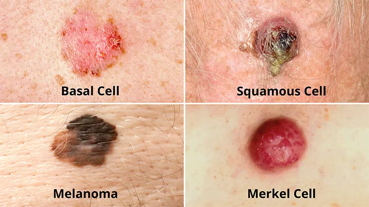
Skin cancer symptoms are the observable manifestations of malignant neoplasms arising from the skin. They represent the external expression of cellular mutations and abnormal growth patterns within the skin’s layers. These symptoms can manifest across three main types of skin cancer:
- Basal Cell Carcinoma (BCC): The most common but least aggressive form
- Squamous Cell Carcinoma (SCC): The second most common type with moderate aggressiveness
- Melanoma: Less common but highly aggressive and potentially lethal
Each type presents with distinctive symptomatic patterns, though overlap exists.
Affected Body Parts/Organs
Skin cancer symptoms primarily manifest on the epidermal (outer) layer of the skin, but can involve deeper skin layers as the disease progresses:
- Epidermis: Where most symptoms first appear and where most skin cancers originate
- Dermis: Involved as cancers invade deeper, leading to more serious symptoms
- Subcutaneous tissue: Affected in advanced cases
- Regional lymph nodes: Show symptoms in cases of metastasis
- Distant organs: May exhibit symptoms in advanced metastatic cases (especially with melanoma)
Common anatomical locations include:
- Sun-exposed areas (face, ears, neck, scalp, shoulders, back, arms, hands)
- Less commonly: palms, soles, beneath fingernails, genital areas
- Mucosal surfaces (in rare cases)
Prevalence and Significance
Skin cancer is the most common malignancy worldwide:
- Global incidence: 2-3 million non-melanoma skin cancers and 132,000 melanoma cases annually
- United States: Over 5 million cases treated yearly
- Australia: Highest rates globally with 2 in 3 Australians developing skin cancer by age 70
- Economic impact: Annual cost exceeding $8 billion in the US alone
The significance of skin cancer symptoms lies in their role as early warning signs:
- Most skin cancers have a high cure rate when detected early through symptom recognition
- Mortality primarily occurs when symptoms are ignored or misinterpreted
- Visual symptoms make skin cancer one of the most detectable cancers, yet delays in recognition remain common
- Symptoms can significantly impact quality of life through disfigurement, pain, and psychological distress
2. History & Discoveries
First Identification of Skin Cancer Symptoms
Documentation of skin cancer symptoms dates back thousands of years:
- Ancient Egypt (1500-2000 BCE): Papyri describe ulcerative skin lesions consistent with skin cancer symptoms
- Hippocratic Corpus (400-370 BCE): Contains descriptions of hard tumors resembling skin cancer, some with black bile (possibly melanoma)
- Celsus (25 BCE-50 CE): Documented different types of skin ulcers and tumors in “De Medicina”
- Medieval period: Various medical texts describe “black moles” that spread and cause death
However, systematic scientific documentation began much later:
- 1787: John Hunter provided the first scientific description of “cancerous ulcers” of the skin
- 1804: René Laennec recognized the distinction between melanoma and other skin cancers
- 1806: Sir Everard Home published detailed case reports of melanoma symptoms
Key Historical Figures
- René Théophile Hyacinthe Laennec (1781-1826): First classified melanoma as a distinct disease entity in 1804
- Sir Robert Carswell (1793-1857): Coined the term “melanoma” in 1838 and documented its visual characteristics
- William Norris (1792-1877): Published the first English-language paper on melanoma symptoms in 1820, noting familial patterns
- Paul Ehrlich (1854-1915): His work on aniline dyes led to early techniques for identifying skin cancer cells
- Frederick Mohs (1910-2002): Developed Mohs micrographic surgery based on observable cancer margins
- Sophie Spitz (1910-1956): Described “juvenile melanoma” (Spitz nevus), a benign condition that mimics melanoma symptoms
- Wallace Clark & Alexander Breslow (20th century): Developed staging systems based on symptom invasion depth
Major Breakthroughs
- 1910s: First systematic descriptions of basal cell and squamous cell carcinoma symptoms
- 1930s: First connection established between UV radiation and skin cancer symptoms
- 1950s: Development of the “ABCD” criteria for evaluating suspicious moles (later expanded to “ABCDE”)
- 1970s: Breslow’s depth measurement system linked symptom appearance to prognosis
- 1980s: First dermoscopy techniques developed to better visualize subtle skin cancer symptoms
- 1985: Foundation of the American Academy of Dermatology’s SPOTme® program, first nationwide skin cancer screening program
- 1990s: Development of the “ugly duckling sign” concept for identifying atypical moles
- 2000s: Advanced imaging technologies (confocal microscopy, optical coherence tomography) to detect subclinical symptoms
- 2010s: Artificial intelligence systems developed to recognize early skin cancer symptoms from images
Evolution of Medical Understanding
The understanding of skin cancer symptoms has evolved dramatically:
- Pre-20th century: Limited to gross visual descriptions without understanding of cellular basis
- Early 20th century: Connection to sun exposure established; basic classification of different types
- Mid-20th century: Development of standardized criteria for evaluating suspicious lesions
- Late 20th century: Recognition of the importance of early detection; public education campaigns
- 21st century: Technological advances allowing detection of pre-symptomatic changes; molecular understanding of symptom development; personalized risk assessment
Key paradigm shifts included:
- From regarding skin lesions as cosmetic concerns to recognizing them as potential cancers
- From treating only obvious symptoms to systematic screening for subtle changes
- From focusing solely on melanoma to recognizing the importance of non-melanoma skin cancers
- From purely visual assessment to multi-modal evaluation with advanced technologies
- From one-size-fits-all to personalized risk assessment based on genetic and environmental factors
3. Symptoms
Early Symptoms by Cancer Type
Basal Cell Carcinoma (BCC)
- Pearly or waxy bump: Small, flesh-colored or pearl-like nodule
- Flat, flesh-colored or brown lesion: Similar to a scar
- Recurrent bleeding or scabbing: Particularly after minor injury
- Subtle depression in the skin’s center: With a rolled border
- Tiny blood vessels visible on the surface (telangiectasias)
- Itching or mild discomfort: Often intermittent
Squamous Cell Carcinoma (SCC)
- Firm, red nodule: Usually rough-textured
- Flat lesion with scaly, crusted surface
- New growth on pre-existing scar or ulcer
- Rough, scaly patch on lip that persists
- Red patch or irritated area on the face, chest, shoulder, arm, or leg
- Mild tenderness or soreness in the affected area
Melanoma
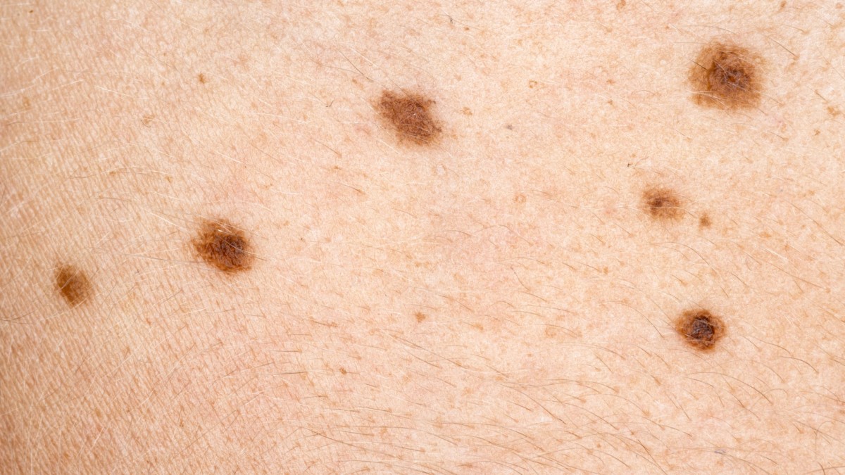
- Asymmetrical mole or lesion
- Border irregularity: Ragged, notched, or blurred edges
- Color variations: Multiple shades of tan, brown, black; may include red, white, or blue
- Diameter larger than 6mm (pencil eraser size)
- Evolving appearance: Change in size, shape, color, or elevation
- “Ugly duckling”: Mole that looks different from other existing moles
Rare Skin Cancers
- Merkel Cell Carcinoma: Painless, firm, shiny nodule on sun-exposed skin
- Cutaneous T-cell Lymphoma: Persistent red patches or plaques that may itch
- Kaposi Sarcoma: Purple, red, or brown blotches or nodules
- Sebaceous Carcinoma: Hard, yellowish nodule, often on the eyelid
Advanced-Stage Symptoms
Basal Cell Carcinoma
- Ulceration: Open sores that won’t heal
- Larger, more prominent nodules
- Crusting and oozing
- Local tissue destruction
- Indentation or depression in skin
- Invasion of surrounding structures (cartilage, bone in extreme cases)
- Pain: Usually mild but can become significant with nerve involvement
Squamous Cell Carcinoma
- Persistent, non-healing ulceration
- Raised edges around ulceration
- Rapid growth over weeks to months
- Deeper invasion causing pain or neurological symptoms
- Satellite lesions appearing around the primary tumor
- Local lymph node enlargement: Suggesting metastasis
- Pain and loss of function in advanced cases
Melanoma
- Ulceration: Breakdown of the skin surface
- Bleeding, oozing, or crusting
- Nodular growth: Raised areas within the lesion
- Satellite lesions: New spots appearing around original melanoma
- Hardening or induration of the surrounding skin
- Systemic symptoms: Fatigue, unexplained weight loss, swollen lymph nodes
- Neurological symptoms: In cases of metastasis to brain
- Respiratory symptoms: In cases of lung metastasis
- Bone pain: In cases of bone metastasis
- Jaundice: In cases of liver metastasis
Common vs. Rare Symptoms
Common Symptoms (>20% of cases)
- Change in appearance of a mole or skin lesion
- New growth on previously normal skin
- Non-healing sore
- Asymmetry in lesions
- Border irregularity
- Color variation within a single lesion
- Itching or mild discomfort
Uncommon Symptoms (5-20% of cases)
- Pain in a skin lesion (more common in advanced stages)
- Ulceration (except in advanced melanoma)
- Bleeding without trauma
- Spread to local lymph nodes
- Neurological symptoms near the lesion (numbness, tingling)
- Significant color changes (turning white, red, or blue)
Rare Symptoms (<5% of cases)
- Paraneoplastic syndromes: Systemic symptoms caused by immune reactions to the cancer
- Multiple primaries appearing simultaneously
- Hyperpigmentation or depigmentation of surrounding skin
- Halo phenomenon: White ring around a lesion
- Regression: Spontaneous partial disappearance of the lesion (especially in melanoma)
- Distant metastatic symptoms as initial presentation
- Eruptive keratoacanthomas: Multiple rapidly growing lesions
Symptom Progression Over Time
Timeline of Progression
Early phase (Weeks to months):
- Subtle changes in color, size, or texture
- Mild intermittent symptoms (itching, tenderness)
- Often dismissed as normal skin changes
Intermediate phase (Months to a year):
- More noticeable growth or change
- Development of characteristic features specific to cancer type
- More persistent symptoms
- Possible early ulceration or bleeding
Advanced phase (1+ years without treatment):
- Significant growth and invasion
- Ulceration, bleeding, and crusting
- Pain from nerve involvement
- Local tissue destruction
- Potential regional or distant spread
Pattern Variations by Cancer Type
- Basal Cell Carcinoma: Typically slow progression over years; rarely metastasizes but can cause significant local destruction
- Squamous Cell Carcinoma: Moderate progression over months to years; higher metastatic potential than BCC
- Melanoma: Highly variable progression; can advance rapidly within months or remain stable for years before suddenly changing
- Merkel Cell Carcinoma: Often rapid progression with high metastatic potential
Warning Signs of Rapid Progression
- Sudden increase in growth rate
- Development of ulceration
- Onset of pain in previously painless lesion
- Satellite lesions appearing around the primary lesion
- Regional lymph node enlargement
- Development of systemic symptoms (fatigue, weight loss)
4. Causes
Biological Causes of Skin Cancer Symptoms
Cellular Mechanisms
- Disrupted cell growth regulation: Cancer cells divide uncontrollably, creating visible masses
- Loss of programmed cell death (apoptosis): Abnormal cells survive instead of being eliminated
- Altered cellular adhesion: Cancer cells detach more easily, leading to irregular borders
- Genomic instability: Accumulation of mutations creates heterogeneity in appearance
- Angiogenesis (new blood vessel formation): Creates visible telangiectasias and feeds tumor growth
- Inflammatory response: Contributes to redness, swelling, and pain
- Invasion of surrounding tissues: Creates ulceration and structural changes
- Disrupted melanin production: Causes color variations in melanoma
Specific Genetic Mutations
- Basal Cell Carcinoma:
- Patched (PTCH) gene mutations in 90% of cases
- Smoothened (SMO) gene mutations
- p53 tumor suppressor gene mutations
- Squamous Cell Carcinoma:
- p53 mutations in 90% of cases
- Notch1 and Notch2 gene mutations
- CDKN2A mutations
- Melanoma:
- BRAF mutations (≈50% of cases)
- NRAS mutations (≈20% of cases)
- CDKN2A mutations (common in familial melanoma)
- c-KIT mutations (especially in acral and mucosal melanomas)
Environmental Causes
Ultraviolet (UV) Radiation
- UVB radiation (290-320 nm): Primarily causes direct DNA damage; most strongly associated with BCC and SCC
- UVA radiation (320-400 nm): Causes oxidative damage and contributes to photoaging; plays a role in melanoma
- Natural sunlight: Contains both UVA and UVB; primary environmental cause
- Artificial UV sources:
- Tanning beds: Emit primarily UVA at high intensity; strongly linked to melanoma
- UV lamps: Used in various industrial applications
- Phototherapy devices: Medical applications
Other Environmental Factors
- Ionizing radiation: X-rays and other medical radiation
- Chemical carcinogens:
- Arsenic: Found in contaminated water and some pesticides
- Coal tar and pitch: Industrial exposures
- Polycyclic aromatic hydrocarbons: Present in air pollution
- Certain pesticides and herbicides
- Viral infections:
- Human papillomavirus (HPV): Associated with some squamous cell carcinomas
- Human herpesvirus 8 (HHV-8): Causes Kaposi sarcoma
- Chronic inflammation: From burns, scars, or long-standing skin conditions
Genetic and Hereditary Factors
Inherited Syndromes
- Xeroderma pigmentosum: Defective DNA repair leads to extreme UV sensitivity
- Basal cell nevus syndrome (Gorlin syndrome): Multiple BCCs, often from young age
- Familial atypical multiple mole melanoma syndrome (FAMMM): Increased risk of melanoma
- Albinism: Lack of protective melanin increases UV damage
- Epidermolysis bullosa: Chronic skin damage increases SCC risk
- Li-Fraumeni syndrome: p53 germline mutations increase risk of various cancers
- Cowden syndrome: PTEN mutations increase risk of multiple cancer types
Hereditary Risk Factors
- Family history:
- First-degree relatives with melanoma increase risk 2-3 fold
- BCC and SCC also show familial clustering
- Inherited pigmentation traits:
- Fair skin, light eyes, and red/blonde hair (MC1R gene variants)
- Freckles and multiple moles
- Inability to tan/tendency to burn
- Genetic polymorphisms: Variations in genes controlling:
- Pigmentation
- DNA repair
- Immune function
- Carcinogen metabolism
Known Triggers and Exposure Risks
Acute Triggers
- Sunburn: Especially blistering sunburns in childhood
- Immunosuppression: Following organ transplantation or due to medications
- Trauma to existing lesions: Can activate dormant cancer cells
- Pregnancy: Hormonal changes may accelerate melanoma growth
Chronic Exposures
- Cumulative sun exposure: Professional or recreational
- Occupational exposures: Outdoor work, industrial chemicals
- Chronic wounds and inflammation: Scars, burns, non-healing ulcers
- Photosensitizing medications: Increase skin’s sensitivity to UV damage
- Geographic location: Proximity to equator increases UV intensity
- Altitude: Higher elevations have increased UV exposure
- Ozone depletion: Reduced atmospheric protection from UV radiation
5. Risk Factors
Demographic Risk Factors
Age
- BCC and SCC: Incidence increases dramatically with age
- Peak incidence: 60-80 years
- Growing incidence in 40-60 age group
- Melanoma: More distributed across age groups
- Most common cancer in young adults (25-29)
- Still has significant incidence in older adults
- Pediatric cases are rare but do occur
Gender
- BCC and SCC: Historically higher in men (2:1), but gap narrowing
- Melanoma:
- Higher incidence in men overall
- Higher incidence in women under age 50
- Men more likely to have trunk and head/neck melanomas
- Women more likely to have extremity melanomas
Race/Ethnicity
- Highest risk: Fair-skinned individuals of Northern European descent
- Moderate risk: Mediterranean and Middle Eastern backgrounds
- Lower risk: Asian, Hispanic/Latino populations
- Lowest risk: African backgrounds
- Special considerations:
- Non-white populations often diagnosed at later stages
- Different distribution of melanoma types by ethnicity
- Acral melanomas more common in darker-skinned populations
Environmental and Lifestyle Risk Factors
Sun Exposure Patterns
- Intermittent intense exposure: Strongly linked to melanoma
- Chronic cumulative exposure: More associated with BCC and SCC
- Sunburns: Even 5+ sunburns doubles melanoma risk
- Tanning bed use: 75% increased risk with use before age 35
Geographic Factors
- Latitude: Closer to equator means higher UV exposure
- Altitude: 4-5% increase in UV intensity per 1000 feet elevation
- Reflective surfaces: Snow, water, sand increase UV exposure
- Cloud cover: Still allows 80% of UV radiation through
Occupational Exposures
- Outdoor workers: Construction, agriculture, landscaping, maritime
- Industrial exposures: Chemical manufacturing, petroleum, mining
- Healthcare workers: Exposure to radiation, certain drugs
- Military personnel: Deployed to high-UV environments
Lifestyle Factors
- Recreational sun exposure: Beach activities, outdoor sports
- Clothing habits: Amount of skin routinely covered
- Sunscreen use: Inconsistent or improper application
- Tanning behaviors: Intentional tanning, use of artificial tanning
- Diet: Low antioxidant intake may increase risk
- Smoking: Associated with increased SCC risk
- Alcohol consumption: Associated with increased BCC and melanoma risk
Pre-Existing Conditions and Medical Factors
Skin Characteristics
- Skin phototype: Fitzpatrick types I and II at highest risk
- Nevus (mole) patterns:
100 common moles increases melanoma risk 7-fold
5 atypical/dysplastic nevi increases risk 6-fold
- Giant congenital melanocytic nevi: significant melanoma risk
- Pre-existing skin damage:
- Actinic keratoses (precancerous lesions)
- Solar lentigines (“age spots” or “liver spots”)
- Actinic cheilitis (precancerous lip condition)
Immune Status
- Organ transplant recipients: 65-100 times increased risk of SCC
- HIV/AIDS: 2-5 times increased skin cancer risk
- Chronic immunosuppressive therapy: For autoimmune conditions
- Primary immunodeficiency disorders
Previous Skin Cancer
- Prior BCC or SCC: 10-fold increased risk of developing another
- Prior melanoma: 9-fold increased risk of developing a second primary melanoma
- Family history of skin cancer: 2-3 fold increased risk
Medical Treatments
- Radiation therapy: Especially for childhood cancers
- Phototherapy: Used for psoriasis and other conditions
- PUVA therapy: Psoralen plus UVA for skin conditions
- Voriconazole and other photosensitizing drugs
6. Complications
Direct Complications of Skin Cancer Symptoms
Local Tissue Complications
- Disfigurement: Especially on cosmetically sensitive areas like the face
- Functional impairment: When cancer affects eyelids, lips, ears, nose, or fingers
- Pain and discomfort: As tumors invade nerves or cause pressure
- Ulceration and bleeding: Leading to infection risk and anemia in extreme cases
- Tissue destruction: Particularly with neglected BCC (“rodent ulcer”)
- Perineural invasion: Cancer spreading along nerve pathways
Regional Complications
- Lymphatic involvement: Swollen, painful lymph nodes
- Regional tissue edema: Especially in extremities with lymphatic obstruction
- Satellite lesions: New tumor deposits near the primary lesion
- In-transit metastases: Tumor deposits between primary site and regional lymph nodes
Systemic Complications
- Distant metastases:
- Melanoma: Lungs, liver, brain, bone, and gastrointestinal tract
- SCC: Lungs, bone, brain, and liver (less common)
- BCC: Extremely rare metastasis to bones, lungs, or lymph nodes
- Paraneoplastic syndromes: Rare systemic effects not due to direct tumor spread
- Cachexia and wasting: In advanced metastatic disease
Long-term Impact on Health
Physical Health Impact
- Chronic pain: From surgical sites or nerve damage
- Lymphedema: Persistent swelling after lymph node removal
- Recurrent disease: Requiring multiple treatments
- Secondary cancers: Increased risk in previously treated areas
- Treatment-related morbidity: Side effects from surgery, radiation, or systemic therapies
Psychological Impact
- Body image concerns: Particularly with visible lesions or surgical scarring
- Anxiety about recurrence: “Scan anxiety” when monitoring for metastasis
- PTSD-like symptoms: Especially in advanced cases
- Depression: 2-3 times more common in skin cancer patients
- Social withdrawal: Due to appearance concerns or physical limitations
Quality of Life Impact
- Sun avoidance: Limiting outdoor activities
- Activity restrictions: Following extensive surgery or in advanced disease
- Financial burden: Direct and indirect costs of ongoing care
- Occupational limitations: Particularly for outdoor workers
- Relationship effects: Sexual and interpersonal impacts
Disability and Fatality Rates
Disability
- Temporary disability: Common after extensive surgery (typically 2-8 weeks)
- Permanent disability rates:
- BCC: <1% (usually from local destruction of critical structures)
- SCC: 1-5% (higher with advanced disease)
- Melanoma: 5-20% (significantly higher with regional or distant spread)
- Factors affecting disability:
- Tumor location (functional vs. non-functional areas)
- Stage at diagnosis
- Treatment extent
- Age and comorbidities
- Access to rehabilitative services
Mortality Rates
- BCC: <0.1% mortality rate; deaths extremely rare
- SCC: Approximately 2% overall mortality rate
- Rises to 20-40% with lymph node involvement
- Melanoma: Highly variable mortality based on stage
- Stage I: 5-year survival >95%
- Stage II: 5-year survival 65-92%
- Stage III: 5-year survival 30-70%
- Stage IV: 5-year survival 15-20% (improving with new therapies)
- Other rare skin cancers:
- Merkel cell carcinoma: 5-year survival 30-64%
- Advanced cutaneous T-cell lymphoma: 5-year survival 24-83%
- Dermatofibrosarcoma protuberans: 5-year survival >90%
Prognostic Factors
- Tumor thickness (Breslow depth for melanoma)
- Ulceration: Significantly worsens prognosis
- Mitotic rate: Higher rate indicates more aggressive disease
- Lymph node involvement: Number and size of involved nodes
- Distant metastasis: Location and number of sites involved
- Age and overall health: Affects treatment options and outcomes
- Immune status: Immunocompromised patients have worse outcomes
- Treatment response: Particularly important with newer immunotherapies
7. Diagnosis & Testing
Initial Clinical Evaluation
Visual Examination
- Naked eye examination: Basic visual inspection under good lighting
- ABCDE criteria for melanoma assessment:
- Asymmetry: One half unlike the other
- Border: Irregular, scalloped, or poorly defined
- Color: Varied from one area to another
- Diameter: Usually greater than 6mm (pencil eraser size)
- Evolving: Changing in size, shape, color, or elevation
- Ugly duckling sign: Identifying lesions that look different from patient’s other moles
- Glasgow 7-point checklist:
- Major features: Change in size, shape, color
- Minor features: Diameter >7mm, inflammation, crusting/bleeding, sensory change
Specialized Clinical Tools
- Dermoscopy (dermatoscopy/epiluminescence microscopy):
- Hand-held device providing 10x magnification with polarized light
- Reveals subsurface structures not visible to naked eye
- Increases diagnostic accuracy by 10-30%
- Shows specific patterns associated with different skin cancers
- Wood’s lamp: Ultraviolet light examination
- Digital photography: For monitoring changes over time
Advanced Diagnostic Techniques
Non-Invasive Imaging
- Confocal laser scanning microscopy:
- Provides cellular-level resolution without biopsy
- 91-97% sensitivity for melanoma
- Particularly useful for facial lesions or multiple suspicious lesions
- Optical coherence tomography (OCT):
- Cross-sectional imaging of skin structures
- Useful for determining depth of invasion
- High-frequency ultrasound:
- Assesses tumor thickness and depth
- Helps plan surgical margins
- Multispectral imaging systems:
- Computer-assisted diagnosis
- Analyzes lesions across multiple wavelengths of light
Biopsy Techniques
- Shave biopsy:
- Removes superficial portion of lesion
- Good for raised lesions when BCC or SCC suspected
- Inadequate for melanoma assessment
- Punch biopsy:
- Removes cylindrical sample of skin
- Provides full thickness for depth assessment
- Limited sample size (2-6mm diameter)
- Excisional biopsy:
- Complete removal of lesion with margin
- Gold standard, especially for suspected melanoma
- Allows full histopathological assessment
- Incisional biopsy:
- Partial removal of large lesions
- Used when complete excision not initially practical
- Fine needle aspiration:
- For sampling suspicious lymph nodes
- Not used for primary skin lesions
Histopathological and Laboratory Testing
Histopathology
- Standard histology:
- Hematoxylin and eosin (H&E) staining
- Assessment of cellular morphology, tissue architecture
- Measurement of invasion depth (Breslow thickness for melanoma)
- Evaluation of surgical margins
- Immunohistochemistry:
- Melanoma markers: S100, HMB-45, Melan-A/MART-1, SOX10
- BCC markers: Bcl-2, Ber-EP4
- SCC markers: Cytokeratins, p63, p40
- Proliferation markers: Ki-67
- Special stains:
- Fontana-Masson: Highlights melanin
- Alcian blue: For mucin in certain variants
Molecular and Genetic Testing
- Mutation analysis:
- BRAF testing (melanoma)
- KIT mutation testing (acral/mucosal melanoma)
- NRAS mutation testing
- Hedgehog pathway mutations (BCC)
- Fluorescence in situ hybridization (FISH):
- Detects chromosomal abnormalities
- Helps distinguish ambiguous melanocytic lesions
- Gene expression profiling:
- 31-gene expression profile (DecisionDx-Melanoma)
- Provides prognostic information independent of stage
- Next-generation sequencing panels:
- Comprehensive genomic profiling
- Identifies therapeutic targets and resistance mechanisms
Staging and Further Evaluation
Clinical Staging
- TNM staging system:
- T: Tumor characteristics (size, thickness, ulceration)
- N: Regional lymph node involvement
- M: Metastasis status
- Sentinel lymph node biopsy (SLNB):
- Identifies first lymph node(s) draining the tumor site
- Standard for melanomas >0.8mm thick or with high-risk features
- Sometimes used for high-risk SCC
Advanced Imaging for Staging
- Regional ultrasound:
- Assessment of regional lymph nodes
- High sensitivity for lymph node metastases
- Computed tomography (CT):
- Evaluates potential metastases in chest, abdomen, pelvis
- Standard for higher-stage skin cancers
- Positron emission tomography (PET):
- Combines functional and anatomic imaging
- Higher sensitivity for distant metastases than CT alone
- Magnetic resonance imaging (MRI):
- Brain MRI standard for high-stage melanoma
- Superior soft tissue resolution
- No radiation exposure
Laboratory Studies
- Lactate dehydrogenase (LDH):
- Elevated in advanced melanoma
- Independent prognostic factor
- Complete blood count and comprehensive metabolic panel:
- Baseline before systemic therapy
- May show abnormalities in advanced disease
- Tumor markers:
- S100B protein: Correlates with melanoma tumor burden
- MIA (Melanoma Inhibitory Activity): Research tool
- No routinely used markers for BCC or SCC
8. Treatment Options
Surgical Treatments
Excisional Surgery
- Wide local excision:
- Gold standard for most skin cancers
- Margins determined by cancer type and stage:
- BCC: 2-4mm margins
- SCC: 4-6mm margins
- Melanoma: 1-2cm margins based on thickness
- Primary closure, flaps, or grafts for wound management
- Mohs micrographic surgery:
- Tissue-sparing technique with 100% margin control
- Highest cure rates (97-99%)
- Preferred for:
- High-risk anatomic locations (face, hands, genitalia)
- Recurrent tumors
- Poorly defined borders
- Aggressive histologic subtypes
- Large tumors
Other Surgical Approaches
- Curettage and electrodesiccation:
- Scraping followed by electrical cautery
- For small, well-defined, low-risk BCC and SCC
- 90-95% cure rate for appropriate lesions
- Cryosurgery:
- Liquid nitrogen destruction
- Good for superficial lesions in low-risk areas
- Less precise than excision
- Lymph node dissection:
- For clinically positive lymph nodes
- Completion lymphadenectomy after positive sentinel node
Non-Surgical Local Treatments
Radiation Therapy
- External beam radiation:
- Primary treatment for inoperable cases
- Adjuvant treatment after surgery for high-risk features
- Palliative treatment for advanced or metastatic disease
- Particularly useful in elderly or poor surgical candidates
- Brachytherapy:
- Localized radiation delivery
- Useful for curved surfaces like the ear or nose
Topical Therapies
- 5-Fluorouracil (5-FU):
- Chemotherapy cream
- Effective for superficial BCC, actinic keratoses
- 70-90% efficacy for appropriate lesions
- Imiquimod:
- Immune response modifier
- Activates toll-like receptor 7
- FDA-approved for superficial BCC
- 80-90% efficacy for appropriate lesions
- Ingenol mebutate:
- Induces rapid lesion necrosis
- For actinic keratoses, sometimes superficial non-melanoma skin cancers
- Photodynamic therapy:
- Application of photosensitizing agent + light activation
- For superficial BCC, Bowen’s disease (SCC in situ)
- 70-90% efficacy for appropriate lesions
Systemic Treatments
Immunotherapy
- Checkpoint inhibitors:
- Anti-PD-1 antibodies (pembrolizumab, nivolumab)
- Anti-CTLA-4 antibody (ipilimumab)
- Revolutionized advanced melanoma treatment
- Response rates 40-60% for melanoma
- Also approved for advanced SCC and MCC
- Cytokines:
- High-dose interleukin-2 (IL-2)
- Interferon-alpha (historical use)
- Oncolytic virus therapy:
- Talimogene laherparepvec (T-VEC)
- Injected directly into melanoma lesions
- Genetically modified herpes virus
Targeted Therapies
- BRAF inhibitors:
- Vemurafenib, dabrafenib, encorafenib
- For BRAF V600-mutant melanoma
- Response rates 50-60%
- Typically combined with MEK inhibitors
- MEK inhibitors:
- Trametinib, cobimetinib, binimetinib
- Used in combination with BRAF inhibitors
- Reduces resistance and side effects
- c-KIT inhibitors:
- Imatinib, nilotinib
- For melanomas with KIT mutations (acral, mucosal)
- Hedgehog pathway inhibitors:
- Vismodegib, sonidegib
- For advanced or metastatic BCC
- Also used for Basal Cell Nevus Syndrome
Chemotherapy
- Dacarbazine/temozolomide:
- Historical standard for melanoma
- Response rates 10-15%
- Limited current use with better options available
- Platinum compounds:
- Cisplatin, carboplatin
- Used for advanced SCC, MCC
- Taxanes:
- Paclitaxel, docetaxel
- For advanced SCC, MCC, angiosarcoma
Emerging Treatments and Clinical Trials
Novel Immunotherapeutic Approaches
- Anti-LAG-3 antibodies:
- Relatlimab approved in combination with nivolumab
- Targets another immune checkpoint
- TIL therapy (Tumor Infiltrating Lymphocytes):
- Harvesting, expanding, and reinfusing patient’s own tumor-fighting cells
- Promising results in advanced melanoma
- Bispecific antibodies:
- Simultaneously target tumor cells and immune cells
- Several in clinical trials
Advanced Targeted Approaches
- Next-generation BRAF/MEK inhibitors:
- Better tolerability and efficacy profiles
- Potential to overcome resistance mechanisms
- CDK4/6 inhibitors:
- For uveal melanoma and other subtypes
- Epigenetic modifiers:
- HDAC inhibitors
- Promising for cutaneous T-cell lymphoma
Combination Strategies
- Dual immunotherapy:
- Anti-PD-1 + anti-CTLA-4 (approved for melanoma)
- Multiple novel combinations in trials
- Immunotherapy + targeted therapy:
- BRAF/MEK inhibitors + checkpoint inhibitors
- Rational sequencing strategies
- Neoadjuvant approaches:
- Therapy before surgery to reduce tumor size and scope of operation
- May provide prognostic information
Other Innovative Approaches
- Intralesional therapies:
- Direct injection of immune stimulants or cytotoxic agents
- PV-10 (Rose Bengal), IL-12 plasmid
- Nanoparticle delivery systems:
- Enhanced drug delivery to tumor sites
- Reduced systemic toxicity
- Gene therapy:
- CRISPR-based approaches
- Early-stage investigation
9. Prevention & Precautionary Measures
Sun Protection Strategies
Physical Sun Protection
- Protective clothing:
- Wide-brimmed hats (brim >3 inches)
- Long-sleeved shirts and pants
- UPF-rated clothing (Ultraviolet Protection Factor)
- Sunglasses with UV protection
- Shade seeking:
- Avoiding sun exposure during peak hours (10am-4pm)
- Using umbrellas, trees, structures for shade
- Remembering that shade reduces but doesn’t eliminate UV exposure
Sunscreen Use
- Sunscreen recommendations:
- Broad-spectrum (UVA and UVB protection)
- SPF 30 or higher
- Water-resistant formulations
- Correct application:
- Apply 15-30 minutes before sun exposure
- Use 1 oz (30mL) for full body coverage
- Reapply every 2 hours and after swimming/sweating
- Types of sunscreen:
- Chemical: Absorbs UV radiation
- Physical: Reflects UV radiation (zinc oxide, titanium dioxide)
- Combination products
Environmental Awareness
- UV Index monitoring:
- Higher index requires more protection
- Available through weather apps and websites
- Seasonal variations:
- Sun protection needed year-round, not just summer
- Snow reflection can nearly double UV exposure
- Water and sand increase reflection
- Geographic considerations:
- Higher UV levels near equator
- Increased exposure at high altitudes
- Reflection from water, snow, sand
Early Detection Practices
Self-Examination
- Monthly skin self-examinations:
- Systematic check of entire body
- Using mirrors or partner assistance for difficult-to-see areas
- Photography to track changes
- Digital apps available to assist documentation
- ABCDE method for self-assessment
- Body mapping systems for monitoring multiple moles
- When to seek medical attention:
- New growths
- Changes in existing lesions
- Sores that don’t heal within 2 weeks
- Unusual symptoms (bleeding, itching, pain)
Professional Screening
- Regular dermatologic examinations:
- Annually for general population
- Every 3-6 months for high-risk individuals
- Full-body examination by trained provider
- Risk-appropriate screening intervals:
- Personal history of skin cancer: 3-6 month intervals
- Family history: 6-12 month intervals
- Multiple atypical moles: 6-12 month intervals
- Immunosuppression: 3-6 month intervals
Technological Screening Aids
- Total body photography:
- Baseline images for comparison
- Particularly useful for patients with multiple moles
- Computer-assisted diagnosis:
- AI-based systems to support clinical decisions
- Some systems showing accuracy comparable to dermatologists
- Teledermatology:
- Remote evaluation for those with limited specialist access
- Store-and-forward or live video consultation
Lifestyle Modifications
Behavioral Changes
- Tanning avoidance:
- No use of indoor tanning beds (increases melanoma risk by 75%)
- Avoiding intentional tanning outdoors
- Using self-tanners as safe alternative
- Outdoor activity timing:
- Scheduling outdoor activities before 10am or after 4pm
- Monitoring UV index for safer times
- Occupational adjustments:
- Rotating outdoor tasks among workers
- Creating shade structures at work sites
- Regular breaks from sun exposure
Nutritional Approaches
- Antioxidant-rich diet:
- Fruits and vegetables with carotenoids
- Foods rich in vitamins C, E, and polyphenols
- Mediterranean diet pattern
- Omega-3 fatty acids:
- May reduce inflammation and photoaging
- Found in fatty fish, flaxseeds, walnuts
- Vitamin D considerations:
- Balancing sun protection with vitamin D needs
- Supplementation or dietary sources when needed
- Limited evidence for dietary supplements:
- Nicotinamide (vitamin B3) may reduce non-melanoma skin cancers
- Polypodium leucotomos extract showing promise in some studies
Special Populations and Considerations
High-Risk Groups
- Organ transplant recipients:
- Intensive screening protocols
- Consideration of preventive medications (acitretin, capecitabine)
- Careful immunosuppression management
- Xeroderma pigmentosum patients:
- Extreme sun avoidance
- Protective clothing and films for windows
- DNA repair enzyme creams
- Previous skin cancer patients:
- Chemoprevention options
- Enhanced surveillance schedules
- Patient education regarding second primary risks
Children and Adolescents
- Sun protection from infancy:
- No sunscreen under 6 months (shade and clothing only)
- Child-specific sunscreen formulations after 6 months
- Role modeling sun-safe behaviors
- School-based interventions:
- Sun safety education programs
- Shade structures on playgrounds
- Scheduled outdoor activities to avoid peak UV times
Occupational Settings
- Workplace policies:
- Sun safety training
- Provision of protective equipment
- Rotation of outdoor tasks
- Scheduling adjustments
- Industry-specific approaches:
- Construction: Temporary shade structures
- Agriculture: UV-protective equipment
- Outdoor recreation: Staff education and client advice
10. Global & Regional Statistics
Global Incidence and Prevalence
Non-Melanoma Skin Cancer
- Global annual incidence: 2-3 million cases
- Prevalence: Most common malignancy worldwide
- Basal Cell Carcinoma: Accounts for 70-80% of non-melanoma skin cancers
- Squamous Cell Carcinoma: Accounts for 20-30% of non-melanoma skin cancers
- Underreporting issue: Many countries don’t include NMSC in cancer registries
Melanoma
- Global annual incidence: 350,000-450,000 cases
- Prevalence: 1.3 million people living with melanoma diagnosis
- Global incidence trend: Increasing 3-7% annually in fair-skinned populations
- 18th most common cancer worldwide, but higher ranking in fair-skinned countries
Rare Skin Cancers
- Merkel Cell Carcinoma: 2,500-3,000 cases annually worldwide
- Dermatofibrosarcoma Protuberans: 4-5 cases per million person-years
- Cutaneous T-cell Lymphoma: 6-8 cases per million person-years
- Kaposi Sarcoma: Dramatic variations based on HIV prevalence
Regional Variations
Highest Incidence Regions
- Australia/New Zealand:
- Melanoma: 33-55 cases per 100,000 person-years
- BCC: 1,000-2,000 cases per 100,000 person-years
- SCC: 250-400 cases per 100,000 person-years
- North America:
- Melanoma: 20-30 cases per 100,000 in US, lower in Canada
- BCC: 450-1,000 cases per 100,000 person-years
- SCC: 100-200 cases per 100,000 person-years
- Northern Europe:
- Melanoma: 15-25 cases per 100,000 person-years
- NMSC: 100-200 cases per 100,000 person-years
Moderate Incidence Regions
- Southern Europe:
- Melanoma: 8-15 cases per 100,000 person-years
- NMSC: 50-150 cases per 100,000 person-years
- Eastern Asia:
- Melanoma: 0.5-3 cases per 100,000 person-years
- NMSC: 20-50 cases per 100,000 person-years
- South America:
- Melanoma: 3-7 cases per 100,000 person-years (higher in southern regions)
- NMSC: 30-100 cases per 100,000 person-years
Lowest Incidence Regions
- Africa:
- Melanoma: 0.5-2 cases per 100,000 person-years
- NMSC: 5-20 cases per 100,000 person-years
- Exception: Higher rates in South Africa among European-descended population
- South Asia:
- Melanoma: <1 case per 100,000 person-years
- NMSC: 2-10 cases per 100,000 person-years
- Southeast Asia:
- Melanoma: 0.5-2 cases per 100,000 person-years
- NMSC: 10-30 cases per 100,000 person-years
Mortality and Survival Statistics
Global Mortality
- Melanoma: Approximately 60,000 deaths annually worldwide
- Non-melanoma skin cancer: Approximately 65,000 deaths annually
- Mortality trends:
- Stabilizing or decreasing in high-income countries with advanced healthcare
- Continuing to increase in many developing regions
Survival Rates by Region
High-income countries:
- Melanoma 5-year survival: 90-92% overall
- Stage-specific melanoma survival:
- Localized: 98-99%
- Regional spread: 65-70%
- Distant metastasis: 25-30% (improving with new therapies)
- NMSC 5-year survival: >95%
Middle-income countries:
- Melanoma 5-year survival: 60-85%
- NMSC 5-year survival: 85-95%
- Greater variation based on healthcare access
Low-income countries:
- Melanoma 5-year survival: 40-60%
- NMSC 5-year survival: 70-85%
- Limited by later-stage diagnosis and treatment access
Demographic Disparities
- Gender differences:
- Men have worse melanoma survival than women at every stage
- Gap of 5-10% in 5-year survival rates
- Racial/ethnic disparities:
- Non-Hispanic whites: Highest incidence but better survival
- Black patients: Lower incidence but 25% lower 5-year survival rate
- Hispanic patients: Intermediate outcomes
- Socioeconomic disparities:
- Lower income associated with later-stage diagnosis
- Education level correlates with melanoma awareness and outcomes
- Insurance status significantly impacts survival
Temporal Trends and Projections
Historical Trends
- 1970s-2000s:
- Dramatic increases in skin cancer rates globally
- Melanoma incidence increased 3-7% annually in many countries
- 2000s-2010s:
- Continued increases but some stabilization in younger cohorts
- Evidence of prevention program impact in Australia and Northern Europe
- 2010s-present:
- Stabilization in some regions (Australia, parts of Europe)
- Continued increases in many other regions
- Dramatic improvements in advanced melanoma survival
Future Projections
- Incidence projections:
- Continued increases expected for 2-3 decades due to aging populations
- Potential long-term decreases from improved prevention in younger cohorts
- Mortality projections:
- Decreasing melanoma mortality in regions with access to new therapies
- Persistent disparities in low-resource settings
- Economic impact projections:
- Increasing costs due to higher incidence and expensive new treatments
- Projected annual costs exceeding $12 billion by 2030
11. Recent Research & Future Prospects
Latest Diagnostic Advances
Artificial Intelligence Applications
- Deep learning algorithms:
- Computer vision systems for lesion classification
- Performance matching or exceeding dermatologists in controlled studies
- Smartphone applications for preliminary screening
- Multimodal AI approaches:
- Combining clinical, dermoscopic, and genetic data
- Enhanced accuracy for difficult-to-diagnose lesions
- Integration into clinical workflow:
- Decision support systems
- Automated risk stratification
- Telehealth triage applications
Advanced Imaging Technology
- Optical coherence tomography (OCT) advancements:
- Higher resolution systems
- Dynamic OCT showing blood vessel patterns
- Correlation with invasive depth
- Reflectance confocal microscopy evolution:
- Hand-held devices for easier clinical use
- Automated feature recognition
- Widening clinical applications
- Multiphoton microscopy:
- Label-free imaging of cellular metabolism
- Potential for “optical biopsy”
- Raman spectroscopy:
- Molecular fingerprinting of skin lesions
- Non-invasive biochemical analysis
Molecular Diagnostic Innovations
- Non-invasive genetic sampling:
- Adhesive patch tests for genetic analysis
- Liquid biopsy approaches for circulating tumor DNA
- Advanced molecular testing:
- Next-generation sequencing panels
- Digital PCR for enhanced sensitivity
- Epigenetic markers for early detection
- Multi-omics approaches:
- Integration of genomics, proteomics, and metabolomics
- Comprehensive biological profiling of tumors
Therapeutic Research Frontiers
Immunotherapy Innovations
- Next-generation checkpoint inhibitors:
- New targets beyond PD-1/PD-L1 and CTLA-4
- Bi-specific and tri-specific antibodies
- Intratumorally administered immunotherapies
- Personalized cancer vaccines:
- Neoantigen-based approaches
- mRNA vaccine platforms
- Combined with checkpoint inhibition
- Cellular therapies:
- Advances in TIL (tumor-infiltrating lymphocyte) therapy
- CAR-T cell approaches for solid tumors
- Natural killer (NK) cell therapies
Targeted Therapy Evolution
- Novel pathway inhibitors:
- SHP2 inhibitors
- CDK inhibitors
- NRAS-targeted approaches
- Resistance mechanisms and countermeasures:
- Triplet therapy for BRAF-mutant melanoma
- Intermittent dosing strategies
- Combination approaches to prevent resistance
- Tumor microenvironment targeting:
- Anti-angiogenic approaches
- Matrix metalloproteinase inhibitors
- Cancer-associated fibroblast modulation
Delivery System Innovations
- Nanoparticle delivery platforms:
- Enhanced penetration of skin barriers
- Targeted delivery to tumor cells
- Reduced systemic side effects
- Transdermal drug delivery:
- Microneedle arrays
- Sonophoresis and iontophoresis
- Magnetophoresis for enhanced delivery
- Local drug delivery devices:
- Implantable sustained-release systems
- On-demand drug release technologies
- Biodegradable drug depots
Ongoing Clinical Trials
Major Melanoma Trials
- Adjuvant therapy optimization:
- Optimal duration of therapy
- Novel combinations
- Risk-stratified approaches
- Neoadjuvant approaches:
- Immunotherapy before surgery
- Pathological complete response as endpoint
- Biomarker development
- Novel combinations:
- Anti-PD-1 + anti-LAG-3
- Targeted therapy + immunotherapy sequencing
- Oncolytic viruses + checkpoint inhibition
Non-Melanoma Skin Cancer Trials
- Hedgehog pathway resistance:
- New inhibitors for resistant BCC
- Combination approaches
- Immunotherapy expansion:
- Checkpoint inhibitors for high-risk SCC
- Novel approaches for BCC
- Field treatment innovations:
- Preventive strategies for actinic damage
- New topical agents and protocols
- Device-based field therapies
Prevention Trials
- Chemoprevention studies:
- Nicotinamide in high-risk populations
- Retinoids for transplant recipients
- COX-2 inhibitors and NSAIDs
- Behavioral intervention trials:
- Technology-enabled prevention strategies
- Community-based intervention programs
- Workplace intervention studies
- Vaccine development:
- HPV vaccination effects on HPV-related skin cancers
- Experimental melanoma prevention vaccines
Future Directions
Preventive Medicine Trends
- Precision prevention:
- Genetic risk assessment
- Personalized screening protocols
- Risk-based intervention strategies
- Smart technology integration:
- UV exposure monitoring devices
- Automated skin self-examination
- Real-time risk assessment applications
- Public health innovations:
- Population-based screening approaches
- Environmental interventions
- Policy-level sun protection initiatives
Emerging Treatment Paradigms
- Curative approaches for advanced disease:
- Complete response with durability
- Treatment discontinuation strategies
- Cure-focused clinical trial endpoints
- Minimally invasive therapies:
- Image-guided interventions
- Focal ablative approaches
- Reduced morbidity treatments
- Aging population considerations:
- Tailored approaches for the elderly
- Comorbidity management
- Quality of life optimization
Future Research Priorities
- Basic science frontiers:
- Tumor microenvironment exploration
- Immune surveillance mechanisms
- Metastatic process understanding
- Translational research:
- Biomarker development for treatment selection
- Resistance mechanism elucidation
- Novel therapeutic target identification
- Health services research:
- Implementation science
- Healthcare delivery optimization
- Disparities reduction strategies
12. Interesting Facts & Lesser-Known Insights
Historical Perspectives
Ancient Observations
- Egyptian mummies: Show evidence of skin lesions consistent with skin cancers
- Hippocratic descriptions: Included “black cancer” that likely represented melanoma
- Celestial connections: Early physicians believed skin lesions were influenced by planetary alignments
- Historical treatments: Included arsenic pastes, caustic compounds, and bloodletting
Notable Historical Cases
- John Wayne: Attributed his lung cancer to filming in Nevada near nuclear test sites, but his heavy smoking and extensive sun exposure on film sets likely contributed to his multiple skin cancers
- Bob Marley: Died from acral lentiginous melanoma that began under his toenail, initially misdiagnosed as a soccer injury
- President Ronald Reagan: Had multiple skin cancers removed during and after his presidency
- First documented melanoma surgery: Performed in 1787 by Scottish surgeon John Hunter
Uncommon Knowledge
Biological Curiosities
- Melanin paradox: While melanin protects against UV damage, the process of producing melanin can create free radicals that potentially damage DNA
- Circadian rhythm effects: Skin cells repair UV damage more effectively at certain times of day; research suggests nighttime sun exposure may be more damaging
- Pregnancy influence: Melanomas may accelerate during pregnancy due to hormonal influences and immune system changes
- Spontaneous regression: Approximately 3-15% of melanomas show evidence of partial spontaneous regression, likely due to immune response
- XP moonlight children: Children with xeroderma pigmentosum in developing countries often live nocturnal lives, only going outdoors after sunset
Geographic and Environmental Factors
- Altitude effect: UV exposure increases 8-10% with every 1,000-meter increase in elevation
- Window glass filtration: Standard window glass blocks UVB but allows 70-80% of UVA to penetrate
- Medication-induced photosensitivity: Over 400 medications can increase sun sensitivity
- Latitude jump effect: Moving from high to low latitudes significantly increases melanoma risk, especially in childhood
- Protective tanning: Some evidence suggests gradual, controlled sun exposure may be protective compared to intermittent intense exposure
Myths vs. Medical Facts
Common Misconceptions
- Myth: Dark-skinned people don’t get skin cancer
- Fact: All skin types can develop skin cancer; darker skin types have lower risk but often present with more advanced disease
- Myth: Tanning beds are safer than natural sunlight
- Fact: Tanning beds emit primarily UVA at intensities 10-15 times greater than midday sun
- Myth: Sunscreen alone provides complete protection
- Fact: Even properly applied sunscreen allows 3-7% of UV radiation through; physical protection is also necessary
- Myth: Skin cancers always present as raised lesions
- Fact: Many early melanomas and BCCs are flat or even depressed
- Myth: Removing a mole prevents it from becoming cancerous
- Fact: Preventive removal is generally not recommended without suspicious features
Surprising Medical Facts
- Sunscreen history: First commercial sunscreen was developed in 1936, but SPF rating wasn’t standardized until the 1970s
- Melanoma heterogeneity: Genomic analysis shows melanoma has one of the highest mutation rates of any cancer
- Nevus count paradox: While having many moles increases melanoma risk, melanomas often arise on previously normal-appearing skin
- Eyelid vulnerability: Eyelids receive significant sun exposure but are commonly missed during sunscreen application
- Cold weather risk: Up to 80% of UV radiation penetrates light cloud cover, and snow reflects up to 80% of UV radiation
Impact on Specific Populations
Occupational Considerations
- Airline pilots and cabin crew: Experience twice the melanoma rate of the general population due to increased cosmic radiation exposure at high altitudes
- Welders: Higher risk of ocular melanoma due to UV radiation exposure
- Glassblowers: Historical high rates of facial skin cancers from infrared radiation
- Nightshift workers: Some evidence suggests disrupted circadian rhythms may increase skin cancer risk
- Professional athletes: Often experience high cumulative sun exposure during practice and competition
Cultural and Ethnic Dimensions
- Albinism in Africa: Persons with albinism in sub-Saharan Africa face extreme skin cancer risk, often dying young without interventions
- Burkini controversy: Full-coverage swimwear developed for religious modesty inadvertently provides excellent sun protection
- Indigenous knowledge: Some traditional cultures developed effective sun-protective practices using natural materials
- Skin whitening dangers: Some skin whitening products contain carcinogens like mercury and hydroquinone
- Stigma factors: In some cultures, skin changes may be hidden due to stigma, leading to diagnostic delays
Special Situations
- Astronaut risk: Space travelers experience increased radiation exposure and altered immune function
- Firefighter connection: Higher rates of both melanoma and non-melanoma skin cancers possibly related to chemical exposures
- Refugee challenges: Displaced populations often lack access to sun protection and skin cancer screening
- Visible difference: Facial tumors and surgical changes can profoundly affect body image and social interaction
- Digital health divide: Telemedicine and AI advances in skin cancer detection may exacerbate healthcare disparities without intentional access efforts
This comprehensive report provides a thorough overview of skin cancer symptoms, covering their definition, history, manifestations, causes, risk factors, complications, diagnosis, treatment, prevention, global statistics, research directions, and interesting insights. While significant advances have been made in detecting and treating skin cancers, continued vigilance through regular skin examinations remains the most important factor in early identification and successful management.