
⚠️ Disclaimer: The information provided in this article is for educational purposes only and does not constitute medical advice. RevisionTown does not provide diagnosis, treatment, or medical recommendations. Always consult a qualified healthcare professional regarding any medical condition, symptoms, or concerns.
Read More – 🏥 Medical Disclaimer
Comprehensive Report on Ringworm (Dermatophytosis)
1. Overview
What is Ringworm?
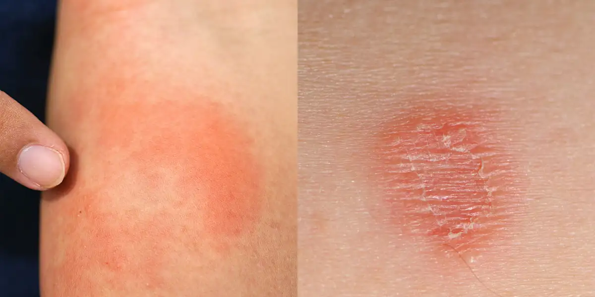
Ringworm, medically known as dermatophytosis or tinea, is a common fungal infection of the skin, hair, and nails. Despite its name, ringworm is not caused by a worm but by a group of fungi called dermatophytes that live on the dead tissues of the skin, hair, and nails. The infection is named “ringworm” because it often causes a ring-shaped rash with a raised, scaly border that resembles a worm curled into a circle.
Dermatophytosis is characterized by its ability to invade the keratinized tissue (the outermost layer of the skin containing the protein keratin) while rarely penetrating into deeper tissues or organs. The fungi responsible for ringworm thrive in warm, moist environments and can be spread through direct contact with infected individuals, animals, or contaminated objects.
Affected Body Parts/Organs
Ringworm can affect various parts of the body, with different clinical presentations depending on the affected site:
Tinea corporis (body ringworm): Affects the trunk, limbs, and face, excluding the beard, scalp, groin, hands, and feet.
Tinea capitis (scalp ringworm): Involves the scalp and hair shafts, common in children.
Tinea pedis (athlete’s foot): Affects the feet, particularly between the toes and on the soles.
Tinea cruris (jock itch): Involves the groin area, inner thighs, and sometimes the buttocks.
Tinea manuum: Affects the hands, particularly the palms and spaces between fingers.
Tinea unguium (onychomycosis): Involves the fingernails or toenails.
Tinea barbae: Affects the facial hair, beard, and mustache areas in men.
Tinea faciei: Appears on the non-bearded facial skin.
Tinea imbricata: A rare form causing concentric rings covering large areas of the body.
Tinea versicolor (technically caused by a different fungus, Malassezia): Causes small, discolored patches of skin.
Prevalence and Significance
Ringworm is one of the most common fungal infections worldwide, affecting an estimated 20-25% of the world’s population at any given time. Its prevalence varies significantly based on geographic location, climate, and socioeconomic factors:
Global impact: Dermatophytosis affects millions of people worldwide, with higher rates in tropical and subtropical regions due to the warm, humid climate that favors fungal growth.
Age distribution: While ringworm can affect people of all ages, certain types show age preferences. Tinea capitis predominantly affects prepubertal children, while tinea pedis and tinea cruris are more common in adolescents and adults.
Economic burden: The annual economic burden associated with dermatophytosis is substantial, encompassing direct medical costs (medications, doctor visits) and indirect costs (work absence, decreased productivity).
Public health significance: Though rarely life-threatening, ringworm represents a significant public health concern due to its high contagiousness, impact on quality of life, and the discomfort it causes.
Veterinary importance: Ringworm also affects domestic animals and livestock, serving as a reservoir for human infections and causing economic losses in animal industries.
In developed countries, improved hygiene, living conditions, and access to antifungal treatments have reduced the prevalence of certain types of ringworm (particularly tinea capitis). However, other forms like tinea pedis remain common, with an estimated lifetime risk of 70% in the general population. In developing countries, the prevalence remains significantly higher due to crowded living conditions, limited access to healthcare, and inadequate awareness about preventive measures.
The clinical significance of ringworm extends beyond its primary symptoms, as untreated infections can lead to secondary bacterial infections, spread to other body parts, or cause permanent scarring and hair loss in severe cases. Additionally, the psychosocial impact of visible skin lesions can lead to embarrassment, reduced self-esteem, and social isolation, particularly in children and adolescents.
2. History & Discoveries
First Identification of Ringworm
The history of ringworm recognition dates back to ancient times, although the fungal nature of the disease wasn’t understood until much later:
Ancient observations: References to skin conditions resembling ringworm appear in early medical texts from various civilizations. Descriptions of circular skin lesions can be found in ancient Egyptian, Greek, and Roman medical writings.
Medieval period: Various skin conditions including ringworm were often grouped under the term “leprosy” or other general classifications for skin diseases.
Early modern period (16th-18th centuries): More specific descriptions of ringworm-like conditions began to appear in medical literature, though the cause remained unknown.
Key Discoveries and Contributors
The scientific understanding of ringworm developed primarily in the 19th century:
1837-1841: Robert Remak, a Polish physician, was among the first to suggest that favus (a severe form of tinea capitis) was caused by a fungus. He observed fungal elements under the microscope from infected scalp samples.
1841: David Gruby, a Hungarian physician working in Paris, is credited with definitively establishing the fungal etiology of ringworm. Between 1841 and 1844, Gruby published a series of papers describing the microscopic appearance and culture characteristics of several dermatophyte species. He is often considered the “father of medical mycology” for this pioneering work.
1845: Ferdinand von Hebra, an Austrian dermatologist, further classified various forms of tinea and helped establish dermatology as a distinct medical specialty.
1860s: Johann Schönlein and Robert Remak further characterized the fungi causing favus.
1892: Raymond Sabouraud, a French dermatologist, published his doctoral thesis on dermatophytes. Sabouraud would later develop specialized culture media for fungal isolation (Sabouraud dextrose agar), which remains in use today.
Major Breakthroughs in Research and Treatment
The 20th century saw significant advances in understanding and treating ringworm:
1910-1920: Raymond Sabouraud published “Les Teignes,” a comprehensive monograph on dermatophytes that established the foundation for modern medical mycology.
1903-1925: Classification systems for dermatophytes were developed, with the genera Microsporum, Trichophyton, and Epidermophyton being established.
1940s: The introduction of griseofulvin, the first effective oral antifungal medication for dermatophytosis, revolutionized treatment. William Brown and colleagues at Imperial Chemical Industries in the UK discovered its antifungal properties in 1939, but it wasn’t until the late 1950s that it became widely used clinically.
1950s-1960s: Development of improved diagnostic techniques, including the use of Wood’s lamp (ultraviolet light) for detecting certain types of dermatophyte infections.
1970s-1980s: Introduction of azole antifungals (like ketoconazole) provided new treatment options with fewer side effects than earlier medications.
1990s: Development of newer azoles (fluconazole, itraconazole) and allylamines (terbinafine) further expanded treatment options with improved efficacy and safety profiles.
1990s-2000s: Molecular techniques allowed for more precise identification of dermatophyte species and better understanding of their epidemiology.
Evolution of Medical Understanding
The understanding of ringworm has evolved significantly over time:
From supernatural to scientific: Early beliefs attributed skin diseases to supernatural causes, divine punishment, or “bad blood.” The recognition of ringworm as a specific, transmissible fungal infection represented a major conceptual shift.
Taxonomic evolution: The classification of dermatophytes has undergone multiple revisions as research techniques advanced. Initially based solely on microscopic appearance, modern classification incorporates molecular genetics, resulting in reclassification of many species.
Ecological understanding: Research revealed the ecological niches of different dermatophytes, categorizing them as anthropophilic (human-loving), zoophilic (animal-loving), or geophilic (soil-dwelling), which helped explain transmission patterns.
Host-pathogen interactions: Advanced research has elucidated the complex interactions between dermatophytes and the human immune system, explaining why some infections resolve spontaneously while others become chronic.
Public health approaches: Recognition of the role of communal facilities like swimming pools, locker rooms, and shared housing in transmission has led to targeted preventive strategies.
One Health concept: Modern understanding acknowledges the interconnection between human, animal, and environmental health in the epidemiology of dermatophytosis, leading to more comprehensive control approaches.
The history of ringworm research illustrates how medical understanding progresses from observation to explanation, with each era building upon previous knowledge and incorporating new technologies to enhance diagnosis, treatment, and prevention of this common fungal infection.
3. Symptoms
Early Symptoms
Ringworm typically begins subtly and may initially be mistaken for other skin conditions. Early manifestations vary depending on the body site affected:
General Early Signs:
- Small, red or pink patch of skin that may be slightly raised
- Mild itching or burning sensation
- Slight scaliness or flakiness of the affected area
- Potential mild discomfort or irritation
Site-Specific Early Symptoms:
Tinea Corporis (Body Ringworm):
- Small, round or oval pinkish-red patch
- Slightly raised border
- Minimal itching
- Gradual enlargement over days
Tinea Capitis (Scalp Ringworm):
- Small areas of scaling on the scalp
- Mild itching
- Subtle hair breakage
- Sometimes small areas of hair loss
Tinea Pedis (Athlete’s Foot):
- Dry, flaky skin between the toes
- Mild itching, especially after removing footwear
- Slight redness between toes
- Minimal discomfort when walking
Tinea Cruris (Jock Itch):
- Mild redness in the groin fold
- Slight itching, especially after sweating
- Minimal scaling or flaking
- Well-defined borders forming on the inner thigh
Tinea Unguium (Nail Ringworm):
- White or yellowish discoloration of the nail edge
- Slight thickening of the nail
- Minor debris under the nail
- No pain initially
Advanced-Stage Symptoms
Without treatment, ringworm progresses and symptoms intensify:
General Advanced Signs:
- Clearly defined circular or ring-shaped lesions
- Prominent raised, red borders with central clearing
- Intense itching that may disturb sleep
- Significant scaling, cracking, or blistering
- Potential spread to adjacent areas
- Secondary bacterial infection risk increases
Site-Specific Advanced Symptoms:
Tinea Corporis:
- Classic ring formation with red, raised borders
- Central clearing creating the characteristic “ring”
- Multiple lesions that may merge
- Vesicles or pustules along the active border
- Intense itching, especially at night
- Potential hyperpigmentation or hypopigmentation
Tinea Capitis:
- Well-defined patches of hair loss
- “Black dot” appearance from broken hair shafts
- Severe scaling and crusting (kerion formation in inflammatory cases)
- Painful, swollen areas in severe inflammatory cases
- Cervical lymphadenopathy (swollen lymph nodes in the neck)
- Potential permanent hair loss in untreated cases
Tinea Pedis:
- Extensive scaling and maceration between toes
- Painful fissures and cracks
- Vesicle formation on the instep or soles
- Extension to the sides and tops of feet
- “Moccasin” distribution in chronic cases
- Potential bacterial superinfection
Tinea Cruris:
- Extensive involvement of groin and upper thighs
- Sharp demarcation of affected vs. unaffected skin
- Intense itching causing excoriation
- Potential extension to buttocks or abdomen
- Secondary changes from scratching (lichenification)
- Maceration in skin folds
Tinea Unguium:
- Complete nail discoloration (white, yellow, or brown)
- Significant thickening and deformity of the nail
- Subungual hyperkeratosis (debris accumulation under nail)
- Brittle, crumbling nail texture
- Potential separation of nail from nail bed (onycholysis)
- Spread to multiple nails
Common vs. Rare Symptoms
Common Symptoms (present in majority of cases):
- Itching (pruritus) – occurs in 90-95% of cutaneous infections
- Circular or ring-shaped lesions with raised borders
- Central clearing with active, scaly periphery
- Redness and inflammation
- Scaling or flaking of the skin
- Clear demarcation between affected and unaffected skin
Uncommon or Rare Symptoms:
- Majocchi’s granuloma: Deep follicular infection causing nodular lesions
- Kerion: Severe inflammatory mass with purulent drainage, typically on scalp
- ID reaction (dermatophytid): Distant, sterile vesicular eruption in response to fungal antigens elsewhere on the body
- Favus: Severe form of tinea capitis with yellow cup-shaped crusts called scutula
- Deep dermatophytosis: Extremely rare invasion of dermal and subcutaneous tissues, typically in immunocompromised patients
- Tinea imbricata: Concentric rings of scale forming elaborate patterns, caused by Trichophyton concentricum in specific geographic areas
Symptom Progression Over Time
Without treatment, ringworm typically follows a predictable course:
Stage 1: Initial Infection (Days 1-7)
- Subtle skin changes may go unnoticed
- Mild redness and minimal scaling
- Slight itching or discomfort
- Often mistaken for other skin conditions
Stage 2: Establishment (Weeks 1-2)
- Development of characteristic features
- Formation of active, raised borders
- Beginning of central clearing
- Increasing itching and discomfort
- Lesions enlarge and multiply
Stage 3: Fully Developed Infection (Weeks 2-4)
- Classic ring-shaped appearance
- Well-defined borders
- Significant scaling and inflammation
- Peak symptom intensity
- Multiple lesions may coalesce
Stage 4: Chronic Phase (Beyond 1 Month)
- Without treatment, transition to chronic infection
- Thickening of skin (lichenification) from scratching
- Potential post-inflammatory pigmentation changes
- Cyclical flaring and partial improvement
- Adaptation of host to fungal presence, potentially resulting in less inflammation but persistent infection
Stage 5: Complications (Variable Timeline)
- Secondary bacterial infections
- Spread to new body areas
- In tinea capitis, potential scarring alopecia
- In tinea unguium, severe nail dystrophy
- Potential systemic effects in severe cases in immunocompromised patients
Factors Affecting Symptom Progression:
- Host immune status (immunocompromised patients experience more rapid and severe progression)
- Dermatophyte species (some species cause more inflammatory reactions)
- Treatment interventions (appropriate therapy halts progression)
- Coexisting conditions (diabetes, eczema can worsen symptoms)
- Secondary bacterial infection (complicates and intensifies symptoms)
- Age (children often show more inflammatory reactions)
- Personal hygiene practices (may accelerate or slow progression)
It’s important to note that symptom progression can be halted at any stage with appropriate antifungal treatment, with earlier intervention generally resulting in faster resolution and fewer complications. However, some forms, particularly tinea unguium (nail infection) and chronic tinea pedis, may persist despite treatment and require longer therapeutic courses.
4. Causes
Biological Causes
Ringworm is caused by a group of fungi known as dermatophytes, which have the unique ability to digest keratin, a protein found in the outer layer of skin, hair, and nails. Three genera of dermatophytes are primarily responsible for ringworm infections in humans:
1. Trichophyton Species:
- T. rubrum: The most common cause of chronic ringworm infections worldwide, particularly tinea pedis, tinea cruris, tinea corporis, and tinea unguium
- T. mentagrophytes: Causes acute inflammatory infections, frequently associated with tinea pedis and tinea corporis
- T. tonsurans: Primary cause of tinea capitis in North and South America
- T. verrucosum: Cattle-associated dermatophyte that causes highly inflammatory lesions in humans
- T. violaceum: Common cause of tinea capitis in Africa, Middle East, and parts of Asia
2. Microsporum Species:
- M. canis: Pet-associated (particularly cats and dogs) dermatophyte, common cause of tinea capitis and tinea corporis
- M. audouinii: Anthropophilic species that was historically a major cause of tinea capitis
- M. gypseum: Soil-dwelling fungus occasionally causing human infections
3. Epidermophyton Species:
- E. floccosum: Causes tinea cruris, tinea corporis, and occasionally tinea pedis
Fungal Biology and Pathogenesis:
Dermatophytes colonize and invade the stratum corneum (outermost layer of skin), hair, and nails through several mechanisms:
Adherence: Fungal arthroconidia (spores) adhere to keratinocytes using specific adhesins
Penetration: The fungi secrete keratinases and other proteolytic enzymes that break down keratin and allow hyphal invasion
Nutrient acquisition: Dermatophytes obtain nutrients from degraded keratin and other proteins in the skin
Survival adaptations: These fungi have evolved to:
- Neutralize the normally acidic skin environment
- Resist host antimicrobial peptides
- Modulate local immune responses
- Adapt to variations in temperature and humidity at different body sites
Host Response:
The interaction between dermatophytes and the human host immune system determines the clinical presentation:
- Innate immunity: Involves the skin barrier, antimicrobial peptides, and neutrophils
- Cell-mediated immunity: T-lymphocytes are crucial for controlling infection
- Inflammatory response: Varies based on fungal species and host factors
- Delayed-type hypersensitivity: Contributes to the inflammatory reaction seen in some infections
Environmental Causes
Environmental factors play a crucial role in the acquisition and persistence of ringworm infections:
1. Climate and Geography:
- Hot, humid climates favor dermatophyte growth and transmission
- Seasonal variations affect incidence, with peaks often occurring during hot, humid months
- Certain species have geographic predilections (e.g., T. concentricum causing tinea imbricata in the South Pacific)
2. Living Conditions:
- Overcrowded living environments increase transmission risk
- Poor sanitation facilitates fungal survival and spread
- Limited access to clean water for hygiene contributes to persistence
3. Reservoirs and Transmission Sources:
- Human sources: Direct skin-to-skin contact with infected individuals
- Animal sources: Contact with infected pets (particularly cats, dogs) or livestock
- Fomites: Contaminated objects such as:
- Shared towels, clothing, and bedding
- Barber shop instruments (for tinea capitis)
- Shower floors and locker room surfaces (for tinea pedis)
- Shared nail care tools (for tinea unguium)
- Soil: Direct contact with contaminated soil (primarily for geophilic species)
4. Occupational and Recreational Environments:
- Communal showers and locker rooms in gyms, pools, and sports facilities
- Occupational settings requiring occlusive footwear or causing excessive sweating
- Agricultural work involving contact with infected animals
- Barefoot activities in public areas
Genetic and Hereditary Factors
While ringworm is not traditionally considered a genetic disease, host genetic factors do influence susceptibility:
1. Host Susceptibility Genes:
- Variations in genes controlling the skin’s antimicrobial peptide production
- Polymorphisms in pattern recognition receptors that detect fungal components
- HLA (human leukocyte antigen) variations affecting T-cell responses to dermatophytes
- Genetic factors influencing the skin microbiome composition
2. Familial Clustering:
- Familial cases often reflect shared environment and exposures rather than genetic predisposition
- However, some families show unusual susceptibility to chronic or recurrent infections
3. Genetic Syndromes Associated with Increased Susceptibility:
- Severe combined immunodeficiency (SCID)
- Chronic mucocutaneous candidiasis
- Autoimmune polyendocrinopathy-candidiasis-ectodermal dystrophy (APECED)
- CARD9 deficiency (increased risk of deep dermatophytosis)
4. Genetic Influence on Clinical Presentation:
- Host genetics partially determine whether infection produces a highly inflammatory or more chronic, indolent course
- Genetic factors influence the efficacy of skin barrier function and repair
Known Triggers and Exposure Risks
Several factors can trigger new infections or exacerbate existing ones:
1. Host-Related Triggers:
- Compromised skin barrier: Microtrauma, cuts, abrasions, or maceration allowing fungal entry
- Occlusion and moisture: Prolonged skin occlusion creating ideal fungal growth conditions
- Altered skin pH: Changes in the skin’s natural acidity can favor fungal invasion
- Excessive sweating (hyperhidrosis): Creates humid microenvironment conducive to fungal growth
2. Exposure Circumstances:
- Direct contact transmission:
- Skin-to-skin contact with infected individuals
- Handling infected pets or livestock
- Sexual transmission (particularly for tinea cruris)
- Indirect transmission:
- Using communal facilities without protective footwear
- Sharing personal items (combs, towels, clothing)
- Contact with contaminated bedding or furniture
- Using unsterilized manicure/pedicure tools
3. Immunomodulating Factors:
- Medical conditions: HIV/AIDS, diabetes, cancer
- Medications: Corticosteroids, immunosuppressants, broad-spectrum antibiotics
- Physiological states: Pregnancy, extremes of age
- Stress and fatigue: Temporary immune suppression
- Nutritional deficiencies: Particularly iron, zinc, and protein malnutrition
4. Behavioral Risk Factors:
- Poor hygiene practices: Infrequent washing, inadequate drying
- Extended wear of occlusive footwear: Creates favorable fungal environment
- Tight-fitting clothing: Particularly synthetic fabrics that trap moisture
- Sharing personal items: Hats, combs, clothing, towels
- Walking barefoot in public areas: Pools, locker rooms, showers
Understanding these causal factors is essential for effective prevention, accurate diagnosis, and comprehensive management of ringworm infections. The interplay between fungal virulence factors, environmental conditions, and host susceptibility determines both the likelihood of infection and its clinical manifestations.
5. Risk Factors
Demographic Risk Factors
Age:
- Children: Particularly susceptible to tinea capitis, with peak incidence between ages 3-7
- Adolescents and young adults: Higher rates of tinea corporis, tinea cruris, and tinea pedis
- Adults: Increasing prevalence of tinea unguium with advancing age
- Elderly: More vulnerable to extensive disease due to immunosenescence and comorbidities
Gender:
- Males: Higher rates of tinea cruris due to anatomical factors and increased sweating
- Females: In some regions, higher rates of tinea corporis
- Tinea capitis: Varies by causative organism and geography; some studies show male predominance, others show equal distribution
- Tinea unguium: Generally more common in males, particularly nail infections associated with tinea pedis
Geographic and Ethnic Variations:
- Tropical/subtropical regions: Higher overall prevalence of dermatophytosis
- Specific ethnic predispositions:
- Tinea capitis caused by T. tonsurans is more common in African American and Hispanic populations in North America
- Tinea imbricata (caused by T. concentricum) primarily affects indigenous populations in Southeast Asia and Pacific Islands
- T. violaceum infections are more prevalent in Africa and the Middle East
- Socioeconomic factors: Prevalence often correlates inversely with socioeconomic status due to living conditions rather than inherent ethnic susceptibility
Environmental Risk Factors
Climate and Seasonality:
- Hot, humid climates: Significantly higher prevalence due to conditions favoring fungal growth
- Seasonal variations: Increases during warmer months in temperate regions
- Monsoon seasons: Peak incidence in tropical regions with monsoon climates
- Indoor heating and reduced ventilation: Can create favorable conditions during winter in colder regions
Living Conditions:
- Population density: Overcrowded living arrangements increase transmission risk
- Shared accommodation: Dormitories, barracks, boarding schools, homeless shelters
- Inadequate sanitation: Limited access to clean water and washing facilities
- Shared bathroom facilities: Particularly for tinea pedis transmission
Contact with Infected Sources:
- Human carriers: Household contacts with active infection
- Animal contact: Pets (particularly cats, dogs, rabbits), livestock, or stray animals
- Contaminated environments: Gymnasium floors, pool decks, shower stalls
- Soil exposure: Gardening, agriculture, or recreational activities in contaminated soil
Occupational Risk Factors
High-Risk Occupations:
- Athletes: Wrestlers, swimmers, martial artists (close physical contact and shared facilities)
- Military personnel: Communal living, shared facilities, physical training
- Healthcare workers: Contact with infected patients
- Veterinarians and animal handlers: Exposure to infected animals
- Agricultural workers: Contact with livestock and soil
- Barbers and hairdressers: Potential exposure to tinea capitis
- Industrial workers: Occlusive footwear and excessive sweating
- Miners: Hot, humid working conditions with poor hygiene facilities
- Housekeeping and laundry staff: Handling potentially contaminated materials
Occupational Conditions Increasing Risk:
- Working in hot, humid environments
- Occupations requiring occlusive clothing or footwear
- Limited access to handwashing or shower facilities during work
- Requirements for shared protective equipment or clothing
- Prolonged standing or excessive sweating
Lifestyle Risk Factors
Personal Habits:
- Hygiene practices: Infrequent bathing, inadequate drying after washing
- Clothing choices: Tight, non-breathable fabrics; infrequent changing of socks and undergarments
- Footwear: Occlusive shoes worn for extended periods; sharing footwear
- Shared personal items: Towels, combs, brushes, clothing
- Nail care practices: Communal nail tools; artificial nails creating spaces for fungal growth
Recreational Activities:
- Contact sports: Wrestling, judo, and other martial arts
- Communal showering: After sports or at fitness facilities
- Swimming pool and public bath use: Barefoot walking in potentially contaminated areas
- Sauna and steam room use: Hot, humid environments
- Martial arts and yoga: Barefoot activities on shared mats
Medical and Physiological Risk Factors
Immune Status:
- Immunodeficiency conditions: HIV/AIDS, primary immunodeficiencies
- Immunosuppressive treatments: Corticosteroids, transplant medications, biologics
- Diabetes mellitus: Impaired immune function and increased skin glucose
- Malnutrition: Particularly protein and micronutrient deficiencies
- Pregnancy: Temporary alterations in immune function
Skin Conditions:
- Hyperhidrosis: Excessive sweating creating favorable fungal environment
- Pre-existing dermatoses: Eczema, psoriasis, ichthyosis disrupting skin barrier
- Peripheral vascular disease: Reduced circulation to extremities
- Obesity: Increased skin folds and sweating
Other Medical Conditions:
- Cushing’s syndrome: Increased susceptibility due to cortisol effects
- Malignancies: Particularly hematological cancers
- Severe burns: Compromised skin barrier and immune function
- Chronic antibiotic use: Disruption of normal skin microbiome
Impact of Pre-existing Conditions
Several pre-existing conditions significantly modify the risk, presentation, and management of ringworm:
Diabetes Mellitus:
- 2-3 fold increased risk of dermatophytosis
- More severe presentations with increased inflammation
- Higher risk of secondary bacterial infections
- More difficult to treat with conventional regimens
- Frequently affects feet, contributing to diabetic foot problems
- May serve as entry point for more serious infections
HIV/AIDS:
- Increased susceptibility to all types of dermatophytosis
- More extensive and atypical presentations
- Higher risk of uncommon manifestations (Majocchi’s granuloma, deep dermatophytosis)
- May be resistant to standard treatment approaches
- More frequent recurrences after treatment
Atopic Dermatitis:
- Impaired skin barrier function increases susceptibility
- Dermatophyte infections may trigger atopic flares
- Difficult differential diagnosis from atopic lesions
- Topical corticosteroid use for atopic dermatitis may mask or exacerbate fungal infections (“tinea incognito”)
Psoriasis:
- Potential diagnostic confusion between conditions
- Koebner phenomenon may lead to psoriatic lesions at sites of fungal infection
- Immunosuppressive psoriasis treatments may predispose to fungal infections
- Nail psoriasis and onychomycosis frequently coexist
Cancer and Chemotherapy:
- Neutropenia increases risk of severe fungal infections
- Mucositis may provide portal of entry
- Altered skin barrier from radiation therapy
- Potential for systemic spread in profound immunosuppression
Organ Transplantation:
- Lifelong immunosuppression increases risk
- More resistant to conventional treatments
- Higher risk of extensive disease and uncommon presentations
- Potential drug interactions between antifungals and immunosuppressants
Chronic Kidney Disease:
- Impaired immunity increases susceptibility
- Dialysis centers can be transmission sites
- Dosage adjustment needed for systemic antifungals
- Pruritus from kidney disease leads to scratching and barrier disruption
Pregnancy:
- Altered immunity and increased body temperature
- Limited treatment options due to safety concerns
- Physiological changes (increased sweating, weight gain) increase risk
- Vaginal pH changes may affect genital area infections
Understanding these risk factors is crucial for identifying high-risk individuals, implementing targeted preventive strategies, and developing appropriate management approaches for those with established infections. Risk factor modification, when possible, should be incorporated into comprehensive treatment plans.
6. Complications
Direct Complications of Ringworm
While ringworm is often considered a benign, self-limited infection, several complications can develop, particularly in untreated or inadequately treated cases:
Secondary Bacterial Infections:
- Prevalence: Occurs in approximately 15-30% of untreated cases
- Common pathogens: Staphylococcus aureus, Streptococcus pyogenes
- Manifestations: Impetigo, cellulitis, folliculitis, or abscesses
- Risk factors: Scratching due to intense itching, poor hygiene, compromised skin barrier
- Consequences: Prolonged healing time, increased risk of scarring, potential for systemic bacterial spread
Spread to Other Body Sites:
- Autoinoculation: Transfer of fungi from one body site to another through scratching or contaminated hands
- Progressive infection: Extension beyond initial borders to involve larger areas
- Multiple-site involvement: Simultaneous infection of different body regions
- Risk of widespread dissemination: Particularly in immunocompromised patients
Dermatophytid (Id) Reaction:
- Prevalence: Occurs in approximately 4-5% of dermatophyte infections
- Nature: Distant, sterile, allergic reaction to fungal antigens
- Typical presentation: Vesicular eruption on hands and fingers in patients with tinea pedis
- Pathophysiology: Delayed hypersensitivity reaction to circulating fungal antigens
- Management challenge: Requires treatment of primary fungal infection to resolve
Site-Specific Complications:
Tinea Capitis:
- Kerion: Severe inflammatory boggy mass with pustules and regional lymphadenopathy
- Scarring alopecia: Permanent hair loss due to destruction of hair follicles
- Favus: Cup-shaped yellow crusts (scutula) with permanent scarring
- Psychological impact: Emotional distress from visible hair loss, particularly in children
Tinea Unguium (Onychomycosis):
- Permanent nail dystrophy: Irreversible nail deformity despite clearing of infection
- Paronychial inflammation: Inflammation of tissues surrounding the nail
- Ingrown nails: Secondary to thickening and deformity
- Pain and functional limitation: Difficulty walking (toenails) or performing fine manual tasks (fingernails)
Tinea Pedis:
- Fissuring and maceration: Painful cracks and breakdown of skin between toes
- Secondary lymphedema: Swelling due to secondary bacterial infection
- Cellulitis: Deep skin infection potentially ascending up the leg
- Recurrent episodes: Chronic, relapsing infection difficult to eradicate
Tinea Corporis/Cruris:
- Post-inflammatory pigmentation changes: Hyper or hypopigmentation persisting after infection clearance
- Lichenification: Thickening of skin due to chronic scratching
- Extensive involvement: Rarely, widespread cutaneous infection
Rare but Serious Complications
In specific populations, particularly severely immunocompromised individuals, rare but serious complications can occur:
Deep Dermatophytosis:
- Definition: Invasion beyond the stratum corneum into deeper skin layers
- Risk factors: Severe immunodeficiency, particularly CARD9 deficiency
- Manifestations: Subcutaneous nodules, abscesses, granulomas
- Management: Requires prolonged systemic antifungal therapy
Majocchi’s Granuloma:
- Definition: Fungal invasion of hair follicles and dermal tissue
- Triggers: Often follows trauma or topical steroid use
- Presentation: Follicular papules and nodules
- Significance: Indicates deeper infection requiring systemic treatment
Systemic Dermatophytosis:
- Extremely rare: Limited to severely immunocompromised patients
- Manifestations: Fungemia, lymph node involvement, internal organ infection
- Mortality: Potentially life-threatening if unrecognized
- Management: Intensive systemic antifungal therapy and immune reconstitution when possible
Dermatophytic Granuloma:
- Definition: Granulomatous reaction to persistent dermatophyte infection
- Prevalence: Predominantly in immunocompromised patients
- Appearance: Firm, non-tender nodules with chronic course
- Treatment challenges: Often resistant to conventional therapy
Long-term Impact on Health
The chronic or recurrent nature of some dermatophyte infections can lead to long-term health effects:
Physical Impacts:
- Chronic skin changes: Lichenification, fibrosis, altered pigmentation
- Permanent hair loss: From scarring forms of tinea capitis
- Nail deformities: Persistent despite mycological cure
- Predisposition to other skin disorders: Disrupted skin barrier increasing risk of other dermatoses
- Lymphatic damage: From recurrent secondary bacterial infections
Functional Impacts:
- Mobility limitations: Severe tinea pedis or onychomycosis affecting walking
- Manual dexterity issues: Fingernail infections impairing fine motor skills
- Occupational limitations: Inability to perform certain jobs due to chronic infection
- Restricted activities: Avoidance of swimming or other activities due to visible lesions or contagion concerns
Psychosocial Impacts:
- Social stigma: Visible skin lesions leading to embarrassment and social withdrawal
- Body image issues: Particularly with facial or scalp involvement
- Psychological distress: Anxiety, depression related to chronic, visible condition
- Relationship difficulties: Concerns about contagion affecting intimate relationships
- Economic burden: Costs of ongoing treatment and potential lost work time
Complications Related to Treatment
Some complications arise not from the infection itself but from its treatment:
Medication Side Effects:
- Topical treatments: Contact dermatitis, skin irritation, burning sensation
- Oral griseofulvin: Headaches, gastrointestinal upset, photosensitivity
- Oral azoles: Hepatotoxicity, drug interactions, hormonal effects
- Oral terbinafine: Taste disturbances, hepatotoxicity, rare Stevens-Johnson syndrome
Treatment Resistance:
- Biofilm formation: Fungal biofilms protecting against antifungal penetration
- Genetic mutations: Development of resistance during prolonged therapy
- Dormant arthroconidia: Survival of fungal spores despite apparently successful treatment
- Reinfection cycles: Environmental sources causing repeated infection episodes
Misdiagnosis Complications:
- Inappropriate treatment: Application of corticosteroids to misdiagnosed fungal infections (“tinea incognito”)
- Delayed proper therapy: Allowing progression to more severe or widespread disease
- Unnecessary treatments: Side effects from medications for incorrectly diagnosed conditions
Mortality and Fatality Rates
Ringworm itself is rarely directly associated with mortality, with some important exceptions:
Mortality Statistics:
- Typical dermatophytosis: Virtually zero direct mortality
- Deep dermatophytosis: Approximately 10% mortality in case series
- Widespread dermatophytosis in severely immunocompromised: Up to 20-40% in limited reports
- Secondary bacterial infections: Rare fatalities from cellulitis progressing to sepsis
Contributing Factors to Severe Outcomes:
- Extreme immunosuppression (e.g., advanced AIDS, leukemia with neutropenia)
- Delayed diagnosis of unusual presentations
- Widespread cutaneous involvement compromising skin barrier function
- Comorbidities limiting treatment options
- Advanced age with multiple organ system compromise
While ringworm is not commonly thought of as a serious health threat, recognizing potential complications allows for appropriate monitoring and early intervention. The impact of complications extends beyond physical health to functional capacity and quality of life, especially in chronic or recurrent cases and those affecting visible areas like the face and scalp.
7. Diagnosis & Testing
Clinical Diagnosis
The diagnosis of ringworm often begins with clinical assessment, as many cases have distinctive presentations:
Key Diagnostic Features:
- Annular (ring-shaped) lesions with central clearing and active, scaly borders
- Pruritus (itching) as a prominent symptom
- Clear demarcation between affected and unaffected skin
- Characteristic distribution patterns based on the type of infection
Site-Specific Clinical Assessment:
Tinea Corporis:
- Classic “ringworm” appearance with scaly, expanding rings
- Erythematous, raised border with central clearing
- Often asymmetrically distributed on exposed areas
Tinea Capitis:
- Patchy hair loss with broken hairs (“black dots”)
- Scaling and erythema of the scalp
- Potential for kerion formation (inflammatory boggy mass)
- Cervical lymphadenopathy (particularly with inflammatory variants)
Tinea Pedis:
- Maceration and scaling between toes
- “Moccasin” distribution on soles in chronic cases
- Vesicle formation on instep in inflammatory types
- Associated toenail changes in chronic cases
Tinea Cruris:
- Erythematous patches in groin extending to inner thighs
- Sharp demarcation with raised, scaly border
- Often asymmetrical
- Sparing of scrotum (unlike candidal intertrigo)
Tinea Unguium:
- Distal-lateral subungual hyperkeratosis
- Yellow or white discoloration of nail plate
- Onycholysis (separation of nail from nail bed)
- Thickening and distortion of nail
Differential Diagnosis Considerations:
- Psoriasis (particularly for tinea corporis and nail infections)
- Eczema/dermatitis (may coexist or be mistaken for fungal infection)
- Pityriasis rosea (herald patch resembles tinea corporis)
- Pityriasis versicolor (different causative organism, different treatment)
- Seborrheic dermatitis (particularly for scalp lesions)
- Alopecia areata (for tinea capitis)
- Contact dermatitis (particularly for foot lesions)
- Bacterial infections (impetigo, cellulitis)
Laboratory Diagnostic Methods
While clinical diagnosis is often suggestive, laboratory confirmation is recommended for definitive diagnosis and appropriate treatment selection:
Direct Microscopic Examination:
KOH (Potassium Hydroxide) Preparation:
- Procedure: Skin scrapings, nail clippings, or hair samples are placed on a slide with 10-20% KOH solution, which dissolves keratin while leaving fungal elements intact
- Finding: Visualization of branching hyphae (in skin and nail specimens) or arthroconidia (in hair)
- Advantages: Rapid (results in minutes), inexpensive, widely available
- Limitations: Examiner experience-dependent, false negatives possible
- Sensitivity: Approximately 70-80% when performed by experienced personnel
Enhanced Visualization Techniques:
- Calcofluor white staining: Fluorescent brightener that binds to fungal cell walls, viewed under fluorescence microscope
- Chicago Sky Blue staining: Improves contrast for better visualization
- Chlorazol black E: Alternative stain for fungal elements
- Advantages: Higher sensitivity (80-90%)
- Limitations: Requires specialized equipment (fluorescence microscope for calcofluor white)
Fungal Culture:
Standard Mycological Culture:
- Medium: Sabouraud dextrose agar, often with antibiotics and cycloheximide
- Incubation: 1-4 weeks at room temperature
- Identification: Based on colonial morphology, microscopic features, and growth characteristics
- Advantages: Species identification, allows antifungal susceptibility testing
- Limitations: Time-consuming, requires expertise for species identification, potential for contamination
- Sensitivity: 50-70% (varies by specimen quality and pre-analytical factors)
Dermatophyte Test Medium (DTM):
- Composition: Nutrient medium with phenol red indicator
- Principle: Dermatophytes produce alkaline metabolites changing medium color from yellow to red
- Advantages: Easier interpretation for non-specialists, faster presumptive identification
- Limitations: Not specific (some non-dermatophytes can cause color change), doesn’t identify species
- Applications: Useful for screening in primary care settings
Histopathological Examination:
Skin Biopsy:
- Indications: Atypical presentations, suspected deep dermatophytosis, diagnostic uncertainty
- Stains: Hematoxylin and eosin (H&E), Periodic acid–Schiff (PAS), Gomori methenamine silver (GMS)
- Findings: Hyperkeratosis, parakeratosis, spongiosis, fungal elements in stratum corneum
- Advantages: Evaluates tissue reaction, identifies deep invasion
- Limitations: Invasive, requires specialized interpretation
- Use cases: Majocchi’s granuloma, kerion, suspicious for non-dermatophyte conditions
Advanced Diagnostic Methods
More sophisticated techniques are increasingly available, especially for difficult cases or epidemiological studies:
Molecular Diagnostic Techniques:
Polymerase Chain Reaction (PCR):
- Target: Dermatophyte-specific DNA sequences (e.g., ribosomal DNA, chitin synthase 1 gene)
- Types: Conventional PCR, real-time PCR, multiplex PCR
- Advantages: Rapid (hours vs. weeks), high sensitivity (90-100%), species identification
- Applications: Onychomycosis, tinea capitis, epidemiological studies
- Limitations: Cost, potential for contamination, not universally available
DNA Sequencing:
- Targets: Internal transcribed spacer (ITS) regions of ribosomal DNA
- Applications: Definitive species identification, taxonomic studies, unusual cases
- Advantages: Gold standard for species identification, detection of new or emerging species
- Limitations: Cost, technical expertise, mainly research applications
Matrix-Assisted Laser Desorption/Ionization Time-of-Flight Mass Spectrometry (MALDI-TOF MS):
- Principle: Analysis of protein profiles specific to fungal species
- Advantages: Rapid identification from cultures, high accuracy, cost-effective long-term
- Limitations: Requires initial culture, reference database quality critical
- Applications: Increasingly used in clinical mycology laboratories
Immunological Methods:
Antibody Detection:
- Techniques: Enzyme-linked immunosorbent assay (ELISA), immunodiffusion
- Applications: Limited clinical utility for typical dermatophytosis, more relevant for deep infections
- Limitations: Cross-reactivity, variable sensitivity
Antigen Detection:
- Development stage: Primarily research applications
- Potential: Point-of-care diagnostics in development
- Limitations: Not widely available for clinical use
Wood’s Lamp Examination
A special ultraviolet light examination with specific applications:
Principle: Certain dermatophyte species produce metabolites that fluoresce under specialized long-wave ultraviolet light (Wood’s lamp)
Applications:
- Microsporum audouinii and M. canis infections: Bright blue-green fluorescence of infected hairs
- Screening: Useful for examining large groups of children for tinea capitis
- Monitoring: Assessing treatment response
Limitations:
- Restricted utility: Only useful for specific Microsporum species
- Trichophyton species: Generally do not produce fluorescence
- False positives: Hair products, soap residues can fluoresce
- Sensitivity: Approximately 50% for tinea capitis overall
Early Detection Methods and Effectiveness
Early detection is crucial for preventing spread and complications:
School Screening Programs:
- Method: Scalp examination, Wood’s lamp screening
- Target: Early detection of tinea capitis in school settings
- Effectiveness: Can reduce outbreaks by 40-60% when combined with prompt treatment
- Challenges: Resource-intensive, requires trained personnel
Self-Examination:
- Education: Teaching high-risk individuals to recognize early signs
- Target areas: Feet, groin, body folds in susceptible individuals
- Effectiveness: Variable, dependent on education quality and compliance
- Applications: Athletes, military personnel, those with previous infections
Point-of-Care Testing Developments:
- Dermatoscopy: Non-invasive skin surface microscopy showing characteristic patterns
- Smartphone-based imaging: Applications using artificial intelligence for preliminary diagnosis
- Rapid molecular tests: Development of faster, office-based PCR systems
- Tape-strip sampling: Less invasive collection method with comparable sensitivity
Comparative Effectiveness of Diagnostic Methods:
| Method | Sensitivity | Specificity | Time to Result | Advantages | Limitations |
|---|---|---|---|---|---|
| Clinical examination | 70-80% | 60-70% | Immediate | No equipment needed, non-invasive | Experience-dependent, mimickers |
| KOH preparation | 70-80% | 80-90% | Minutes | Rapid, inexpensive | Examiner-dependent, false negatives |
| Fungal culture | 50-70% | >95% | 1-4 weeks | Species identification, gold standard | Slow, expertise required |
| PCR | 90-100% | 95-100% | Hours | High sensitivity, species-specific | Cost, specialized equipment |
| Wood’s lamp | 50% (for M. species) | 80-90% | Immediate | Non-invasive, rapid | Limited to certain species |
| Histopathology | 85-90% | 90-95% | Days | Tissue reaction assessment | Invasive, cost |
Early and accurate diagnosis is essential for appropriate management of ringworm infections. While traditional methods like KOH preparation and culture remain mainstays, molecular techniques are increasingly available for difficult cases. Clinical suspicion remains the first step, and the choice of confirmatory test should be guided by the clinical scenario, available resources, and need for species identification.
8. Treatment Options
Standard Treatment Protocols
The management of ringworm infections typically follows established protocols based on the infection site, extent, and patient factors:
General Treatment Principles:
- Treatment duration should extend at least one week beyond clinical resolution
- All potentially infected individuals in a household should be evaluated
- Environmental sources (pets, fomites) should be addressed
- Treatment failure should prompt reassessment of diagnosis and compliance
Site-Specific Standard Protocols:
Tinea Corporis and Tinea Cruris:
Localized, Mild to Moderate:
- First-line: Topical antifungals for 2-4 weeks
- Azoles: Clotrimazole 1%, miconazole 2%, econazole 1% (apply twice daily)
- Allylamines: Terbinafine 1%, naftifine 1% (apply once or twice daily)
- Other: Ciclopirox olamine 1% (apply twice daily)
Extensive, Severe, or Recalcitrant:
- Oral therapy: 1-2 weeks
- Terbinafine: 250mg daily (adults)
- Itraconazole: 200mg daily
- Fluconazole: 150-300mg weekly
Tinea Capitis:
Always requires oral therapy due to hair follicle involvement:
- First-line:
- Griseofulvin: 10-20mg/kg/day for 6-12 weeks (microsize formulation)
- Terbinafine: Weight-based dosing for 4-6 weeks
- <20kg: 62.5mg daily
- 20-40kg: 125mg daily
40kg: 250mg daily
- Itraconazole: 3-5mg/kg/day for 4-6 weeks
Adjunctive measures:
- Antifungal shampoo (ketoconazole 2% or selenium sulfide 2.5%)
- Anti-inflammatory agents for kerion if needed
Tinea Pedis:
Mild to Moderate:
- Topical therapy for 4 weeks:
- Allylamines (terbinafine, naftifine)
- Azoles (clotrimazole, miconazole)
- Undecylenic acid preparations
- Tolnaftate 1%
Severe or Chronic:
- Oral therapy:
- Terbinafine: 250mg daily for 2 weeks
- Itraconazole: 200mg daily for 1-2 weeks
- Fluconazole: 150-300mg weekly for 2-4 weeks
Preventive measures:
- Antifungal powders or sprays
- Barrier socks, alternate footwear
Tinea Unguium (Onychomycosis):
Fingernail Infections:
- Oral therapy:
- Terbinafine: 250mg daily for 6 weeks
- Itraconazole: 200mg twice daily for 1 week per month, for 2 months
- Fluconazole: 150-300mg weekly for 3-6 months
Toenail Infections:
- Oral therapy:
- Terbinafine: 250mg daily for 12 weeks
- Itraconazole: 200mg twice daily for 1 week per month, for 3-4 months
- Fluconazole: 150-300mg weekly for 6-12 months
Topical options for mild cases or those unable to take oral therapy:
- Ciclopirox 8% nail lacquer
- Efinaconazole 10% solution
- Tavaborole 5% solution
- Amorolfine 5% nail lacquer (not available in US)
Detailed Medication Information
Topical Antifungals:
Azole Antifungals:
- Mechanism: Inhibit ergosterol synthesis, disrupting fungal cell membrane
- Examples: Clotrimazole, miconazole, econazole, ketoconazole, sertaconazole
- Efficacy: 70-80% clinical cure for localized dermatophytosis
- Side effects: Minimal; occasional contact dermatitis, burning sensation
- Application: Typically twice daily application to affected area and 2cm beyond visible border
Allylamine Antifungals:
- Mechanism: Inhibit squalene epoxidase, essential for ergosterol synthesis
- Examples: Terbinafine, naftifine, butenafine
- Efficacy: 80-90% cure rates, generally superior to azoles for dermatophytes
- Side effects: Minimal; occasional irritation or burning
- Application: Once or twice daily, potentially shorter treatment duration than azoles
Other Topical Agents:
- Ciclopirox olamine: Broad-spectrum agent disrupting cell membrane transport
- Tolnaftate: Effective for most tinea infections except nail
- Undecylenic acid: Natural fatty acid with antifungal properties
- Amorolfine: Morpholine derivative (nail lacquer formulation)
- Selenium sulfide: Used primarily as adjunctive shampoo for tinea capitis
Systemic (Oral) Antifungals:
Allylamines:
- Terbinafine:
- Dosage: 250mg daily for adults
- Efficacy: 70-95% cure rates depending on infection site
- Advantages: Fungicidal, shorter treatment courses, fewer drug interactions
- Side effects: Gastrointestinal disturbances, taste disturbances, headache, rare severe cutaneous reactions, hepatotoxicity
- Monitoring: Baseline liver function tests; consider follow-up for extended treatment
Azoles:
Itraconazole:
- Dosage: 100-200mg daily or pulse therapy
- Efficacy: 70-90% cure rates
- Advantages: Broad spectrum, good for Candida co-infection
- Side effects: Gastrointestinal effects, hepatotoxicity, heart failure exacerbation
- Drug interactions: Significant with many medications through CYP3A4 inhibition
- Monitoring: Liver function tests, avoid in heart failure
Fluconazole:
- Dosage: 150-300mg weekly
- Efficacy: 60-80% cure rates
- Advantages: Convenient weekly dosing, good CNS penetration
- Side effects: Generally well-tolerated; headache, GI effects, rare hepatotoxicity
- Drug interactions: Fewer than itraconazole but still significant
- Monitoring: Consider liver function tests for extended treatment
Other Systemic Agents:
- Griseofulvin:
- Dosage: 500-1000mg daily (microsize) or 330-750mg daily (ultramicrosize)
- Primary indication: Tinea capitis, particularly in children
- Efficacy: 70-80% cure rates for tinea capitis
- Side effects: Headache, gastrointestinal effects, photosensitivity
- Limitations: Long treatment duration, less effective for non-scalp infections
- Note: First oral antifungal specifically for dermatophytosis
Adjunctive Therapies and Approaches
Physical Approaches:
- Nail debridement: Mechanical or chemical removal of infected nail material
- Nail avulsion: Partial or complete removal of severely infected nails
- Laser therapy: Nd:YAG and other laser systems showing variable results
- Photodynamic therapy: Using light-activated compounds (primarily investigational)
- Occlusive dressings: Enhancing penetration of topical agents
Combination Approaches:
- Topical + oral therapy: For extensive cutaneous disease or onychomycosis
- Sequential therapy: Different agents used at different phases of treatment
- Antifungal + keratolytic agents: Salicylic acid or urea to enhance penetration
- Antifungal + anti-inflammatory: For highly inflammatory lesions like kerion
Supportive Measures:
- Anti-pruritic agents: For symptomatic relief of itching
- Antibiotics: For secondary bacterial infections
- Hygienic measures: Proper drying, absorbent powders, moisture-wicking socks
- Environmental management: Disinfection of fomites, treatment of pets
Emerging Treatments and Clinical Trials
Novel Antifungal Agents:
- New azoles: Luliconazole, efinaconazole (primarily for onychomycosis)
- ME1111: Investigational topical agent with enhanced nail penetration
- VT-1161 (Oteseconazole): Selective CYP51 inhibitor with fewer side effects
- F901318 (Olorofim): Novel mechanism targeting dihydroorotate dehydrogenase
Advanced Delivery Systems:
- Nanoparticle formulations: Enhanced penetration through skin and nail
- Transungual delivery systems: Improving drug delivery across nail plate
- Iontophoresis: Using mild electrical current to enhance drug penetration
- Fixed-dose combinations: Antifungal with keratolytic agents in single product
Biological Approaches:
- Antimicrobial peptides: Natural or synthetic peptides with antifungal activity
- Immunomodulators: Targeting host response to enhance fungal clearance
- Essential oils and natural compounds: Tea tree oil, oregano oil in standardized formulations
Notable Clinical Trials:
- Phase III trials of VT-1161 for onychomycosis showing >50% complete cure rates
- Studies of topical luliconazole for tinea cruris with 1-week treatment regimens
- Combination studies of oral terbinafine with novel topical agents for resistant onychomycosis
- Evaluations of medicated nail polishes for female patients with onychomycosis
- Prophylactic regimens for high-risk populations like athletes and military personnel
Special Population Considerations
Pediatric Treatment:
- Safety considerations: Limited approval of many agents in young children
- First-line agents for tinea capitis: Griseofulvin, terbinafine
- Formulation challenges: Limited availability of pediatric formulations
- Weight-based dosing: Critical for efficacy and safety
- Compliance strategies: Flavored suspensions, once-daily regimens
Pregnant and Breastfeeding Women:
- FDA pregnancy categories:
- Topical azoles: Generally considered safe
- Oral agents: Most are contraindicated or used only when benefit outweighs risk
- Terbinafine and griseofulvin: Contraindicated in pregnancy
- Fluconazole: Associated with birth defects at high doses
- Breastfeeding considerations: Limited data; topical agents preferred
Elderly Patients:
- Pharmacokinetic changes: Altered drug metabolism and clearance
- Drug interactions: Increased risk with polypharmacy
- Nail changes: Age-related changes complicating diagnosis and treatment
- Dose adjustments: May be needed for renal impairment
Immunocompromised Patients:
- Extended treatment durations: Often needed for full resolution
- Higher relapse rates: Requiring maintenance therapy in some cases
- Potential for unusual presentations: Requiring specialized approaches
- Drug interactions: Particular concern with HIV and transplant medications
Treatment Success Rates and Prognosis:
| Infection Type | Topical Therapy Cure Rate | Oral Therapy Cure Rate | Recurrence Rate | Typical Duration to Clinical Improvement |
|---|---|---|---|---|
| Tinea corporis/cruris | 70-90% | 90-95% | 10-15% | 1-2 weeks |
| Tinea pedis | 60-80% | 80-90% | 20-40% | 2-4 weeks |
| Tinea capitis | <10% (ineffective) | 80-95% | 5-15% | 2-4 weeks |
| Tinea unguium (fingernails) | 35-50%* | 70-80% | 10-20% | 3-6 months |
| Tinea unguium (toenails) | 25-40%* | 60-70% | 20-50% | 6-12 months |
*For limited distal disease with newer nail lacquer formulations
Successful management of ringworm requires a multifaceted approach tailored to the specific infection type, patient factors, and extent of disease. While established treatment protocols exist, ongoing research continues to develop more effective and convenient therapeutic options.
9. Prevention & Precautionary Measures
Personal Prevention Strategies
Preventing ringworm infections relies heavily on individual hygiene practices and behavioral modifications:
Skin Hygiene:
- Regular washing: Daily cleansing with soap and water, particularly after potential exposure
- Thorough drying: Special attention to body folds, between toes, and groin area
- Post-activity showering: Immediately after sweating or contact sports
- Hand hygiene: Frequent handwashing, especially after touching pets or communal surfaces
Clothing and Footwear Practices:
- Clean clothing: Daily change of undergarments and socks
- Breathable fabrics: Natural fibers that reduce moisture accumulation
- Proper fit: Avoiding tight clothing that causes friction and excessive sweating
- Alternating shoes: Allowing footwear to dry completely between uses
- Protective footwear: Wearing sandals or shower shoes in communal bathing areas and locker rooms
Personal Item Management:
- No sharing: Avoiding shared use of towels, clothing, combs, brushes, and sports equipment
- Regular laundering: Washing potentially contaminated items in hot water with fungicidal laundry additives
- Segregation of items: Keeping potentially contaminated items separate from other belongings
- Disinfection practices: Cleaning personal items with appropriate antifungal solutions
Prophylactic Measures for High-Risk Individuals:
- Antifungal powders/sprays: Preventive application to susceptible areas
- Barrier methods: Moisture-wicking socks, specialized undergarments
- Antifungal shampoos: Periodic use for those at risk of tinea capitis
- Nail care: Regular trimming, avoiding artificial nails in susceptible individuals
Environmental Prevention Strategies
Controlling environmental sources of infection is essential for comprehensive prevention:
Home Environment:
- Regular cleaning: Disinfection of bathroom surfaces, particularly floors
- Humidity control: Dehumidifiers in damp areas to reduce fungal growth
- Ventilation: Adequate airflow in bathrooms, closets, and other potentially humid areas
- Bedding management: Frequent washing of sheets and pillowcases, especially for those with scalp infections
Public Facilities:
- Proper disinfection protocols: For gyms, pools, locker rooms
- Adequate ventilation: In communal shower and changing areas
- Floor cleaning: Regular disinfection with effective antifungal solutions
- User education: Signage promoting preventive behaviors
Pet Management:
- Regular veterinary checks: For pets, particularly for skin lesions
- Prompt treatment: Of infected animals to prevent human transmission
- Limitation of contact: With animals suspected of infection
- Environmental cleaning: Of pet bedding, toys, and living areas
Institutional Settings:
- School protocols: Screening programs in outbreak situations
- Sports team policies: Equipment cleaning, towel management
- Healthcare settings: Isolation measures for infectious cases
- Childcare facilities: Toy disinfection, regular cleaning of play areas
Lifestyle Modifications
Long-term prevention often requires sustainable lifestyle changes:
Activity-Related Modifications:
- Post-exercise practices: Changing out of sweaty clothes promptly
- Sports participation: Adherence to team hygiene protocols
- Recreational choices: Caution with high-risk activities in public facilities
- Travel considerations: Preventive measures when using shared accommodations
Clothing Choices:
- Material selection: Moisture-wicking fabrics for exercise
- Footwear characteristics: Well-ventilated shoes, moisture-absorbing insoles
- Laundering habits: Appropriate washing temperatures and frequency
- Occupational clothing: Considerations for those in hot or humid work environments
Environmental Adaptations:
- Home design: Bathroom ventilation, appropriate flooring choices
- Climate considerations: Additional preventive measures in hot, humid climates
- Seasonal awareness: Heightened vigilance during summer months or rainy seasons
- Living space arrangements: Particular attention to shared housing situations
Special Prevention Considerations for High-Risk Groups
Targeted strategies are necessary for populations with elevated infection risk:
Athletes:
- Sport-specific protocols: Wrestling mats disinfection, laundry procedures
- Equipment management: Regular cleaning of protective gear
- Surveillance programs: Team screening during outbreaks
- Education initiatives: Focused on early recognition and reporting
Children:
- Parental education: Recognition of early signs, proper hygiene supervision
- School-based interventions: No sharing policies, education campaigns
- Play area management: Disinfection of shared toys and equipment
- Hair care practices: Particular attention to shared brushes, hair accessories
Immunocompromised Individuals:
- Enhanced vigilance: More frequent self-examination
- Environmental controls: Stricter household cleaning protocols
- Prophylactic regimens: As recommended by healthcare providers
- Contact precautions: Avoiding high-risk environments or activities
Occupational Risk Groups:
- Workplace policies: For agricultural workers, childcare providers, healthcare workers
- Protective equipment: Appropriate gloves, footwear for high-risk occupations
- Scheduled breaks: For proper hygiene maintenance during workday
- Monitoring programs: Regular skin checks in occupational health settings
Public Health Approaches
Comprehensive prevention requires coordinated public health strategies:
Community Education:
- Public awareness campaigns: Recognizing signs and symptoms
- School-based education: Age-appropriate hygiene instruction
- Media engagement: Accurate information dissemination
- Cultural considerations: Tailored messaging for diverse populations
Outbreak Management:
- Case identification: Prompt diagnosis and treatment of index cases
- Contact tracing: Evaluation of potentially exposed individuals
- Environmental investigation: Identifying common sources
- Containment strategies: Temporary closure of facilities if necessary
Policy Development:
- Institutional guidelines: For schools, daycare centers, sports facilities
- Occupational standards: For high-risk workplace settings
- Housing regulations: Particularly for congregate living facilities
- Animal control measures: For zoonotic transmission prevention
Surveillance Systems:
- Reporting mechanisms: For clusters or unusual presentations
- Data collection: Tracking epidemiological trends
- Sentinel monitoring: In high-risk settings
- Research initiatives: Evaluating effectiveness of preventive strategies
Effectiveness of Prevention Strategies
Evidence supports the value of preventive interventions:
Documented Prevention Impact:
- Personal hygiene: 30-50% reduction in individual risk with consistent practices
- Footwear in public facilities: 40-60% reduction in tinea pedis acquisition
- Pet treatment programs: Up to 90% reduction in household transmission
- School-based initiatives: 40-70% reduction in outbreaks with comprehensive approaches
- Prophylactic antifungal use: 50-80% reduction in recurrence among high-risk individuals
Cost-effectiveness:
- Primary prevention: More cost-effective than treatment, with estimates suggesting $3-5 saved in treatment costs for every $1 spent on effective prevention
- Targeted interventions: Particularly cost-effective when focused on high-risk groups
- Integrated approaches: Combined strategies show synergistic effects on prevention
Implementation Challenges:
- Adherence issues: Difficulty maintaining long-term preventive behaviors
- Resource limitations: Particularly in underserved communities
- Knowledge gaps: Among both public and some healthcare providers
- Cultural barriers: Affecting acceptance of certain preventive recommendations
Comprehensive prevention of ringworm infections requires a multi-level approach combining individual practices, environmental controls, targeted interventions for high-risk groups, and supportive public health policies. While no single strategy can eliminate risk entirely, consistent application of evidence-based preventive measures significantly reduces both initial infections and recurrences.
10. Global & Regional Statistics
Global Incidence and Prevalence
Ringworm infections represent one of the most common fungal diseases worldwide, though precise global statistics are challenging to compile due to non-mandatory reporting in most countries:
Global Prevalence Estimates:
- Estimated total affected population: 20-25% of the world’s population at any given time
- Lifetime risk of acquiring at least one dermatophyte infection: 70-80%
- Annual incidence of new cases: Approximately 200-250 million globally
Distribution by Infection Type:
- Tinea pedis: Most common form, affecting 10-15% of global population
- Tinea unguium: 5-10% global prevalence, increasing with age
- Tinea corporis: 3-8% prevalence, higher in tropical regions
- Tinea capitis: 1-5% of children globally, with significant regional variation
- Tinea cruris: 3-5% overall, predominantly affecting males
Temporal Trends:
- Overall increasing global prevalence over past 50 years
- Shift in causative organisms (T. rubrum now predominant in many regions)
- Increasing antifungal resistance rates
- Growing recognition of impact in specific populations
Regional Variations
The epidemiology of ringworm shows marked geographic differences:
North America:
- Prevalence: 5-10% overall population affected at any time
- Common types: Tinea pedis, tinea unguium, tinea corporis
- Causative agents: T. rubrum most common (70% of cases), followed by T. mentagrophytes
- Tinea capitis patterns: T. tonsurans predominant (90% of cases)
- Trends: Stable overall rates, increasing onychomycosis in aging population
Europe:
- Prevalence: 7-15% average, with north-south gradient
- Common types: Tinea pedis and onychomycosis in Northern Europe; more tinea corporis in Southern Europe
- Causative agents: T. rubrum predominant, M. canis significant for tinea capitis in Mediterranean regions
- Notable features: High rates of tinea capitis in immigrant communities
- Trends: Increasing rates of T. tonsurans tinea capitis, previously uncommon
Asia:
- Prevalence: Highly variable (5-30%) depending on region
- Common types: Tinea corporis, tinea cruris in tropical zones; tinea pedis in industrialized areas
- Causative agents: T. rubrum, T. mentagrophytes, regional variations with T. concentricum in specific areas
- Unique features: Tinea imbricata endemic in parts of Southeast Asia and Pacific Islands
- Trends: Rising rates with urbanization and lifestyle changes
Africa:
- Prevalence: 15-30%, among highest globally
- Common types: Tinea capitis (highest worldwide rates), tinea corporis
- Causative agents: M. audouinii, T. soudanense, T. violaceum for tinea capitis; regional variation
- Childhood impact: 30-50% of children affected by tinea capitis in some regions
- Trends: Persistent high rates despite intervention efforts in some areas
Latin America:
- Prevalence: 10-20% overall
- Common types: Varied by climate zone; tinea capitis, corporis in tropical areas
- Causative agents: T. tonsurans, M. canis, regional distributions
- Notable features: High rates in indigenous populations
- Trends: Changing patterns with urbanization
Australia/Oceania:
- Prevalence: 10-15% overall
- Common types: Tinea pedis, tinea corporis
- Causative agents: T. rubrum, M. canis, T. mentagrophytes
- Unique features: Endemic T. concentricum in some Pacific islands
- Indigenous populations: Higher rates and distinct patterns
Socioeconomic Factors
The burden of ringworm shows clear socioeconomic patterns globally:
Income-Related Disparities:
- Low-income countries: 3-5 times higher prevalence than high-income countries
- Within-country disparities: Lower income communities show significantly higher rates
- Access to treatment: Directly correlates with socioeconomic status
- Persistence of infection: Longer duration in resource-limited settings
Housing Impact:
- Overcrowded living conditions: Strong predictor of transmission
- Shared sleeping spaces: Particularly for tinea capitis and tinea corporis
- Bathroom facilities: Limited access associated with higher tinea pedis rates
- Urban vs. rural patterns: Different predominant types and causative agents
Healthcare Access Factors:
- Diagnosis delays: Average 1-3 months longer in underserved communities
- Treatment completion: Lower rates in economically disadvantaged populations
- Preventive care: Limited availability of preventive education
- Follow-up: Inconsistent in resource-limited settings
Age and Gender Distribution
Ringworm infections show distinct demographic patterns:
Age-Specific Patterns:
Tinea capitis:
- Peak age: 3-7 years
- Rare after puberty except in specific populations
- Global incidence: 200-300 per 100,000 children annually
Tinea corporis:
- Bimodal distribution: Children and older adults
- Highest rates: School-age children and those >60 years
- All-age incidence: 100-150 per 100,000 annually
Tinea pedis and tinea cruris:
- Predominantly post-pubertal
- Peak age: 20-40 years
- Increasing with age until 50-60 years
Tinea unguium:
- Prevalence increases with age
- Rare in children (<1%)
- 20% prevalence in those >60 years in some populations
Gender Distribution:
- Tinea cruris: Male predominance (3:1 male-to-female ratio)
- Tinea capitis: Varies by region and causative organism
- Tinea pedis: Male predominance (1.5-2:1 ratio)
- Tinea corporis: Generally equal distribution
- Tinea unguium: Slight male predominance for toenail; female predominance for fingernail
Economic Impact
The financial burden of ringworm is substantial:
Direct Healthcare Costs:
- Global annual expenditure: Estimated $5-7 billion USD
- Cost breakdown: Medications (40%), clinical visits (35%), diagnostic tests (15%), other (10%)
- Onychomycosis treatment: Highest per-case cost, averaging $1,000-2,500 per complete treatment course
- Regional variations: 10-fold difference between high and low-income countries
Indirect Costs:
- Productivity loss: Estimated 250-300 million workdays annually
- Quality of life impact: Significant but difficult to monetize
- School absenteeism: Particularly with inflammatory tinea capitis
- Psychosocial costs: Social stigma, reduced participation
Cost-Effectiveness Analysis:
- Prevention programs: Highly cost-effective with ROI of 3:1 to 5:1
- Early treatment: More cost-effective than delayed intervention
- School-based programs: Particularly cost-effective for tinea capitis
Mortality and Morbidity
While rarely fatal, ringworm infections cause significant morbidity:
Mortality:
- Direct mortality: Extremely rare, essentially zero for typical infections
- Invasive infections: <1% mortality in immunocompromised hosts
- Indirect contribution: Extremely rare cases of severe secondary bacterial infection
Morbidity Measures:
- Disability-Adjusted Life Years (DALYs): Estimated 2-3 million globally
- Quality of Life Impact: Moderate to severe in 30-40% of cases
- Duration of symptoms: Average 3-6 months without treatment
- Recurrence rates: 15-40% depending on infection type
- Permanent sequelae: Primarily with scarring tinea capitis (3-5% of cases)
Special Population Impact:
- Children: Psychological impact of visible infections, particularly tinea capitis
- Athletes: Performance impairment, competition restrictions
- Immunocompromised: More severe, extensive, and persistent infections
- Elderly: Reduced mobility from severe tinea pedis and onychomycosis
Changing Epidemiological Patterns
Several notable epidemiological shifts have been observed globally:
Emerging Trends:
- Causative organisms: Shift from zoophilic to anthropophilic species in many regions
- Drug resistance: Increasing treatment failures with standard regimens
- New risk groups: Emerging in previously low-prevalence populations
- Atypical presentations: Increasing reports of unusual clinical manifestations
Factors Driving Changes:
- Globalization: International travel spreading previously geographically restricted species
- Migration patterns: Changing regional epidemiology
- Climate change: Altering fungal distribution and viability
- Urbanization: Creating new transmission dynamics
- Healthcare practices: Antibiotic and corticosteroid use affecting presentation
Surveillance Challenges:
- Non-reportable status: Most jurisdictions don’t require reporting
- Diagnostic inconsistency: Variable access to mycological confirmation
- Research limitations: Few population-based studies
- Data integration: Lack of coordinated international tracking
The global burden of ringworm represents a significant public health challenge, with marked disparities in prevalence, causative organisms, and access to effective treatment. While not life-threatening, these infections cause substantial morbidity, economic impact, and quality of life reduction for millions worldwide. Understanding regional variations and changing patterns is essential for developing targeted interventions and appropriate resource allocation.
11. Recent Research & Future Prospects
Latest Advancements in Treatment
Recent years have seen significant innovations in ringworm treatment approaches and technologies:
Novel Pharmaceutical Approaches:
New Topical Agents:
- Luliconazole 1% cream: Enhanced potency against dermatophytes, once-daily application, shorter treatment courses
- Efinaconazole 10% solution: Specifically designed for onychomycosis with improved nail penetration
- Tavaborole 5% solution: Novel boron-based antifungal for nail infections with different mechanism of action
- ME1111: Investigational agent with superior nail penetration currently in late-stage trials
Systemic Therapy Innovations:
- VT-1161 (Oteseconazole): Highly selective fungal CYP51 inhibitor with reduced side effects and drug interactions
- F901318 (Olorofim): Novel mechanism targeting dihydroorotate dehydrogenase, active against resistant strains
- APX001 (Fosmanogepix): First-in-class Gwt1 inhibitor with novel mechanism of action
- Ibrexafungerp (SCY-078): First of a new class of triterpenoid antifungal compounds
Delivery System Advancements:
Enhanced Penetration Technologies:
- Nanoparticle formulations: Improved delivery of existing agents through stratum corneum and nail plate
- Microemulsions and liposomal delivery: Enhanced bioavailability and tissue penetration
- Iontophoresis systems: Using mild electrical current to drive medication into tissue
- Transungual delivery systems: Novel methods for improving drug penetration across nail plate
Controlled Release Systems:
- Polymer-based topical matrices: Providing sustained drug release
- Medicated nail polishes: Combining cosmetic acceptability with therapeutic delivery
- Adhesive patches: For continuous drug delivery to affected skin
- Film-forming solutions: Creating invisible drug-containing film on skin or nail
Combination Approaches:
- Fixed-dose combinations: Antifungal agents with keratolytic compounds
- Sequential therapy protocols: Different mechanisms used at different treatment phases
- Combination oral and topical regimens: Synergistic approaches for difficult infections
- Multi-mechanism formulations: Addressing both fungal growth and environmental factors
Biological and Alternative Approaches
Research into non-traditional approaches has expanded treatment possibilities:
Biological Therapies:
- Antimicrobial peptides: Natural and synthetic peptides with specific antifungal activity
- Bacteriotherapy: Beneficial microorganisms to compete with dermatophytes
- Fungal vaccines: Therapeutic vaccines to enhance immune response to infection
- Immunomodulators: Targeting host response to enhance fungal clearance
Physical Modalities:
- Laser therapy: Nd:YAG, diode, and fractional lasers showing promise for onychomycosis
- Photodynamic therapy: Light-activated compounds selectively damaging fungal cells
- Plasma technology: Cold atmospheric plasma with antifungal properties
- Ultrasound-enhanced delivery: Improving penetration of topical agents
Natural Product Research:
- Essential oils: Tea tree, oregano, thyme oils in standardized formulations
- Plant extracts: Standardized extracts with demonstrated antifungal activity
- Propolis and honey-based products: Particularly for resistant infections
- Chitosan derivatives: Natural polymers with antifungal properties
Emerging Technologies:
- CRISPR-based approaches: Targeting essential fungal genes
- RNA interference: Silencing critical fungal growth genes
- 3D bioprinting: Custom nail overlays with sustained drug release
- Smart delivery systems: Environmental-responsive drug release
Ongoing Studies and Clinical Trials
Numerous studies are advancing our understanding and treatment capabilities:
Key Clinical Trials:
- Phase III trials of VT-1161 showing >70% complete cure rates for onychomycosis
- Comparative studies of shortened treatment protocols with newer azoles
- Evaluation of prophylactic regimens for high-risk populations
- Combination studies of oral terbinafine with novel topical agents
Diagnostic Research:
- Point-of-care molecular testing for rapid species identification
- Artificial intelligence algorithms for clinical image diagnosis
- Non-invasive sampling techniques with improved sensitivity
- Biomarkers for predicting treatment response and recurrence risk
Epidemiological Studies:
- Large-scale surveillance of changing causative organisms
- Monitoring of antifungal resistance patterns
- Risk factor identification in emerging populations
- Assessment of climate change impact on dermatophyte distribution
Basic Science Research:
- Dermatophyte genome sequencing projects
- Fungal-host interaction studies
- Biofilm formation and resistance mechanisms
- Virulence factor identification and targeting
Future Medical Possibilities
Several promising directions may transform ringworm management in coming years:
Precision Medicine Approaches:
- Genetic susceptibility testing: Identifying high-risk individuals for targeted prevention
- Pathogen genomics: Rapid identification and resistance profiling
- Tailored therapy selection: Based on species, strain, and resistance patterns
- Personalized dosing regimens: Accounting for patient-specific metabolism
Novel Therapeutic Targets:
- Fungal biofilm disruptors: Addressing a key resistance mechanism
- Quorum sensing inhibitors: Disrupting fungal communication
- Cell wall synthesis inhibitors: New classes targeting fungal-specific pathways
- Efflux pump inhibitors: Overcoming a common resistance mechanism
Preventive Innovations:
- Antimicrobial textiles: Clothing and linens with antifungal properties
- Persistent environmental treatments: Long-lasting disinfection for high-risk areas
- Engineered skin microbiome: Protective microbial communities
- Vaccination approaches: For high-risk populations
Technology Integration:
- Telemedicine platforms: Specialized for dermatophyte diagnosis and monitoring
- Smartphone-based monitoring: Applications tracking treatment response
- Wearable sensors: Detecting environmental conditions conducive to infection
- Smart footwear: Incorporating moisture control and antifungal properties
Barriers and Challenges to Progress
Despite promising advances, several challenges require addressing:
Scientific Challenges:
- Antifungal resistance: Increasing reports of treatment failure
- Biofilm formation: Protecting fungi from treatment effects
- Limited targets: Few fungal-specific pathways for drug development
- Penetration barriers: Difficulty delivering agents to infection site
Clinical Implementation Barriers:
- Cost considerations: Many newer treatments prohibitively expensive
- Regulatory hurdles: Complex approval process for novel agents
- Diagnostic limitations: Suboptimal testing availability in many settings
- Treatment adherence: Lengthy regimens reducing compliance
Public Health Challenges:
- Limited surveillance: Inadequate monitoring of changing patterns
- Education gaps: Both public and provider knowledge deficits
- Access inequities: Disparities in availability of newer treatments
- Prevention prioritization: Inadequate focus on preventive strategies
Timeline for Future Developments
Based on current research trajectories, several advances can be anticipated:
Short-term Horizon (1-3 years):
- Approval of several new topical and systemic antifungals
- Improved diagnostic tests with faster turnaround
- Enhanced delivery systems for existing drugs
- Standardized protocols for laser and photodynamic therapies
Medium-term Horizon (3-7 years):
- Novel mechanism antifungals entering clinical practice
- Point-of-care molecular diagnostics becoming routine
- Combination therapy approaches with established evidence base
- Personalized risk assessment tools for prevention
Long-term Horizon (7-15 years):
- Biological therapies targeting fungal-host interaction
- Preventive strategies based on microbiome modulation
- Genetically informed therapeutic selection
- Possible vaccination for high-risk populations
The future of ringworm management looks promising, with diversification of therapeutic approaches, improved diagnostic capabilities, and more personalized treatment strategies. While challenges remain, the integration of novel pharmaceutical, biological, and technological innovations offers potential for significantly improved outcomes and reduced disease burden globally.
12. Interesting Facts & Lesser-Known Insights
Uncommon Knowledge About Ringworm
Despite being a common infection, ringworm harbors many fascinating and little-known aspects:
Historical Curiosities:
- The term “ringworm” dates back to the 14th century, when the circular lesions were thought to be caused by a worm curled up under the skin
- Ancient Egyptians considered ringworm infections to be divine punishment and treated them with applications of excrement from various animals
- In Medieval Europe, ringworm was sometimes treated by applying a freshly cut pigeon to the affected area
- Native American healers used soils containing fungi that produced primitive antifungals to treat skin infections, inadvertently predating modern antifungal discovery
Biological Peculiarities:
- Dermatophytes are among the few organisms that can digest keratin, a protein too tough for most microbes to break down
- Some dermatophytes produce fluorescent compounds visible under Wood’s lamp, essentially “glowing in the dark”
- Certain dermatophyte species can survive in soil or on surfaces for over a year in the form of arthroconidia
- Dermatophytes can alter their metabolism based on available nutrients, adapting to different body sites
- Some species produce pigments as a stress response, changing color when exposed to certain environments
Transmission Oddities:
- The longest documented case of viable dermatophyte recovery was from a 75-year-old museum artifact (a contaminated hairbrush)
- Wrestling mats can harbor viable dermatophytes for up to 12 weeks, even with regular cleaning
- Fungal elements can become airborne during vacuuming of contaminated carpets, potentially causing respiratory exposure
- Shared virtual reality headsets have emerged as a modern transmission vector for facial ringworm
- Public transportation grab rails have been identified as unexpected transmission sources
Unexpected Associations:
- Tinea pedis is so common among sumo wrestlers in Japan that it’s considered an occupational hazard, with nearly 100% prevalence in active competitors
- Certain professions show unusual species distributions, like astronomical rates of T. verrucosum in dairy farmers, sometimes called “milker’s ringworm”
- Some patients develop a “two-foot-one-hand syndrome” where infection appears on both feet and one hand (usually the dominant hand used to scratch the feet)
- Dermatophyte infections show seasonal variations that differ by geographic region, with some areas seeing summer peaks and others winter increases
Myths and Misconceptions vs. Medical Facts
Numerous misconceptions persist about ringworm, often affecting prevention and treatment:
Myth: Ringworm is caused by a worm or parasite. Fact: Despite its name, ringworm is caused by fungi called dermatophytes, not by any type of worm or animal parasite.
Myth: Ringworm only affects people with poor hygiene. Fact: While good hygiene helps prevent infection, ringworm can affect anyone regardless of hygiene practices. Athletes, children, and people with certain occupations are at higher risk despite good hygiene.
Myth: Ringworm always appears as a perfect circle. Fact: While the classic presentation is ring-shaped, many infections appear as irregular patches, especially early in development or in certain body locations.
Myth: Natural remedies like vinegar or garlic are as effective as antifungal medications. Fact: While some natural substances have mild antifungal properties, their efficacy is generally much lower than proven medical treatments. Relying solely on these remedies often leads to persistent infection.
Myth: Once the rash disappears, the infection is cured. Fact: Visible symptoms often improve before the infection is fully eradicated. Stopping treatment prematurely when symptoms resolve is a major cause of recurrence.
Myth: Ringworm can transform into more serious conditions like cancer. Fact: Ringworm remains a superficial fungal infection and does not transform into cancer or other diseases, though secondary bacterial infections can complicate untreated cases.
Myth: You can catch ringworm from contaminated water. Fact: Dermatophytes don’t survive well in water. Swimming pool-associated cases typically come from contaminated surfaces around pools, not the water itself.
Myth: Tinea versicolor is a type of ringworm. Fact: Despite sometimes being called “tinea,” versicolor is caused by the Malassezia fungus, not by dermatophytes, and requires different treatment approaches.
Myth: Over-the-counter antifungal creams are always adequate for treatment. Fact: While effective for superficial skin infections, OTC products are insufficient for hair, nail, or extensive infections, which require prescription-strength medications.
Myth: Once you’ve had ringworm, you develop immunity to future infections. Fact: Unlike many viral infections, recovering from ringworm does not confer immunity. Reinfection is common, especially in susceptible individuals.
Impact on Specific Populations or Professions
Certain groups experience unique challenges related to ringworm infections:
Athletic Communities:
Wrestlers:
- Prevalence rates of 20-40% during season
- Specialized protocols including daily prophylaxis
- Competition restrictions during active infection
- “Mat herpes” (herpes gladiatorum) often confused with ringworm
- Required skin checks before competitions
Judo and Jiu-Jitsu Practitioners:
- Close contact on shared mats creates ideal transmission conditions
- Cultural factors in some dojos regarding barefoot practice
- Gi (uniform) as potential fomite requiring special laundering
- Belt rank testing sometimes delayed due to active infections
Swimmers and Water Sport Athletes:
- Locker room exposure rather than pool water as primary risk
- Foot protection protocols becoming standard in elite teams
- Chemical pool environment potentially altering clinical presentation
- Occlusion from competition swimwear creating favorable conditions
Occupational Impacts:
Healthcare Workers:
- Increased exposure, particularly in dermatology and pediatrics
- Hand dermatitis from frequent washing creating susceptibility
- Occupational health screening and monitoring programs
- Potential for transmission to vulnerable patients
Agricultural Workers:
- Up to 30% prevalence in some livestock farming communities
- Zoophilic species causing highly inflammatory reactions
- Economic impact from work restrictions during treatment
- Occupational skin disease classification affecting workers’ compensation
Barbers and Hairstylists:
- Historical role in spreading tinea capitis leading to licensing regulations
- Modern sterilization requirements specifically targeting fungal transmission
- Client screening responsibilities in some jurisdictions
- Professional liability considerations regarding transmission
Cultural and Ethnic Considerations:
Indigenous Populations:
- Unique species distribution in some communities
- Traditional healing practices sometimes effective due to natural antifungals
- Cultural factors affecting healthcare seeking behavior
- Housing conditions in some communities facilitating transmission
Immigrant Communities:
- Introduction of geographically restricted species to new regions
- Cultural practices regarding shared items affecting transmission
- Language barriers impacting prevention education
- Economic factors limiting access to effective treatments
Religious Communities:
- Shared ritual bathing facilities as potential transmission sites
- Head coverings creating favorable microenvironments in some traditions
- Religious restrictions potentially affecting treatment options
- Community gathering spaces as transmission hubs
Special Circumstances:
Military Populations:
- “Jungle rot” during tropical deployments (often mixed fungal/bacterial infections)
- Barracks living arrangements facilitating transmission
- Combat conditions limiting hygiene opportunities
- Specialized prevention protocols in elite units
Incarcerated Populations:
- Prevalence rates 4-7 times higher than general population
- Limited access to prevention resources
- Shared facilities creating transmission opportunities
- Challenges in treatment adherence monitoring
Disaster and Refugee Settings:
- Outbreak potential in temporary shelters
- Disrupted hygiene infrastructure
- Resource limitations affecting treatment access
- Challenging conditions for infection control
The diverse impacts of ringworm across different populations highlight the complex interplay between biological, environmental, occupational, and cultural factors in fungal skin infections. Understanding these unique considerations is essential for developing targeted prevention strategies and effective management approaches for specific high-risk groups.
Understanding ringworm’s unusual aspects, clarifying persistent misconceptions, and recognizing its unique impact on specific populations adds depth to our comprehension of this common but fascinating fungal infection. These insights not only provide interesting perspective but also inform more effective prevention and management strategies tailored to diverse populations and contexts.