
Osteogenesis Imperfecta (OI) – A Comprehensive Report
Overview
Definition: Osteogenesis imperfecta (OI) is a group of genetic disorders characterized by extremely fragile bones that break easily. The name literally means “imperfect bone formation.” It is a heritable connective-tissue disorder caused by defects in collagen, the protein that gives bones strength . Because of its hallmark feature, OI is commonly called “brittle bone disease.”
Affected Body Parts: OI primarily affects the skeletal system, leading to low bone mass and frequent fractures. However, it is a systemic connective tissue disorder, so other organs and tissues can be involved. Common additional features include:
•Eyes: Blue sclerae (a blue or gray tint to the whites of the eyes) due to thin collagen in the sclera .
•Teeth: Dentinogenesis imperfecta – brittle, discolored teeth prone to wear .
•Ears: Hearing loss often developing in adulthood (due to abnormalities in middle ear bones) .
•Joints & Skin: Joint hyperlaxity (unusually loose joints), ligamentous weakness, and skin that may bruise easily or be thin .
•Stature & Muscles: Short stature is common, and infants may have hypotonia (low muscle tone) . In severe cases, limb and spinal deformities can impair mobility.
•Cardiopulmonary: In rare instances, the heart and lungs are affected – for example, weak connective tissue can lead to heart valve insufficiency or aortic root dilation , and severe spinal curvature can restrict lung function.
Prevalence: OI is a rare disease. Global incidence is estimated at roughly 1 in 15,000–20,000 births . This translates to an estimated prevalence of around 5–7 per 100,000 people. In the United States, approximately 25,000 to 50,000 individuals are living with OI . Similar incidence rates are reported worldwide across all ethnic groups; OI affects males and females of all races equally .
Significance: While uncommon, OI is the most prevalent genetic bone fragility disorder . Its impact on patients’ quality of life is significant—children and adults with OI may suffer repeated fractures, chronic pain, deformities, and disability. The disease often requires lifelong medical management and multidisciplinary care. Early diagnosis and intervention can improve outcomes, making awareness of OI important for medical professionals (to manage and differentiate from other causes of fractures), the general public (for support and understanding of those affected), and policymakers (to ensure resources for rare diseases and disability accommodations).
History & Discoveries
Early Observations: OI has been recognized for centuries. One of the earliest likely descriptions comes from 1674, when a French priest named Bernard Père Mélabreuil (often cited simply as “Melabranche”) reported a 20-year-old man with an unusually fragile skeleton . In 1788, Swedish surgeon Olaus Jakob Ekman gave one of the first scientific descriptions of OI, although he initially thought the condition was a form of rickets or osteomalacia . By the early 19th century, physicians had identified key clinical features: in 1831, Edmond Axman highlighted four classic signs of OI – fragile body build, easily broken bones, hypermobile joints prone to dislocation, and blue sclerae . A few years later, in 1833, Johann Lobstein recognized the hereditary nature of the disease, noting that it could run in families .
Origin of the Term: The term “osteogenesis imperfecta” was first coined in 1849 by Dutch anatomist Willem Vrolik . Translated from Latin, it means “imperfect bone formation,” reflecting the core defect in bone development. Prior to Vrolik’s terminology, the condition was sometimes referred to by names like “fragilitas ossium” (fragility of the bones) in medical literature. The identification of OI as a distinct condition in the 1800s helped distinguish it from other bone diseases like rickets.
20th Century Advances: A major breakthrough in understanding OI came in 1979 when Australian medical geneticist David Sillence (along with Alison Senn and David Danks) proposed the first widely used classification system for OI . This system categorized OI into four types (Type I–IV) based on clinical severity and radiographic findings. For example, Sillence’s Type I was the mildest form (with blue sclerae and normal stature), Type II was perinatally lethal (fractures present at birth), Type III was severe and progressively deforming, and Type IV was moderate in severity. The Sillence classification (1979) became a cornerstone in clinical diagnosis and is still referenced today , though it has been expanded as new forms of OI were discovered.
Around the same time, researchers began to unravel the biochemical basis of OI. In the 1970s, studies of bone and collagen set the stage for genetic breakthroughs . The early 1980s saw a series of seminal discoveries linking OI to mutations in the gene for type I collagen. By 1983-1984, several groups had demonstrated that dominantly inherited OI is caused by defects in the genes coding for collagen type I (the major protein in bone) . This was a pivotal discovery: it was one of the first times scientists connected specific gene mutations (in COL1A1/COL1A2) to the clinical manifestations of OI . It confirmed that OI is fundamentally a collagen-related disorder, explaining the diffuse connective tissue symptoms.
Recent Discoveries: In the 21st century, OI research experienced another leap forward. Starting in 2006, advances in genetic sequencing allowed the identification of numerous new genes associated with OI, especially for cases that did not have collagen mutations . Scientists have since discovered over 15 different genes (in addition to COL1A1 and COL1A2) that can cause rare subtypes of OI . These genes are often involved in collagen processing, bone mineralization, or osteoblast function. This ongoing discovery of new genetic forms led to an expanded classification (OI Types V and above) and a deeper understanding that OI is genetically heterogeneous. For example, Type V OI (identified in the 2000s) is caused by a mutation in the IFITM5 gene and is known for unique features like calcification of forearm membranes and “hyperplastic callus” formation . The evolution of OI knowledge from ancient case reports to molecular genetics is a remarkable journey, offering insights not only into OI but also into bone biology in general.
Medical Breakthroughs: Treatment advances also mark OI’s history. Historically, care was limited to fracture management and supportive measures. In the late 20th century, however, doctors found that drugs used for osteoporosis could help OI patients. In the mid-1990s, bisphosphonates (medications that slow bone loss) were introduced for OI in children. Pioneering trials – such as those by Dr. Frank Glorieux in Montreal – showed that intravenous pamidronate could increase bone density and reduce fracture rates in children with moderate to severe OI . This was a major therapeutic breakthrough and bisphosphonates soon became standard care for growing children with OI. Surgical techniques also improved over time: the use of telescopic intramedullary rods (first introduced in the 1960s and refined in subsequent decades) in long bones helps support the bones as a child grows, greatly reducing deformities and fracture risk.
In the late 1990s, doctors even attempted the first stem cell (bone marrow) transplant for OI, aiming to introduce healthy osteoblast cells – an approach that had very limited success but opened the door to thinking about biological and genetic therapies. Today, OI research is very active, with ongoing clinical trials for novel drugs and exploration of gene editing (these developments are detailed in later sections). Each discovery, from Vrolik’s naming of the disease to Sillence’s classification and collagen genetics, has contributed to the current understanding and management of OI.
Symptoms
OI has a wide spectrum of symptoms, varying from mild cases with only a few fractures to severe cases involving numerous complications. The symptoms can be considered in terms of early-life presentation versus later-life progression, as well as distinguishing common symptoms from rarer ones.
Early-Stage vs. Advanced-Stage Symptoms:
•Infancy and Childhood: In moderate to severe OI, symptoms often appear at birth or soon after. Infants with severe OI (Type II or III) may be born with multiple fractures, soft skull bones, and bent or shortened limbs due to in-utero fractures. For example, a newborn with OI may present with crumpled ribcage, a fragile cranium, and long bone fractures incompatible with life in the most extreme cases . Less severe forms might not show fractures at birth but can still have early signs such as blue sclerae or an unusually pliable body. In milder OI (Type I), a child might have relatively normal appearance at birth aside from perhaps blue sclerae. Fractures typically begin when the child starts moving and walking – for instance, toddlers learning to walk may suffer broken bones from minimal trauma. As the child grows, they may experience frequent fractures, especially in the long bones of arms and legs, or compression fractures in the spine from normal activities. Early skeletal deformities can occur if fractures heal improperly; bowed legs or scoliosis (curvature of the spine) might start to become evident in childhood for severe OI.
•Adulthood: As individuals with OI age, the symptom profile can change. In some patients, fracture frequency decreases after puberty, especially for Type I OI. (During puberty, hormonal changes and growth often increase bone mass, which can temporarily strengthen bones.) Many adults with mild OI lead relatively normal lives with only occasional fractures. However, certain issues tend to arise in adulthood or later stages of life: hearing loss is a classic late-onset symptom – often beginning in the 20s or 30s for Type I OI . This hearing loss is due to abnormalities or otosclerosis (fixation) of the tiny middle ear bones. Another later-life issue can be the development of early osteoarthritis or chronic pain in joints that have been repeatedly injured. Some women with mild OI experience more fractures after menopause, when bone density naturally decreases , indicating a second peak of fracture risk in later adulthood. In contrast, individuals with severe deforming OI (Type III) may be wheelchair-bound by adulthood and can experience progressive skeletal deformities, respiratory difficulties from curved spines, and even neurological symptoms from skull base malformations. In summary, early-stage OI is often dominated by fractures and acute injury events, while advanced-stage (later life) is characterized by cumulative effects like chronic pain, hearing impairment, and mobility limitations.
Common Symptoms:
Regardless of OI type, there are several hallmark symptoms that appear in most cases:
•Bone Fragility and Fractures: Frequent fractures from minimal trauma are the signature symptom of OI. Fractures can occur in any bone, but most often in the long bones, ribs, and vertebrae. In milder Type I OI, a person might have only a handful of fractures in their lifetime, whereas in severe Type III OI, dozens of fractures may occur, sometimes even before birth (detected on prenatal ultrasound) . Fractures can happen from everyday activities – for example, rolling over in bed or a gentle fall might break a bone. Often, multiple fractures are in various stages of healing at the same time.
•Bone Deformities: Due to repeated fractures or soft bone structure, deformities of limbs and spine are common. Bowed legs or arms, a twisted long bone from improperly healed fractures, or a scoliosis/kyphosis of the spine can develop. Compressed vertebrae in the spine may lead to a loss of height or a hunched posture. In severe cases, the limb bones can become progressively bent and shortened over time (e.g., children with Type III OI often have very short stature with curved bones due to constant fracture and healing).
•Short Stature: Many OI patients, especially with Type III or IV, have significantly shorter than average height due to both the genetic effect on bone growth and damage from fractures. Even Type I OI often results in a final adult height a bit below average, although it can be near-normal in some individuals .
•Blue Sclerae: The normally white part of the eyes (sclera) often has a blue or gray tint in OI, particularly in Types I and III. This is a classic diagnostic feature. It occurs because the sclera is thinner than normal (due to collagen defect), allowing the underlying choroidal tissue (which is bluish) to show through. Blue sclerae are present from infancy and can persist or sometimes become less noticeable in adulthood. In mild OI (Type I, “blue sclera” type), this symptom is very common, whereas in moderate Type IV OI, the sclerae might be normal colored .
•Dental Fragility (Dentinogenesis Imperfecta, DI): About 50% of OI cases (especially Types III and IV, and some Type I) have dentinogenesis imperfecta, a condition where the teeth are discolored (blue-gray or yellow-brown), translucent, and prone to wear and breakage . The tooth enamel is normal, but the dentin beneath is abnormal, leading to easy chipping, cavities, and early tooth loss. Children with OI may have baby teeth that wear down rapidly or break; adults can have severe dental issues requiring crowns or dentures.
•Hearing Loss: Hearing is typically normal in childhood, but many individuals with OI develop hearing impairment in early adulthood (often by the 20s or 30s) . This usually starts as loss of hearing for high-pitched sounds and can progress to substantial hearing loss or deafness if not treated (e.g., with hearing aids or surgery). The cause is often fixation or fracture of the ossicles (tiny bones) in the middle ear, or less commonly nerve-related hearing loss. By middle age, the majority of people with Type I OI have some degree of hearing loss.
•Joint Laxity and Muscle Weakness: Many OI patients have hypermobile joints, meaning joints that move beyond normal range due to loose ligaments . This can manifest as frequent sprains or dislocations, flat feet, and a general flexibility. Infants may appear “floppy” due to low muscle tone (hypotonia) and ligamentous laxity. Muscle development can be affected secondarily – children may have weaker muscles because pain and fractures limit their physical activity, leading to disuse muscle atrophy.
•Easy Bruising: Because collagen is a component of blood vessel walls, some people with OI bruise easily or have more fragile blood vessels. They might have frequent bruises from minor bumps.
Rare or Severe Symptoms:
OI can also produce less common complications, especially in moderate-to-severe forms:
•Spinal and Skull Problems: In very severe OI, the bones of the spine and skull base can be malformed. A notable rare complication is basilar invagination, where the top of the spine pushes upward into the skull due to a soft or misshapen base of the skull. This can compress the brainstem, leading to neurological symptoms like headaches, weakness, or even life-threatening neural impairment . OI patients must be monitored for signs of spinal cord compression. Additionally, cranial bone softening can cause an unusually large head (relative macrocephaly) or a triangular face shape. Wormian bones (extra small bones) in the skull sutures are often seen on X-rays – not a complication per se, but a radiographic sign of OI.
•Respiratory Issues: Fractured or deformed ribs and severe scoliosis can reduce lung capacity. Infants with the gravest form (Type II) often have underdeveloped lungs and a small ribcage, leading to respiratory failure at birth . Older patients with severe spine curvature or chest deformities may develop restrictive lung disease, meaning they cannot fully expand their lungs. This can cause shortness of breath, especially on exertion, and make them more susceptible to pneumonia. Asthma and other breathing problems can be exacerbated by the skeletal issues. In a study, respiratory ailments were a leading cause of death in OI patients, underlining how serious lung complications can be .
•Cardiovascular Symptoms: Though not common, OI can affect the heart and blood vessels. The same type I collagen present in bone also contributes to the structure of vessels and heart valves. Valve prolapse or insufficiency (like mitral valve prolapse) and aortic root dilation (enlargement of the base of the aorta) have been reported in some OI patients . These conditions can potentially lead to heart murmurs or, rarely, heart failure if severe. An extremely rare but dangerous complication in severe OI is aortic dissection – a tear in the layers of the aorta – which has been noted in medical literature in OI patients . Regular cardiac monitoring is often advised for older patients with OI, especially if they have chest deformities or a family history of aortic problems.
•Excessive Sweating and Heat Intolerance: Some individuals with OI report hyperhidrosis (excess sweating) and difficulty regulating body temperature . The cause isn’t fully understood, but it may relate to autonomic nervous system differences in connective tissue disorders.
•Hernias: There is a slightly increased incidence of hernias (such as inguinal hernias) in OI, thought to be due to the connective tissue weakness in the abdominal wall .
•Vision Problems: The blue sclerae themselves do not usually affect vision, but there may be a higher rate of myopia (nearsightedness) in OI. In some severe cases, fragile blood vessels in the eye could lead to retinal detachment, though this is quite rare.
Symptom Progression: The progression and severity of symptoms depend greatly on the type of OI and the individual. OI is often described along a spectrum: Type II is perinatally lethal, Type III is severe and progressively deforming, Type IV is moderate, and Type I is mild . Generally:
•Patients with mild OI (Type I) may have the most symptoms in childhood (fractures during growth) and then see improvement in fracture frequency in adulthood. They often live essentially normal lives with some adjustments. After middle age, they might experience a slight increase in fractures again due to age-related osteoporosis superimposed on OI. Hearing loss often progresses in mid-life, but other complications are rare. Many with Type I OI reach normal life expectancy with manageable symptoms .
•Those with moderate OI (Type IV) have significant fractures in childhood and may develop moderate bone deformities. They often require surgeries (like rods in bones) during childhood or adolescence to prevent worsening deformities. In adulthood, they might walk with assistance (braces or crutches) and have short stature. They can have a near-normal lifespan but with chronic orthopedic issues.
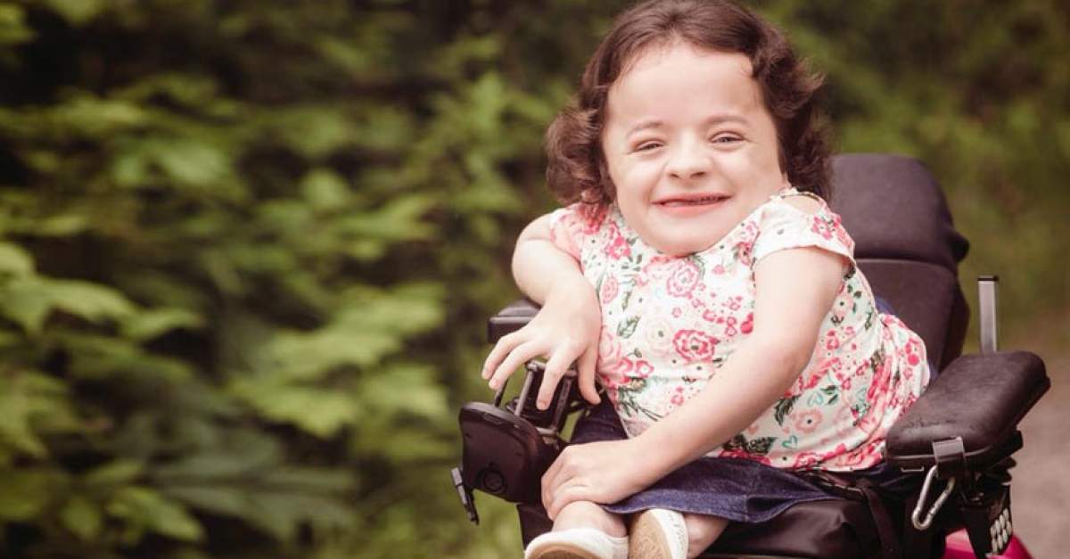
•Individuals with severe OI (Type III) experience a continuous cycle of fractures and bone deformity from infancy onward. They usually have noticeable bowing of limbs by early childhood, and many lose the ability to walk as they get older (if they ever acquire it – some never walk independently). They are particularly prone to complications like scoliosis, chest deformities, and basilar invagination. Respiratory problems can emerge in teenage or adult years because of severe scoliosis compressing the lungs. With modern interventions, many Type III OI patients now survive into adulthood (in contrast to past eras where frequent infections and respiratory failure led to earlier mortality). Their quality of life greatly hinges on surgical interventions, physical therapy, and assistive devices to maximize mobility and independence.
•In lethal OI (Type II), infants often do not survive beyond birth or the first year of life due to the severity of fractures and underdeveloped lungs. This type is evident on prenatal ultrasound or at birth with the infant having a very small, fragile ribcage and numerous fractures. Perinatal mortality is extremely high for Type II OI – the term “perinatally lethal” indicates most affected newborns succumb shortly after birth.
Despite these challenges, it’s important to note that OI does not affect intelligence or cognitive development. Children with OI are mentally normal and, with appropriate medical care, can attend school and participate in many activities. Over time, proactive management (surgical rodding, medications like bisphosphonates, and physical therapy) has significantly improved the outlook for OI patients, allowing many to lead full lives with controlled symptoms.
Causes
OI is fundamentally a genetic disorder caused by mutations that affect the body’s ability to make strong connective tissue (particularly bone collagen). Both biological (genetic) and hereditary factors are central, while environmental causes or triggers do not play a significant role in causing OI.
Genetic Causes: The vast majority of OI cases are caused by a mutation in one of the genes that are critical for producing Type I collagen. Collagen is the primary protein “building block” of bone (as well as skin, tendons, and other tissues), forming a fiber framework on which minerals like calcium are deposited. In normal bone, sturdy collagen fibers give the bone flexibility and resilience. In OI, a genetic defect disrupts this process.
•Collagen Gene Mutations: Approximately 85–90% of all OI cases result from mutations in the COL1A1 or COL1A2 genes, which encode the two chains of Type I procollagen . These are typically autosomal dominant mutations – meaning a single mutated copy of the gene inherited from one parent (or arising new in the child) can cause the disease. COL1A1/COL1A2 mutations come in two major types:
•Quantitative (Null) mutations: These mutations cause one of the two COL1A1 genes to produce no collagen chain (haploinsufficiency). The result is a reduced amount of normal collagen. This usually causes milder OI (Type I), since the collagen that is made is of normal quality, there’s just less of it . People with these mutations have half the normal collagen production, leading to bone fragility but often fewer deformities .
•Qualitative (Structural) mutations: These are mutations that change the structure of the collagen protein (often by substituting one amino acid for another in the collagen triple helix, classically a glycine substitution) . These abnormal collagen molecules have a dominant-negative effect: they get incorporated into collagen fibers and make them weak or dysfunctional. Structural mutations typically cause moderate to severe OI (Types II, III, or IV) . For example, a single glycine substitution in the collagen chain can prevent the triple helix from forming properly, leading to weak, misfolded collagen that the cell has trouble processing . The collagen quantity may be normal or near-normal, but its quality is poor, which severely weakens bones . This is why these mutations often result in more severe clinical outcomes, including perinatal lethal OI in the worst cases. Over 1,500 unique mutations in COL1A1/COL1A2 have been identified in OI patients , explaining the wide range of severity observed.
The immediate effect of these collagen mutations is that bone tissue is improperly formed. A defect in type I collagen “weakens connective tissues, particularly bone, resulting in the characteristic features” of OI . In practical terms, bones are brittle because either there isn’t enough collagen scaffold or the scaffold is faulty.
•Other Gene Mutations: About 10–15% of OI cases are caused by mutations in other genes – often inherited in recessive or X-linked patterns (meaning both copies of a gene are defective, or the gene is on the X chromosome) . To date, scientists have identified at least 19 distinct forms (types I through XIX) of OI based on different genetic causes . These rarer types of OI typically involve genes that play a role in collagen processing or bone formation. For instance:
•Some genes code for proteins that help modify or fold the collagen properly inside cells. Examples include CRTAP and P3H1 (also known as LEPRE1), which work together in a complex that hydroxylates the collagen protein during its synthesis. Mutations in these can cause recessive OI (former Types VII and VIII), leading to severe OI despite normal collagen genes .
•Other genes affect osteoblast function (the cells that build bone). For example, mutations in WNT1 (involved in a signaling pathway for bone formation) or SP7 (a transcription factor for bone development) can cause OI or OI-like syndromes . Mutations in IFITM5 cause Type V OI (unique features like calcified forearm membrane and normal teeth but bone fragility). An X-linked gene PLS3 can cause a form of OI in boys (since males have only one X chromosome).
•There are also genes like SERPINF1, FKBP10, SPARC, BMP1, etc., each associated with particular OI variants . These mutations disrupt different steps of collagen maturation (e.g., SERPINH1 encodes HSP47, a collagen chaperone) or bone mineralization processes. The end result of these non-collagen gene mutations is still brittle bones, but often with some clinical differences (for instance, some recessive forms do not have blue sclera or have atypical features).
In summary, what all these genetic causes have in common is that they impair the structural integrity of bone. Whether by faulty collagen or by disturbed bone cell function, the biological cause of OI is always tied to the proteins that build and maintain bone tissue. This is why OI is considered a prototype “collagen-related” genetic disorder.
Hereditary Patterns:
OI can be inherited or arise de novo:
•The classic inheritance for most OI (Types I–IV) is autosomal dominant. A person with OI has a 50% chance of passing the mutant gene to each child. Many families have multiple generations affected. For instance, an individual with Type I OI will often have a parent and perhaps siblings with the condition. However, in some cases the gene mutates for the first time in the egg or sperm (a spontaneous mutation), so a child can be born with OI to unaffected parents.
•The rarer types (VII and above, and a few cases of Type II/III) can be autosomal recessive, where both parents are carriers without symptoms and have a 25% chance with each pregnancy to have an affected child . Recessive OI might be suspected if the baby has OI but neither parent has any signs; genetic testing can confirm if both parents carry a mutation in a recessive OI gene.
•There is also an X-linked form (very rare) involving the PLS3 gene on the X chromosome, which typically affects males (as females have a second X that can compensate).
It’s worth noting that a significant fraction of OI cases are due to new mutations rather than inherited ones. For example, most cases of the lethal OI Type II are new dominant mutations (the parents are healthy). In such scenarios, the recurrence risk in the family is low, but not zero. A phenomenon called germline mosaicism can occur, where a parent carries the mutation in a subset of their reproductive cells without showing OI in their own body. In fact, if a couple has one child with a new dominant OI mutation, there’s an estimated 2–7% chance of another child with OI due to mosaicism in a parent . This is why genetic counseling is important even for seemingly isolated cases.
Environmental Factors: There are no known environmental causes or triggers for OI. One cannot “catch” OI or develop it from external factors; it is present from the moment of conception when the genetic mutation is in place. Things like nutrition, environment, or trauma do not cause OI – however, they can influence its severity. For example, poor nutrition (especially insufficient vitamin D or calcium) can exacerbate bone fragility in someone who already has OI, and certain activities or injuries obviously can trigger fractures in OI patients. But in a person without the genetic mutation, environmental factors alone will not create OI.
OI is not preventable through lifestyle changes by the parents or avoidance of certain exposures during pregnancy – it is fundamentally a genetic issue. That said, once a person has OI, a healthy lifestyle (good diet, supplements, physical therapy) can help optimize their bone health, which we will discuss in “Prevention & Precautionary Measures.”
In summary, OI is caused by inborn genetic mutations affecting collagen or bone biology, with COL1A1 and COL1A2 mutations accounting for most cases . It is typically inherited in families in a dominant pattern, though new mutations are common. Environmental or external factors do not cause OI, though they may affect outcomes for those with the condition.
Risk Factors
Because OI is a genetic disorder, the “risk factors” for OI largely revolve around genetics and heredity rather than the typical lifestyle or demographic risk factors we consider for many diseases. Unlike conditions such as heart disease or diabetes, factors like diet, smoking, or occupation are not relevant to whether a person will be born with OI. However, there are certain factors that increase the likelihood of OI occurring in a family or population:
•Family History and Genetics: The single biggest risk factor for OI is having a parent with OI or a family history of the disorder. If one parent has an autosomal dominant form of OI, each child has a 50% chance of inheriting the mutated gene and having OI . For recessive OI, if both parents are carriers of a defective gene (and typically unaffected themselves), there is a 25% chance with each pregnancy for the child to have OI . Thus, OI can cluster in families across generations or among siblings. Any couple who have one child with OI (especially if neither parent is affected) has a slightly elevated risk in future pregnancies due to possible germline mosaicism, as mentioned earlier . Families known to carry OI mutations (either dominant or recessive) are clearly at high risk of having affected offspring.
•Demographic Factors (Age, Gender, Ethnicity): OI does not discriminate by sex or ethnicity. Males and females are affected equally since most forms are autosomal (not sex-linked) . OI occurs in all racial and ethnic groups with similar frequency. There are some rare subtypes that have appeared more frequently in certain isolated populations due to founder mutations (for example, a particular recessive OI mutation may be more common in a specific geographic or ethnic group that had a common ancestor) . For instance, Type VI OI (caused by SERPINF1 mutations) was initially identified in a consanguineous community; Type V OI has been noted in certain regions of Canada (First Nations) with a founder effect . But these are exceptions – broadly, OI’s incidence is uniform worldwide. Parental age is not a well-established factor, although some studies in genetic disorders suggest that older paternal age can be associated with a higher rate of new dominant mutations. If this holds for OI, older fathers might have a slightly increased chance of having a child with a spontaneous OI mutation, but concrete data specific to OI is limited. Maternal age does not seem to influence OI risk. In summary, being born to a parent with OI or into a family with the gene is the primary demographic risk factor; otherwise, everyone has roughly the same baseline chance of a random mutation causing OI (~1 in 15,000 births).
•Consanguinity: Marriages or unions between relatives (even distant cousins) increase the risk of recessive genetic conditions. In populations or cultures where consanguineous marriage is common, there may be a slightly higher prevalence of rare recessive OI types. This is because carriers of the same rare mutation are more likely to pair. If two carriers of a rare OI gene variant have children, the risk of OI in their child is 25%. For example, recessive OI due to mutations in genes like FKBP10 or BMP1 might appear in such contexts more than in the general outbred population .
•Existing Bone Disorders: Having a pre-existing condition is generally not applicable to OI risk, since OI is present from birth. OI is sometimes confused with juvenile osteoporosis, but that is a different condition that arises in childhood without the collagen gene mutations. There is no evidence that conditions like childhood osteoporosis, rickets, or any metabolic bone disease will turn into OI – they are separate diagnoses. However, one important consideration: infants and children who present with multiple fractures need careful evaluation to rule out OI versus other causes (like abuse or other bone diseases). A history of multiple unexplained fractures in siblings could be a clue to OI as a risk factor within that family.
•Lifestyle and Occupation: These do not cause OI. No lifestyle factor of the parents (before or during pregnancy) has been linked to causing OI in a child. For the person with OI, lifestyle can affect how often they get injured (for instance, contact sports will raise their risk of fractures, high-impact occupations would be risky), but these are risk factors for fractures, not for having the disease itself. Therefore, from a causation perspective, lifestyle/occupation aren’t risk factors for acquiring OI (one is born with it), though they are considerations in managing OI (e.g., someone with OI should avoid jobs with a high fall risk).
In summary, the primary risk factor for OI is genetic inheritance. If OI “runs in the family,” the risk to offspring is high. In the general population, OI is rare and occurs sporadically due to spontaneous mutations. Everyone has a small baseline risk of having a child with OI due to a new mutation, but it’s not linked to any modifiable factor. This makes OI quite different from common diseases – here the emphasis is on genetic counseling and family history, rather than prevention via risk factor modification.
Important context: Because OI is rare and often involves new mutations, many affected children are born into families with no history of the disorder. This sometimes leads to initial suspicions of child abuse when unexplained fractures are found, rather than recognizing a genetic condition. It underscores that any infant with recurrent fractures should be evaluated for OI (via history, exam for blue sclera or other signs, and genetic testing) even if no family risk factors are apparent. The absence of risk factors does not mean absence of disease – OI can truly appear out of the blue in a family due to a de novo mutation.
Complications
OI’s complications arise from its multi-system involvement and the cumulative damage from fractures and deformities. These complications can affect the musculoskeletal system, dental and auditory systems, respiratory and cardiovascular systems, and overall longevity and quality of life. Below we outline key potential complications, long-term effects, and outcomes:
•Musculoskeletal Complications: The most direct complications of OI are orthopedic. Repeated fractures can lead to chronic bone deformities. For example, multiple fractures in a limb, especially if inadequately treated, can result in malunions (bones healed in improper alignment) or pseudoarthrosis (a “false joint” where a bone did not heal and forms a fibrous connection) . Long-term, these issues cause reduced mobility or function of the limb. Joint problems are also common: malformed bones can cause abnormal joint loading and early-onset osteoarthritis. Many adults with OI experience joint pain and stiffness from their 30s or 40s onward. Contractures (permanent joint stiffness) can occur in severe cases due to muscle imbalance around fragile bones . Spinal deformities like scoliosis (sideways curvature) and kyphosis (forward curvature, e.g., a hunched upper back) can become severe, especially in wheelchair-bound patients. A severe scoliosis (>60 degrees curve) can compress the lungs and abdominal organs, leading to secondary issues (respiratory compromise and gastrointestinal issues like acid reflux from pressure). Vertebral compression fractures in the spine can cause chronic back pain and height loss. In severe OI, the combination of short stature and bone deformities can result in a markedly reduced arm span or leg length, making self-care activities challenging. Many individuals with deforming OI (Type III) rely on wheelchairs or crutches due to lower limb deformities and fracture risk – this in turn can cause complications like muscle atrophy or pressure sores if not well managed.
•Permanent Disability: Due to the bone and joint complications, OI can lead to physical disabilities. These range from mild (like a slight limp or moderate hearing loss) to severe (complete dependence on a wheelchair for mobility, or need for personal assistance in daily activities). Mobility impairment is a major long-term effect – some children with severe OI lose the ability to walk as fractures and deformities accumulate. However, with modern rodding surgeries and rehab, many can maintain some level of ambulation or at least independent transfer (moving between bed and chair, etc.). Fine motor issues may arise if arm bones are severely deformed or if multiple arm fractures limit function (though the arms are often less affected than legs in OI). Despite these challenges, with adaptive devices and environmental accommodations, many OI patients finish school, work, and lead productive lives. Disability in OI is highly variable; importantly, cognitive function is normal, so individuals can intellectually engage fully even if physically limited.
•Dental Complications: In patients with dentinogenesis imperfecta, dental health can be a lifelong challenge. Teeth may wear down to the gum-line, break easily, or fall out. This can lead to difficulties with chewing and nutrition, as well as self-esteem issues due to appearance. Early dental intervention (crowns, overlays) is often needed to preserve teeth. Even with good dental care, many OI patients require dentures or implants at a relatively young age. Dental infections or abscesses can also occur if brittle teeth crack and expose the pulp. Good oral hygiene and regular dentist visits are crucial to manage these issues.
•Hearing Complications: Hearing loss in OI (usually conductive hearing loss from ossicle issues) can progress to significant levels, impacting communication. Many adults with OI benefit from hearing aids. In some cases, surgical stapedectomy (replacing the stapes bone in the middle ear) can improve hearing. Without these interventions, hearing loss can be a serious social disability. It’s not life-threatening, but it affects quality of life and independence (e.g., difficulty talking on phone, hearing alarms, etc.). Regular hearing evaluations are recommended starting in the teenage years for those with OI, so that any decline can be caught early and addressed.
•Neurological Complications: Basilar invagination and atlantoaxial instability (instability of the first two cervical vertebrae) are rare but serious in severe OI. Basilar invagination can cause compression of the brainstem, leading to symptoms like headaches, vertigo, weakness or tingling in the limbs, and in severe cases, problems with breathing or swallowing. It may require neurosurgical intervention (such as spinal fusion or decompression surgery) to prevent fatal outcomes. Spinal cord compression from severe scoliosis or vertebral fractures can also cause neurological deficits (like paraplegia, though this is uncommon in OI compared to other conditions). Additionally, chronic pain due to nerve compression (for instance, nerves pinched by collapsed vertebrae or by deformities) is a complication requiring pain management. Hydrocephalus (fluid build-up in the brain) has been reported in some OI patients with skull abnormalities, but it’s not common. Overall, most neurological complications in OI stem from skeletal abnormalities impacting the nervous system mechanically.
•Respiratory Complications: Perhaps the most consequential complications in terms of mortality are those involving breathing. As mentioned, restrictive lung disease from severe scoliosis or chest wall deformities can reduce lung capacity. This predisposes patients to frequent respiratory infections (pneumonia) and can lead to chronic respiratory failure in adulthood. Some individuals with severe OI require nocturnal ventilation support (like CPAP or BiPAP machines at night) if their lung function is low, or even tracheostomy with ventilator support in extreme cases. Asthma and bronchitis may also be more severe in OI patients because of already reduced pulmonary reserve. In fact, a long-term Danish study found that OI patients had a higher risk of death from respiratory diseases compared to the general population . For infants with Type II OI, underdeveloped lungs are usually the immediate cause of death at birth. For survivors with Type III, careful monitoring of lung function and early intervention (bracing or spinal surgery to slow scoliosis progression, pulmonary physiotherapy, etc.) is important to mitigate respiratory issues.
•Cardiovascular Complications: While not as prevalent as respiratory issues, heart and vessel problems can occur. Connective tissue weakness can cause mitral valve prolapse or aortic dilation as noted earlier. Over years, aortic dilation can progress and risk dissection (tear). OI patients are also slightly more prone to arterial ruptures or aneurysms; for example, there are case reports of aortic dissection in people in their 20s or 30s with OI . If an OI patient complains of acute chest pain or back pain, physicians must consider these possibilities. Preventative cardiology (use of beta-blockers or blood pressure control) might be advised if aortic root enlargement is detected. Additionally, the Danish study noted an increased risk of death from cardiovascular causes in the OI population (though not as high as respiratory causes) . Regular echocardiograms may be recommended in moderate-severe OI to monitor the heart.
•Renal and Other Organ Issues: Generally, OI doesn’t directly cause kidney or liver problems. However, long-term use of certain medications (like bisphosphonates) requires monitoring of kidney function. Also, immobilization from fractures can cause kidney stones or hypercalciuria (excess calcium in urine) in some cases. There have been some reports of OI patients developing bone-related tumors like osteosarcoma at fracture sites in adulthood, but it’s unclear if OI increases cancer risk or if those were coincidental.
•Psychosocial Impact: A softer aspect of “complications” is the psychological toll. Children growing up with frequent fractures may develop anxiety or fear of new activities. They might also face social challenges due to short stature or using a wheelchair, which can affect their mental health. Chronic pain in adults with OI can lead to depression if not properly managed. These aren’t medical complications per se, but they are important long-term issues. Nonetheless, many individuals with OI are remarkably resilient and adapt well with strong family and community support.
•Mortality: OI has a broad range of mortality outcomes depending on type. Type II OI is typically fatal in the perinatal period – most infants with Type II die within days to weeks of birth due to respiratory failure or intracranial hemorrhage from skull trauma during delivery . For Types III and IV, historically life expectancy was reduced, but with modern care many live into middle age or beyond. For Type I (mild), life expectancy is often normal or only slightly reduced. A comprehensive study in Denmark (which followed 687 OI patients over 35 years) found that overall, OI patients have a higher mortality rate than the general population at all ages . The study reported that the median survival was 72.4 years for men with OI vs. 81.9 years in the general male population, and 77.4 years for women with OI vs. 84.5 years in the general female population . That indicates an average reduction in lifespan by a few years (mostly attributable to severe cases). The hazard ratio for premature death in OI was about 2.9 times that of the normal population in this study . The leading causes of excess mortality were respiratory issues (like chronic lung disease and pneumonia), gastrointestinal causes (possibly related to surgery complications or severe scoliosis compressing GI tract), and trauma (fatal fractures or head injuries from falls) . Encouragingly, with improved treatments, more people with OI (even severe forms) are surviving longer than ever before. Many with milder OI have near-normal lifespans , and even those with moderate OI often live into late adulthood, especially if they avoid serious complications.
In summary, OI can lead to significant complications affecting multiple organ systems:
•Bones/joints: chronic deformities, arthritis, pain, disability.
•Teeth: severe dental issues requiring major dental work.
•Hearing: loss treatable with aids or surgery.
•Spine/neurology: risk of spinal cord or brainstem compression in severe cases.
•Lungs: reduced capacity, higher infection risk.
•Heart: occasional valve or vessel problems.
•Life expectancy: slightly reduced on average, especially in severe cases, due to the above complications.
Early and proactive management of OI (orthopedic surgeries, dental care, hearing checks, pulmonary monitoring) can prevent or lessen many of these complications. For instance, rodding bones early can prevent crippling deformities; spinal fusion can halt a worsening scoliosis; bisphosphonate treatment can improve bone density and reduce fracture rates (thus reducing deformity and pain complications) . Thus, while OI poses many potential complications, attentive medical care and supportive therapies have greatly improved the outlook, enabling individuals with OI to manage complications and lead fulfilling lives into older age.
Diagnosis & Testing
Diagnosing osteogenesis imperfecta typically involves a combination of clinical evaluation, family history, and specialized testing. Because OI shares the symptom of fractures with more common conditions (like child abuse or other bone disorders), an accurate diagnosis is critical for proper management. Below are the common diagnostic approaches and tests for OI, as well as methods for early detection:
Clinical Evaluation:
A thorough medical history and physical examination are the first steps. Doctors will note the history of fractures (frequency, circumstances, and whether they were disproportionate to the level of trauma). They will ask about family history of OI or of relatives with frequent fractures or skeletal issues. On exam, clinicians look for classic OI signs: blue sclerae, tooth abnormalities, bone deformities, short stature, joint laxity, and hearing loss. Often, the presence of multiple signs makes OI apparent. For example, a child with recurrent fractures, blue sclerae, and dentinogenesis imperfecta has a very high likelihood of OI.
Importantly, doctors also evaluate for signs that might suggest alternative diagnoses – for instance, in a child with fractures, they’ll look for bruising patterns or other injuries that could indicate non-accidental trauma (child abuse). Ruling out abuse is a key part of the initial evaluation when OI is suspected, given the serious implications . Typically, certain features (like the presence of blue sclerae, family history, or multiple fractures in various healing stages without soft tissue injury) point toward OI rather than abuse.
Clinically, OI has a set of “Major clinical features” that aid diagnosis:
•Generalized decreased bone mass and bone fragility (leading to fractures) – this is usually evident from the history .
•Blue sclerae .
•Dentinogenesis imperfecta (opalescent teeth) .
•Hearing loss (especially in adults or a family history of early hearing loss) .
•Other supportive features include ligamentous laxity, short stature, and a positive family history of OI .
If a patient presents with these hallmark features, clinical diagnosis of OI is strongly supported even before any genetic test is done.
Radiological Imaging:
Imaging studies are crucial in diagnosing and assessing OI:
•X-rays (Radiographs): Plain X-rays of affected bones can show signs characteristic of OI. These include:
•Multiple fractures in different stages of healing.
•Bone deformities like bowing of long bones.
•Osteopenia or generalized low bone density (bones appear thinner or less opaque than normal).
•Wormian bones in the skull – extra small bones in the cranial sutures – which are often seen in OI .
•Vertebral compression fractures or shape abnormalities (vertebrae may be biconcave or flattened, known as “codfish” vertebrae).
•In some types (like OI Type V), hyperplastic callus formation is visible – an exuberant, enlarged callus at fracture sites that can look like a bone tumor on X-ray .
•Thin cortices (outer bone layer) and wide bone shafts (as bones remodel oddly after fractures) are common findings .
•“Popcorn calcifications” at the ends of long bones in children – irregular, spotty calcifications in cartilage – are sometimes noted in OI .
Radiologists experienced with OI can often recognize the bone “pattern” on X-rays. For example, “codfish” vertebrae, wormian skull bones, and multiple old rib fractures in a toddler strongly suggest OI over abuse or other conditions.
•Bone Density Scan (DXA): A dual-energy X-ray absorptiometry (DXA) scan can measure bone mineral density. In OI patients (even infants), bone density is usually significantly below average for age . While a DXA alone can’t diagnose OI, it supports it by showing osteoporosis in a young person. It’s also useful for tracking response to treatments like bisphosphonates (expecting an increase in density).
•Ultrasound: In some cases, prenatal ultrasound can detect severe OI. As early as 16–18 weeks’ gestation, signs like short, bowed or fractured limbs, a small chest, and undermineralized skull bones (which may compress easily with probe pressure) can be seen in OI Type II . Milder OI may not be evident until later in pregnancy (or not at all on ultrasound). Obstetricians may note polyhydramnios (excess amniotic fluid) in pregnancies with an OI fetus because the fetus moves less or has a weak suck (in severe cases) . Ultrasound is very effective for detecting the lethal OI forms before birth, enabling early counseling of the parents.
•CT/MRI: These are not routine for diagnosing OI, but have specific uses:
•A CT scan of the skull can show wormian bones or basilar invagination in detail .
•High-resolution CT of the ear bones may be done if planning surgery for hearing loss (to see the ossicle structures).
•MRI of the craniocervical junction is critical if basilar invagination is suspected, to assess brainstem compression . MRI can also help evaluate spinal cord compression from scoliosis or gauge the integrity of spinal ligaments if someone with OI has a neck injury.
Overall, imaging builds the case for OI by revealing the bone abnormalities consistent with the disease and by excluding other pathologies (like rickets, which has different bone changes).
Laboratory Tests:
There is no simple blood test that “checks for OI” in the way one might test for an infection or a metabolic disease. Routine bloodwork (blood counts, electrolytes, calcium, phosphorus) in OI patients is usually normal. However, some lab aspects include:
•Bone Turnover Markers: Alkaline phosphatase (ALP), an enzyme associated with bone activity, might be mildly elevated in children with OI because their bones are constantly repairing fractures . In adults, ALP is often normal unless there’s a healing fracture. Other markers (osteocalcin, PINP, CTX) could be abnormal, but these are not specific or routinely used for diagnosis.
•Collagen Biochemical Testing: Before genetic tests became widely available, an older diagnostic method was a skin biopsy to examine type I collagen production. A small skin sample can be cultured into fibroblast cells, and those cells will produce collagen. By analyzing the collagen’s structure (electrophoresis or mass spectrometry), about 85-90% of individuals with dominant OI show an abnormal collagen pattern (either a smaller amount or abnormal mobility of collagen chains) . For example, in Type I OI, one would see about half the normal amount of type I collagen produced. In structural mutation OI, one might see two types of collagen molecules (normal and abnormal) being made. This test can confirm the collagen defect in uncertain cases. It’s noted that this method is positive in ~80% of Type I–IV OI (some mosaic or mild cases might be missed). Today, DNA testing has largely supplanted this because genetic testing can directly pinpoint the mutation.
•Genetic Testing: This is the definitive test for OI now. A gene panel test can analyze the genes known to cause OI (COL1A1, COL1A2, and all the others associated with OI types V–XIX). If a causative mutation is found, it confirms the diagnosis at the molecular level. Genetic testing is particularly useful in:
•Mild cases where OI signs are subtle (to differentiate from just being accident-prone or having early osteoporosis).
•Atypical cases or when other diagnoses are possible (like inherited rickets vs OI).
•Prenatal or preimplantation diagnosis (testing a fetus or embryo for the family’s known OI mutation).
For most patients with a clear clinical picture, genetic testing primarily provides confirmation and can guide family planning (knowing the exact mutation allows testing of relatives or prenatal diagnosis in future pregnancies). Notably, no commercially available single “blood test” fully diagnoses OI without genetic analysis . OI’s genetic heterogeneity means a multi-gene panel or complete sequencing is often needed to catch unusual variants.
•Differential Diagnosis Exclusion: Sometimes additional tests are done to exclude other conditions:
•Metabolic tests (vitamin D levels, parathyroid hormone) to rule out nutritional rickets or other metabolic bone diseases.
•Blood calcium/phosphate (which are normal in OI, but abnormal in some metabolic bone diseases).
•Tests for child abuse: unfortunately there’s no test for abuse, but doctors may involve social services to investigate when fractures are unexplained. If OI is suspected, finding a genetic mutation greatly helps differentiate from abuse. Doctors must proceed carefully and gather all evidence.
•Biopsy of bone: rarely done, but a bone biopsy could show thin, woven bone with abnormal histology in OI. This is seldom necessary if genetic tests confirm OI.
Early Detection:
•Prenatal Testing: If there is a family history of OI or a known parental mutation, prenatal diagnosis is possible. Chorionic villus sampling (CVS) (at ~10-12 weeks of pregnancy) or amniocentesis (at ~15-18 weeks) can obtain fetal DNA to test for the OI mutation . This can definitively tell if a fetus has inherited the mutation. Families with severe OI sometimes choose this route to prepare for management or consider pregnancy options. As mentioned, ultrasound can also signal OI in severe cases (Type II or III) as early as the second trimester . Ultrasound is non-invasive but not 100% – it might miss mild OI or confuse OI with other skeletal dysplasias. A very early clue on ultrasound for severe OI is “compressible skull” (the skull can be easily indented by the ultrasound probe due to poor ossification).
•Newborn Screening: There is no routine newborn screening test for OI (like a blood spot test) because it’s rare and not easily detectable by biochemical markers. However, a neonatologist or pediatrician may notice certain things at birth: for instance, if a newborn has bone fractures at birth, very blue sclerae, and limb deformities, they will suspect OI immediately. In mild OI Type I, a baby might not have fractures at birth, so it could escape detection until the first fracture occurs months or years later.
•Early Childhood: Pediatricians are trained to watch for patterns like repeated fractures. If a toddler has had multiple fractures with relatively low-impact injuries, OI should be considered. Some milder OI cases get diagnosed around age 2-5 when the child starts ambulatory activities that reveal bone fragility. A clue could be a delay in walking or crawling due to fractures.
•Genetic Counseling: If OI is known in a family, genetic counseling and possibly preimplantation genetic diagnosis (PGD) can be used. For example, with IVF, embryos can be tested for the family’s OI mutation and only unaffected embryos implanted . This is a form of prevention of passing on OI (discussed more in the Prevention section), but it’s also an early detection method at the embryonic stage.
In practice, diagnosing OI is often straightforward when classic features are present, but can be challenging in mild cases or when it must be distinguished from other causes of fractures. The approach is multidisciplinary: orthopedists, geneticists, radiologists, and sometimes endocrinologists (for bone metabolism expertise) all play a role. A confirmed diagnosis is crucial because it changes management – for example, if a child is diagnosed with OI, clinicians will avoid certain treatments (like high-force manipulation of bones) and will institute fracture prevention strategies and medications.
To summarize, diagnosis of OI involves:
•Clinical signs (fragile bones, blue sclerae, etc.) and family history.
•Radiographic evidence of diffuse bone fragility and old fractures.
•Genetic testing to confirm mutations in collagen or related genes.
•Excluding look-alike conditions (especially non-accidental trauma in children).
Early and accurate diagnosis allows for timely treatment (like starting bisphosphonates to strengthen bones in infancy if needed) and counseling of the family. Given that OI is a lifelong condition, diagnosing it even in the first year of life can significantly improve the child’s long-term outcome by guiding proper management and preventing harmful assumptions (such as false allegations of abuse).
Treatment Options
There is currently no cure for osteogenesis imperfecta, but there are many treatment options to manage symptoms, strengthen bones, prevent fractures, and improve quality of life. Treatment of OI is highly individualized, based on the patient’s age, disease severity, and specific needs. It requires a multidisciplinary approach including orthopedists, endocrinologists, physical therapists, dentists, audiologists, and others. Below we cover standard therapies and emerging treatments:
Supportive Care & Lifestyle Management:
Regardless of disease severity, patient and family education is fundamental. Caregivers are taught safe handling techniques for infants (to avoid causing fractures inadvertently) – for example, never lifting an OI baby by the underarms, and instead supporting their buttocks and back with the body well-supported . As children grow, they are encouraged to stay active within safe limits. Physical therapy is important to build muscle strength (strong muscles help support and protect bones) and to improve gross motor skills. Low-impact exercise like swimming is often recommended. Activities that put excessive stress or risk high impact (like jumping from heights, contact sports) are typically avoided . Parents may need to make home modifications (ramps instead of stairs, extra padding on hard corners, etc.) to create a safe environment. Nutrition plays a role – ensuring adequate intake of calcium, vitamin D, and calories for bone health and to support growth. Some children with OI have difficulty gaining weight due to energy spent on fracture healing or limited mobility; nutritional supplements might be needed.
Orthopedic Interventions:
Managing fractures and bone deformities is a core aspect:
•Casting/Splinting: Fractures in OI usually heal at a normal rate (often surprisingly fast in children) , but the bone may be fragile during healing. Casts or splints are used to immobilize broken bones, though lighter weight immobilization (like bracing) is preferred over heavy casts that can cause stress on neighboring bones. A caution: overly aggressive reduction (setting) of fractures can itself cause new fractures, so gentle handling is key.
•Intramedullary Rodding: This surgical procedure is one of the most effective orthopedic treatments for OI. In rodding, a metal rod is inserted into the marrow canal of long bones (such as the femur or tibia) to provide internal support. Telescopic rods (like the Fassier-Duval rod) are especially useful in growing children, as they elongate as the bone grows. Rodding stabilizes the bone, prevents or corrects deformities, and reduces fracture incidence (because the rod acts as an internal splint) . For instance, a child with OI who has recurrent femur fractures and bowing will often get rods placed in the femurs and possibly tibiae. Post-surgery, the child usually gains mobility and has fewer fractures in those bones. Rodding can be done in early childhood and rods may be replaced or updated as the child grows.
•Spinal Surgery: If severe scoliosis develops, spinal fusion surgery may be needed to straighten and stabilize the spine. This can prevent respiratory compromise and further curvature. Spinal surgery in OI is delicate due to bone fragility but can be lifesaving in terms of preserving lung function.
•Bracing: Braces (orthoses) for the legs, ankles, knees, or spine can help support weak bones or prevent deformity. For example, a thoracolumbar spinal brace might slow progression of scoliosis in a growing child. Leg braces can add stability for walking. Custom mobility aids (like standing frames) help children with severe OI bear weight safely, which is beneficial for bone strength.
•Surgery for specific fractures or deformities: Besides rodding, OI patients might require surgeries to fix bowed bones (osteotomy to cut and realign a bone, followed by rod insertion), to stabilize joint subluxations, or to address basilar invagination (which may need a neurosurgical decompression and fusion).
•Amputation: This is rarely done in OI, but in a few cases where a limb is non-functional due to multiple pseudoarthroses, an amputation and a prosthetic limb might provide better function. This is very uncommon and usually a last resort.
Orthopedic management aims to maximize function – enabling the patient to sit, stand, and walk if possible, and to use their hands and arms effectively. Regular follow-ups with an orthopedic surgeon are essential, since as the child grows, rods might need extending or replacing, and new issues might arise.
Pharmacological Treatments:
Two main categories of medications have been used: anti-resorptives (to reduce bone breakdown) and anabolic agents (to stimulate bone formation).
•Bisphosphonates: These are anti-resorptive drugs widely used in OI. They bind to bone and inhibit osteoclasts (the cells that break down bone tissue). In children with OI, intravenous bisphosphonates like Pamidronate or Zoledronic acid have become standard for moderate to severe OI. Studies have shown that bisphosphonate therapy can increase bone density, reduce bone pain, and decrease fracture rates in OI . For example, a child on pamidronate might go from having a fracture every month to maybe one or two a year. It also often improves mobility – children report less bone pain and can participate more in physical therapy. Bisphosphonates are usually started in infancy or early childhood for severe cases (Type III, severe Type IV) and given periodically (IV infusions every 3–6 months). Oral bisphosphonates (like alendronate) are sometimes used in milder cases or in adults. Long-term use of bisphosphonates in growing children is still being studied, but many will continue therapy through the growth years and sometimes into adulthood. Side effects include temporary bone pain or flu-like symptoms after infusions; there is concern about over-suppressing bone turnover, so doctors monitor dosage carefully. Overall, bisphosphonates are considered a mainstay of OI treatment and have significantly improved outcomes .
•Teriparatide (PTH Analog): This is an anabolic (bone-building) agent, basically a form of parathyroid hormone given by daily injection. It’s normally used for osteoporosis in adults to stimulate new bone formation. There has been a small trial of teriparatide in adults with OI, which showed increased bone density, but its use is not yet standard and it’s not approved specifically for OI . It may be considered in adult OI patients (particularly Type I or IV) who have persistent fracture issues, since children cannot use teriparatide (it’s contraindicated in open growth plates due to risk of osteosarcoma in rats). Research is ongoing in this area.
•Denosumab: This is a monoclonal antibody that targets RANKL, another way to inhibit osteoclasts (sort of like bisphosphonates but via a different mechanism). Denosumab has been tried in some OI patients in research settings and has shown improvement in bone density in small studies . It’s not yet an approved standard treatment for OI. One challenge is that denosumab’s effects wear off quickly after each dose and it can cause rebound high bone turnover if not continued, so its use requires careful monitoring of calcium and bone metabolism. It may become an alternative for patients who do not respond to or cannot take bisphosphonates.
•Growth Hormone: In some children with OI (particularly Type I), recombinant human growth hormone has been tried to improve growth and possibly bone density. Results have been mixed; it might increase height velocity modestly but is not broadly used.
•Calcitonin, Fluoride, Magnesium, Vitamins: Historically, various supplements and drugs were attempted (like calcitonin, sodium fluoride, magnesium, vitamin C) but they showed no significant long-term benefit for OI . Ensuring normal vitamin D levels is important, but megadoses or special formulas beyond standard nutrition aren’t curative.
Pain Management: Chronic pain from fractures or deformities is addressed with medications like acetaminophen or ibuprofen for mild pain, and sometimes opioids for severe acute fracture pain. However, caution with long-term opioid use is needed. Adjunct therapies like nerve blocks or muscle relaxants can help if muscle spasms or nerve pain is present.
Rehabilitation Therapies:
•Physical Therapy: PT is tailored to each patient. In infants, it might focus on gentle range-of-motion exercises and positioning to prevent flattening of the skull or tightness in hips. In toddlers and older children, PT includes muscle strengthening (low resistance exercises), weight-bearing activities (like supported standing, which encourages bone mineralization), and training in use of mobility aids (walkers, crutches). Aquatic therapy (hydrotherapy) is wonderful for OI patients – water allows movement with less gravitational stress on bones.
•Occupational Therapy: OT helps with fine motor skills and adaptation for daily activities. For example, learning how to dress with fragile limbs, use modified tools or pencils if one has deformities in arms, etc.
•Assistive Devices: Wheelchairs (often custom-made lightweight chairs), walkers, crutches, and canes are commonly used at different stages. Braces and splints may be used chronically to support ankles or knees for stability. Adaptive equipment at home (bath chairs, transfer boards) ensures safety. As patients get older, powered mobility scooters or electric wheelchairs can give independence if they cannot push a manual wheelchair easily.
•Orthotics: Bracing we mentioned in orthopedic, but foot orthotics or ankle-foot orthoses can help stabilize ankles for those who can walk. Spinal orthoses can be used for mild scoliosis or to support sitting posture.
Audiological and Dental Care:
•Hearing Aids: If hearing loss is present, early use of hearing aids can dramatically improve quality of life. Patients are monitored annually for hearing changes, and when thresholds drop, digital hearing aids are fitted. In cases of conductive hearing loss where hearing aids aren’t sufficient, surgical options like stapedectomy (replacement of the stapes bone in the ear with a prosthesis) may restore hearing.
•Dental Treatments: Children with dentinogenesis imperfecta need to see pediatric dentists familiar with OI. Baby teeth might need crowns to prevent wear. As permanent teeth come, they often crown the teeth or use bonded resin fillings to reinforce them. Orthodontic treatment can be complex if jaws are misaligned, but it’s often still possible with caution. Many patients eventually need full dental implants or dentures; with modern implantology, even OI patients can sometimes get dental implants (though bone fragility can be a challenge in the jaw as well). Maintaining oral hygiene is vital to avoid additional stresses like infections.
Emerging and Experimental Treatments:
Research is actively ongoing to find better treatments, aiming to improve bone quality beyond what bisphosphonates can do. Some exciting areas include:
•Sclerostin Inhibitors: Sclerostin is a protein that inhibits bone formation. Antibodies against sclerostin (like Romosozumab and Setrusumab) have been developed to treat osteoporosis by boosting bone formation. In OI, these drugs are being tested in clinical trials. Animal models of OI showed that sclerostin antibody treatment increased bone mass and strength . As of now, romosozumab has shown beneficial effects on bone in OI mice and is undergoing trials in humans, and setrusumab (UX143) is in Phase 3 clinical trials for OI . These drugs could become the first targeted anabolic therapy specifically for OI if proven safe and effective.
•TGF-beta Inhibition: Transforming growth factor beta (TGF-β) is another pathway of interest. Excess TGF-β activity is thought to contribute to bone fragility. An antibody drug, Fresolimumab, which neutralizes TGF-β, was tested in a small trial in adults with OI. Early results indicated some improvements in bone density, but research is ongoing . Animal studies (using a mouse OI model) with TGF-β antibodies showed promising results with stronger bones . This approach is still experimental.
•Gene Therapy and Gene Editing: Given that OI is monogenic (caused by single gene mutations), it’s a candidate for gene therapy. The challenges are immense because to “cure” OI, one would need to fix or silence the defective collagen gene in the majority of bone-producing cells. Researchers have experimented with strategies like siRNA to silence mutant collagen alleles, or using CRISPR gene editing in cells. So far, these are in preclinical or very early phases . No human gene therapy trial for OI is underway yet, but this is a hopeful avenue for the future. One concept is to use induced pluripotent stem cells from the patient, correct the mutation in the lab, and then infuse them back to produce healthy bone cells.
•Stem Cell Transplantation: In 1999, a landmark case was published where mesenchymal stem cells from a healthy donor were transplanted into a child with severe OI. The child showed some initial improvement in growth and reduced fractures. This opened exploration into bone marrow transplantation or mesenchymal stem cell (MSC) therapy for OI. A few subsequent attempts had mixed results – any engraftment of donor cells was low, and the effect tended to wane over time. However, research continues: there are ongoing trials using prenatal transplantation of stem cells (treating the baby while still in the womb) aiming to give OI fetuses a population of healthy bone-forming cells before birth. This is highly experimental.
•Other Medications: Various other pathways are being looked at. For example, some studies suggest that drugs like beta-blockers might reduce fracture rates by affecting sympathetic nervous regulation of bone (this is not mainstream). Another area is imperfect bone signaling correction – understanding how the mutant collagen leads to bone cell dysfunction and targeting those signals. Some have suggested that colony-stimulating factors or other hematologic drugs might boost bone repair in OI, but evidence is scant.
•Clinical Trials: As of now, the most active clinical trials for OI involve sclerostin inhibitors (like setrusumab) and growth stimulators. The OI community and organizations (like the Osteogenesis Imperfecta Foundation) are closely watching these developments.
Therapy by OI Type: A summary of typical management:
•Type I (mild): Focus on safety, physical therapy, and possibly periodic bisphosphonate if there are many fractures. Many Type I patients may not need surgical intervention except perhaps dental or hearing-related in adulthood.
•Type II (perinatal lethal): Intensive neonatal care is often not successful due to lung failure. Treatment is usually supportive/comfort care. In any subsequent pregnancy, prenatal diagnosis is offered due to the severe recurrence risk.
•Type III (severe): Aggressive management – start IV bisphosphonates in infancy, multiple rodding surgeries in early childhood, bracing, wheelchair mobility, etc. Essentially all modalities are used. Despite interventions, these patients have significant residual deformity, but treatments greatly improve their ability to sit, stand with support, and live longer.
•Type IV (moderate): Intermediate approach – often bisphosphonates and maybe a few rodding surgeries for the more problematic bones, physical therapy, etc. Many Type IV children can walk (with or without aids) after treatment, whereas they might not have without it.
Monitoring: OI treatments require regular monitoring. Bone density scans track improvement on medications. Blood tests monitor for any drug side effects (e.g., kidney function with bisphosphonate, calcium levels with new drugs like denosumab). X-rays check the status of rods or progression of deformities. Hearing tests annually, dental exams biannually, etc., are all part of comprehensive care.
In conclusion, the management of OI is about maximizing bone strength and minimizing injury:
•Medications like bisphosphonates fortify the bones from within by slowing breakdown.
•Surgeries like rodding mechanically reinforce bones.
•Therapies improve muscle support and skills.
•Adaptations and assistive devices help the person live as normally as possible while accommodating their fragility.
•Experimental therapies on the horizon aim to directly improve the bone’s biology further (e.g., by increasing formation or fixing the genetic defect).
With these treatments, outcomes for OI patients today are better than ever. Children who once might have been bedridden can now often walk with braces or a walker. Pain can be managed, and complications prevented or delayed. The overarching goal is to enable individuals with OI to achieve independence and a good quality of life, despite the challenges posed by their “imperfect” bones.
Prevention & Precautionary Measures
Because OI is a genetic disorder present from conception, true “prevention” of the disease is not straightforward in the way one can prevent infectious diseases or lifestyle diseases. However, there are measures to prevent transmission of OI to the next generation (genetic counseling) and numerous precautions to prevent fractures and complications in individuals who have OI. We will discuss these in turn:
Genetic Counseling and Family Planning:
For families affected by OI, particularly autosomal dominant forms, the primary preventive strategy is through informed family planning:
•Genetic Counseling: Families with OI are advised to see a genetic counselor who can explain the inheritance patterns and recurrence risks. Since each child of an affected parent has a 50% chance of inheriting OI (for dominant types) , some families consider limiting family size or exploring reproductive options. Counselors can also discuss the variability of OI – even if a parent has a mild form, the child’s presentation could range from mild to severe (due to phenomena like germline mosaicism or genetic modifiers).
•Preimplantation Genetic Diagnosis (PGD): This is a high-tech option available to those who want to avoid passing on OI. It involves IVF (in vitro fertilization) where the parents’ embryos are created in the lab and then tested at a very early stage for the OI mutation. Only embryos free of the mutation are implanted into the uterus. This essentially “guarantees” a child without OI from affected parents . PGD has ethical and financial considerations, but it is an option many OI families pursue to break the cycle of inheritance.
•Prenatal Diagnosis: If a pregnancy is already underway, couples can opt for prenatal testing (CVS or amniocentesis) to determine if the fetus has OI . If the fetus is affected, the family then faces the decision of whether to continue the pregnancy. Some may prepare for the birth of a child with OI (lining up specialists, etc.), while others may consider termination, especially if a lethal form is diagnosed. These are deeply personal decisions. Prenatal diagnosis is more about planning than prevention, but in a way, it can prevent the birth of an affected child if the parents choose that route. It’s worth noting that such choices raise ethical discussions; the OI community is sensitive to the notion of “selecting against” OI, with some disability advocates cautioning against viewing lives with OI as undesirable . Genetic counseling aims to support whatever decision is right for the family.
•Avoiding Known Risk Matings: In recessive forms, if a family is identified as carriers (say they had an affected child and discovered both parents carry a recessive OI gene), any further children have a 25% risk. Some families in that situation might use donor sperm or donor eggs to avoid the combination, or opt for adoption rather than risk another affected child. In communities with common recessive OI, carrier screening might be considered to avoid marriages where both individuals are carriers (though OI is so rare that this is not commonly done like it is for, say, thalassemia or Tay-Sachs in certain groups).
In summary, “prevention” of having a child with OI is possible only via reproductive technologies or decisions. This is a form of primary prevention at the family level, but it doesn’t cure the gene in the population; it’s an individual choice to not pass it on. Importantly, none of these methods can prevent or change OI in someone already born with it.
Precautionary Measures for Individuals with OI:
For those who have OI, the focus is on preventing injuries (fractures) and preventing secondary complications. Precautionary measures include:
•Safe Handling (Infants and Children): Parents and caregivers are taught specific techniques to handle babies with severe OI. As outlined by the Osteogenesis Imperfecta Foundation guidelines, all movements should be slow, deliberate, and gentle . Never yank or twist an infant’s limbs. When picking up an infant, one should support the head, neck, and torso with one hand and the buttocks with the other, effectively “scooping” the baby instead of lifting by the arms . Do not lift under the armpits, as this can fracture the ribs or shoulders . When changing diapers, rather than lifting the legs by the ankles (which is common with regular babies), one should roll the baby gently to the side or lift under the hips with a hand . Dressing the baby involves sliding sleeves or pant legs on without pulling limbs; choose clothing that opens in front or has snaps to avoid forceful tugging on arms or legs . These specialized handling techniques greatly reduce fracture incidents in infancy.
•Protective Aids: As children grow, they may use protective gear. For example, some toddlers with OI wear a protective helmet when learning to walk to guard against head injury in case of falls. Padding or splints on lower legs can help prevent fractures during crawling. Later, wheelchair seating systems are cushioned to prevent bumps. In cars, it’s crucial to have proper car seats that support the child well; avoid front seat placement (airbags deploying can be harmful) . Some OI children have custom molded car seats or inserts that fit their small, delicate frames.
•Home and Environment Modifications: The living space can be adjusted to minimize fall risk and trauma. Examples: installing ramps or stair lifts (stairs are a hazard); using carpeting or foam flooring in play areas (softens impact if a fall happens); padding furniture edges; using shower chairs and grab bars in bathrooms to avoid slips. Even simple measures like keeping floors free of clutter and using gate guards can prevent accidents.
•Activity Precautions: While exercise is encouraged, it must be tailored. Contact sports or high-impact sports (football, hockey, trampoline jumping, gymnastics) are generally off-limits. Even in school physical education, adaptations are needed – e.g., an OI child might do swimming or light exercise instead of contact games. Bicycle riding often requires consideration; many can ride a bike carefully, but some use adaptive tricycles with seat belts for extra stability. Avoiding falls is a lifelong concern: as adults, OI patients may use canes or walkers if unsteady. Using non-slip mats, well-fitting shoes, and being cautious on icy or wet surfaces are important. If an adult with OI has balance issues due to deformities, physical therapy can train them in fall prevention strategies.
•Medical Precautions: Healthcare providers take special care with OI patients. For example, during anesthesia for surgery, intubation must be done gently because the jaw or neck can be unstable; blood pressure cuffs are used cautiously to not fracture the arm; IM injections are avoided or done carefully to not stress the bone. In general, any procedure is done with fracture risk in mind.
•Preventing Bone Loss: Secondary osteoporosis can worsen OI outcomes. So lifestyle advice includes adequate calcium and vitamin D, avoiding smoking (smoking can reduce bone density), and avoiding medications that can thin bones (like long-term steroids, if possible). Post-menopausal women with OI are often advised to continue some therapy (like bisphosphonates or other) to stave off age-related bone loss on top of OI.
•Vaccinations and Infections: There are no specific vaccines for OI, since it’s not an infection. However, it’s wise for OI patients to get routine vaccines (like flu and pneumonia shots) to prevent respiratory infections, which can be dangerous if their lung function is compromised by scoliosis. Also, maintaining dental hygiene and treating dental infections promptly can prevent complications that might require extractions (extractions can be tricky if jaw is fragile).
•Monitoring and Screening: Regular monitoring acts as a precaution by catching complications early. For example, periodic spine X-rays to monitor scoliosis progression (treat early if needed), periodic hearing tests to fit hearing aids early, and eye exams to check for any retinal issues. Screening for basilar invagination via imaging if any symptoms arise can catch it before it causes severe problems.
•Fall-back plans: Families often have an emergency plan. For instance, knowing which hospital has physicians familiar with OI, or having a care letter that explains the child’s condition to avoid confusion with abuse in an ER setting, is a form of precaution to ensure proper care if a fracture occurs. Some patients carry information cards or wear medical alert bracelets indicating “Fragile bones – Osteogenesis Imperfecta” so that first responders handle them carefully in an accident.
Precautions Against Injury in Adulthood: Adults with OI continue many of the same strategies: keeping fit to maintain muscle (reduces falls), using assistive devices as needed, home modifications (like one-level living space to avoid stairs). In older age, if an OI patient develops balance issues, they might use hip protectors (padded garments) to prevent hip fractures, similar to those used in osteoporotic elderly patients.
Community and Social Precautions: Schools need to be prepared if they have a student with OI – like having an emergency fracture plan, perhaps allowing class changes earlier to avoid crowded hallways, etc. Employers of an adult with OI might need to adjust the work environment (e.g., ensure the workspace is safe, duties do not involve heavy physical tasks that could risk injury). These aren’t exactly “preventive medicine,” but they are preventive measures in daily life to avoid injury triggers.
In terms of preventing the progression of complications, early treatment is key. For instance, using bisphosphonates early can prevent some future fractures; rodding a bone early can prevent a future severe deformity. So following the treatment protocols outlined in the prior section is itself a form of prevention (secondary prevention).
Summary: While one cannot prevent having OI in an individual post-conception, it is possible to prevent the birth of an affected child through advanced reproductive techniques and possible prenatal choices . For those with OI, the emphasis is on preventing fractures and complications through lifestyle modifications, protective measures, and vigilant medical care. The goal is to minimize the negative impacts of OI on the person’s life. Many individuals with OI who adhere to these precautionary strategies are able to avoid many potential injuries – for example, some adults with milder OI have had long fracture-free periods by being cautious without being totally sedentary.
Finally, an important aspect of precaution is education of those around the patient. Family, friends, teachers, coaches – everyone should understand that the person has fragile bones. This avoids accidental rough play or handling that could cause harm. Educating the patient themselves as they grow (so they understand their limits and how to stay safe) is also crucial. Children with OI often learn to say “no” to certain activities and advocate for their safety needs from a young age, which is an effective personal precaution.
In essence, managing OI is as much about living wisely with the condition as it is about medical treatment – and that means a lifetime of careful but not disabling precautions.
Global & Regional Statistics
OI is a rare disorder, and gathering precise global statistics is challenging due to its rarity and sometimes underdiagnosis. However, various studies and registries give us insight into its incidence, prevalence, and outcomes across different regions. Below is an overview of key statistics:
Incidence (New Cases): Globally, OI is estimated to occur in roughly 1 in 10,000 to 20,000 live births . A commonly cited figure is ~1 in 15,000. This indicates that in a country with 1.5 million births a year, one might expect around 100 new OI cases annually. Incidence can vary slightly by region but is generally in that range:
•In the USA and Europe, studies have found incidence near the middle of that range. For example, a European epidemiological study cited ~1 in 16,000 births.
•Some small-scale studies: one report from India suggested an incidence closer to 1 in 20,000 (which could be underestimation due to underreporting), whereas a report from a region in Africa found ~1 in 12,000 in their hospital-based data.
•Type-specific incidence: Type I (mild) is the most common form. It’s been reported that Type I OI occurs in about 2.5 to 4.5 per 100,000 people worldwide . Type II (lethal) is rarer, roughly 1 in 40,000 live births (which aligns with the general incidence of OI since Type II often results in neonatal death) . Types III and IV are less common than Type I, but exact incidence is not well-established due to their rarity and varying definitions in older studies.
•It’s important to note some infants with lethal Type II might not always be recorded in registries if they die soon after birth, potentially making incidence figures slightly underestimated in regions without good neonatal records.
Prevalence (Existing Cases): Prevalence is often given per population at a given time. Because OI patients (especially mild forms) can live a normal lifespan, prevalence accumulates over time.
•A commonly cited prevalence is approximately 6 to 7 per 100,000 population when all types are included. In other terms, about 0.006–0.007% of people.
•The United States, with ~330 million people, has an estimated 20,000 to 50,000 individuals living with OI . This wide range (25k–50k) reflects uncertainty and the inclusion of undiagnosed mild cases.
•A detailed study in Denmark (which has good national health registers) found a point prevalence of 10.3 per 100,000 (about 1 in 10,000) in the Danish population as of 2012 . They identified 575 people with OI in a country of 5.6 million . This relatively higher prevalence (compared to 6-7 per 100k elsewhere) could be due to comprehensive case finding and the inclusion of mild cases that might be missed in other countries.
•In Canada and Western Europe, prevalence likely ranges between 8-12 per 100k for similar reasons (good healthcare identification). In developing countries, prevalence data are sparse, but possibly lower numbers are reported simply due to underdiagnosis and lower survival of severe cases in early years.
Population Distribution: OI occurs worldwide. There is no evidence of higher prevalence by ethnic background in general, though certain remote communities have higher localized prevalence due to founder effects:
•For instance, a particular First Nations community in Manitoba, Canada, has a higher occurrence of a recessive OI (Type VII) from a founder mutation .
•In the Yup’ik Eskimo population of Southwest Alaska, a condition called Kuskokwim syndrome was identified, which is essentially a form of OI (later linked to an FKBP10 mutation, similar to Type XI) that had a higher prevalence in that isolated gene pool .
•In these pockets, prevalence can be many times the global average. But these are exceptions and in small populations.
Gender Distribution: As noted, males and females are affected equally overall . Some statistics might show a slight male predominance in severe cases, but that’s not due to genetics (since most OI is autosomal) – it might be an artifact or possibly that severe OI (Type III) has marginally higher male survival in utero? Not conclusively known. For practical purposes, no gender difference.
Mortality and Survival:
•Perinatal mortality for Type II OI is extremely high (approaching 100% within the first year). Modern neonatal intensive care can sometimes prolong life for a few months, but true long-term survivors of Type II are exceedingly rare (some that were reclassified may have actually had a severe Type III instead).
•For Types I, III, IV, survival into adulthood is the norm now. Historical data suggested reduced life expectancy for severe OI, but contemporary data is more optimistic. For example, as referenced before, a study found median survival ~75 years for OI overall (combining all types), compared to ~81 years median in the general population . This indicates many OI patients live to old age, but also that some die earlier (bringing the median down).
•The childhood mortality for OI (beyond Type II) is mainly in very severe Type III cases that may succumb to respiratory issues or complications in infancy/childhood. With current treatments, childhood mortality for Type III has dropped significantly. There are children born in the 1990s with Type III who are now in their late 20s or 30s, thanks to medical advances.
•Survival rates can depend on access to care. In countries with advanced pediatric orthopedic and intensive care services, even very severe OI kids get aggressive care and can survive. In less resourced settings, they might not. Thus, regional differences exist: e.g., a severe OI newborn in a rural part of a low-income country might not survive long due to lack of ventilatory support for underdeveloped lungs, whereas the same newborn in a major city hospital might get care that prolongs life. These differences are hard to quantify but likely contribute to lower observed prevalence in poor regions (because those patients don’t survive to be counted).
Country-wise Comparisons:
•United States: Prevalence roughly 50-150 per million (using 25k-50k for 330 million pop). The OI Foundation in the U.S. maintains a registry but it’s voluntary.
•Europe: Similar prevalence. For instance, Germany’s OI association estimates about 3,000 people with OI in Germany (prevalence ~37 per million). The UK estimates around 4,000 individuals (prevalence ~60 per million).
•Denmark/Sweden: Studies there give ~100 per million (as mentioned, Denmark ~103 per million).
•Asia: Data is limited. OI is presumably underdiagnosed in some regions. One study in southern China identified about 50 cases in a population of ~20 million over some years, likely undercounting mild cases. In India, no national registry, but given the birth rate, new cases should number in the hundreds per year theoretically.
•Africa: Very little data; OI might be mistaken for other conditions or not diagnosed. But since it’s genetic and not ethnic-specific, one can assume similar incidence if the mutations are truly random. Some African countries might have fewer diagnosed due to healthcare access.
•Oceania: Australia and New Zealand have small populations but have OI support groups. Prevalence likely similar to Europe/North America in those countries.
Trends:
•There is no indication that OI incidence is changing over time due to environmental factors (since it’s genetic). However, trends in survival are positive – more OI babies (especially severe ones) are surviving now than in previous decades. This means prevalence is gradually increasing because these individuals live longer. For example, 50 years ago many Type III patients might have died by early adulthood; now they may live to middle age or beyond, thus adding to prevalence counts.
•Another trend is diagnosis rates – with better genetic testing and awareness, mild cases that might have gone undiagnosed historically (people who just thought they had “bad luck” with fractures or were just short and blue-sclera’d without knowing why) are now being identified as OI. This can also increase reported prevalence.
•Registries and Networks: Many countries have started rare bone disease networks that keep better track of OI cases, improving data accuracy. For example, the European Rare Bone Network (ERN-BOND) collects data on OI among other conditions.
Economic and Healthcare Impact: While not exactly “statistics,” it’s worth noting regionally the impact:
•In countries with specialized OI clinics (like the USA, Canada, many European nations), patients benefit from expert care and the latest treatments. These healthcare systems must budget for expensive treatments (e.g., bisphosphonate infusions, surgeries). OI is often covered by rare disease policies or support because of its life-long nature.
•In contrast, in countries without such infrastructure, families with OI may struggle to get even basic care (like casting a fracture correctly, or getting a wheelchair). Polio eradication programs left behind skilled orthopedic surgeons in some developing countries who now also treat OI and other disabilities – but resources vary widely.
Quality of Life Metrics: Some studies have looked beyond mortality to things like mobility rates:
•For instance, historically it was said that about 50% of people with OI Type III could ambulate (walk) with or without aids at least household distances, whereas the other half were wheelchair-dependent for mobility. With better rodding and therapy, these numbers might be improving.
•The ambulation rate for Type IV is higher, most can walk albeit often with braces or crutches.
•Education and Employment: In developed countries, most individuals with OI (if given appropriate support) complete schooling. Many go to college and work in a variety of fields. There is no formal global statistic on this, but anecdotal evidence from OI foundations shows a trend towards higher educational attainment as physical barriers are reduced by technology and laws (like the ADA in the US) ensure access.
To encapsulate some key numbers:
•Incidence: ~1:15,000 births (global average) .
•Prevalence: ~5-10 per 100,000 globally; higher (up to 10-12/100k) in countries with better detection .
•Type distribution: Type I (mild) ~ 60-70% of OI cases; Type II ~ 10% (though many die and are not in prevalence counts), Type III ~ 10-15%, Type IV ~ 10-15%. New types (V and beyond) collectively probably <5% of cases (they are very rare each).
•Survival: >90% of those who survive infancy will reach adulthood. Median survival ~75 years (vs ~80 in general population) .
• Gender ratio: ~1:1.
•Geographical: Found worldwide, no strong geographic hotspots except micro-populations with founder mutations.
OI being rare means that even at “higher” prevalence of 10 per 100k, a given medium-sized city might only have a handful of individuals with OI. This rarity underlines the need for specialized centers of excellence where expertise can be concentrated.
In summary, from a global perspective, OI is uncommon but not ultra-rare among genetic disorders – to put it in context, it’s more common than conditions like progeria or certain enzyme disorders, but less common than say cystic fibrosis (which is ~1 in 2500 in Caucasian populations). Countries differ in their ability to count and care for OI, but the condition uniformly affects people around the world at roughly the same base rate. Continued improvements in care and data collection are likely to show slight increases in prevalence (due to better survival) in coming years, a positive sign that patients are living longer and being recognized.
Recent Research & Future Prospects
Research into osteogenesis imperfecta is a dynamic field, spanning from basic science on collagen biology to cutting-edge genetic therapies. Here we will highlight some of the latest advancements in understanding and treating OI, as well as promising avenues that could shape the future for patients:
Genetic Discoveries and Implications:
As mentioned earlier, since 2006 there has been a steady stream of new gene discoveries related to OI . By 2021, researchers had identified at least 19 types of OI with distinct genetic causes . The ongoing effort to map all genetic causes of OI has several implications:
•It expands the definition of OI beyond just a collagen disorder. We now know OI can result from defects in collagen chaperone proteins, bone mineralization pathways, etc. This broadens possible treatment targets.
•It allows for gene-specific therapies in the future. For example, IFITM5 (Type V OI) is a small mutation that creates a novel binding site – researchers are studying if a molecule could block that specific mutant effect. Similarly, knowing a patient’s specific gene can help tailor their monitoring (e.g., some recessive OI types have higher risk of certain eye issues or might respond differently to treatments).
•The genetic knowledge has prompted proposals for a new classification combining clinical and molecular info , which in future will help doctors better predict outcomes and management needs for each genetic subtype.
Bone Biology & Mechanisms:
OI research has shed light on bone formation processes. Studies have shown that not only do collagen mutations cause brittle bones, but they also create a kind of cellular stress in osteoblasts (the bone-building cells). Misfolded collagen can accumulate in the endoplasmic reticulum of these cells, triggering something called ER stress . This can actually impair the function of osteoblasts, making them less effective at building bone. Understanding this has led scientists to consider treatments that reduce cellular stress or enhance protein folding. For instance, certain chemical chaperones or drugs that modulate the unfolded protein response in cells are being explored in lab models of OI.
Additionally, OI research has intersected with studies on muscle-bone interactions, Wnt signaling, TGF-beta signaling, etc., because OI patients often have changes in these pathways secondary to the collagen defect. This cross-talk understanding might allow repurposing of osteoporosis drugs for OI (like sclerostin inhibitors, as discussed).
Pharmacological Advances:
•Bisphosphonate Era and Beyond: The introduction of bisphosphonates in the late 90s dramatically improved OI care. Now, researchers are evaluating how to optimize this therapy. Questions like: How early should it start? What is the ideal dosage schedule? When should it be stopped (if ever)? There are ongoing studies following children who started bisphosphonates in infancy and are now adults, to see the long-term effects on bone quality. One finding is that bisphosphonate-treated OI bones have different microarchitecture – they’re denser, but some worry about brittleness if turned over too slowly. So research is examining balancing anti-resorptives with perhaps anabolics for the best outcome.
•New Anabolic Treatments: The move now is to find drugs that actively stimulate bone formation in OI. We talked about teriparatide (PTH analog) and its trial showing increased bone density in OI . There’s interest in whether a similar drug could be used in growing children (maybe a safer analog or intermittent dosing of PTH-related peptide). Also, romosozumab (sclerostin antibody) is a big hope – it both increases bone formation and decreases resorption. A Phase I study in adults with OI showed it increased bone density significantly over a year. A larger trial (sponsored by Mereo BioPharma/Ultragenyx with drug setrusumab) is underway, and early Phase II data indicated improved bone density as well . If Phase III confirms reduction in fractures, this could become an approved therapy in a few years, representing the first new major drug class for OI since bisphosphonates.
•Biological Agents: The anti-TGF-beta antibody (like fresolimumab) trial in adults with moderate/severe OI is noteworthy. In that small study, some patients showed increased bone density and mobility. However, TGF-beta is a very potent pathway with many effects (including immunity), so any therapy must be cautious about side effects. Nonetheless, this novel approach addresses an upstream issue – in OI mice, high TGF-beta activity was found to correlate with bone fragility, and blocking it improved bone mass . That research has even broader implications because it might apply to other osteoporosis conditions too.
•RANKL inhibitors (Denosumab): Although denosumab is similar in outcome to bisphosphonates (both stop bone resorption), it’s being studied as an alternative with a different mechanism. Case reports and small series in children who took denosumab showed increased bone density but when the drug wears off there can be a rebound of bone turnover requiring careful follow-up . Research continues to refine how denosumab might be safely used in OI (possibly short-term or in combination with something else). As of now, it’s not standard, but it’s a good example of how treatments from general osteoporosis research are filtering into OI trials.
• Gene-based Therapy:
•Nonsense suppression: Some OI mutations are “nonsense” (premature stop codons). There’s research into drugs that allow the cell to ignore premature stop signals (like the medication ataluren, used in Duchenne muscular dystrophy, which reads through stop codons). If such a drug could make a cell ignore a COL1A1 premature stop codon, the full collagen protein might be made (even if slightly faulty) rather than none at all, converting a severe OI into a milder one. This is still theoretical for OI, but the concept exists.
•Gene Editing: The CRISPR/Cas9 revolution opens the door to possibly correcting OI mutations. The challenge is delivery – how to edit genes in the billions of bone cells in a person. One idea is ex-vivo editing: harvest the patient’s own stem cells, correct the mutation with CRISPR in the lab, then transplant the cells back. This has been done in mice models for other diseases. For OI, there are encouraging mouse studies: researchers have used gene editing in mouse embryos to correct an OI mutation and produced mice with stronger bones. While human application will require leaps in technology (especially to avoid off-target effects and to get enough cells homing to bone), gene therapy/editing is a promising potential cure in the long run.
•mRNA Therapy: Another future approach could be giving modified mRNA (messenger RNA) that encodes correct collagen, to cells. This is the technology behind some vaccines and is being explored for genetic diseases like cystic fibrosis. One could imagine an injection of mRNA that tells bone cells to make normal collagen – if repeated regularly, perhaps it could supplement the defective production. This is speculative, but within the realm of current biomedical engineering research.
•Stem Cells: Building on the initial stem cell transplants, recent work has tried to improve engraftment. One approach is using gene-corrected stem cells (a combination of gene therapy and stem cells). Another is in utero transplantation: There is an ongoing clinical trial treating fetuses with OI by giving stem cell infusions during pregnancy (when the fetus’s immune system is more tolerant and bones are still developing). Early reports showed a baby treated in utero and again after birth had improved bone condition and fewer fractures in the first year of life than expected. If these trials succeed, it could become a standard for severe OI: treating before birth to give the child a better starting point. Still, these are complex and resource-intensive procedures far from general use.
Holistic and Long-term Studies:
As more patients live longer, research is focusing on adult OI care and issues: such as how OI bones behave in menopause or in advanced age, how to manage joint replacements in OI (since bones are small and soft, regular prostheses might not fit or hold well), and the best practices for rehabilitation over a lifetime.
Also, quality of life studies are being done to see how OI impacts mental health, what social interventions help, etc. For example, a recent survey study might look at the employment rate among adults with OI and identify barriers to work – information that can shape policy or support services.
Patient Perspective and Advocacy Research:
The OI community has active patient organizations (like OIF in the US, BBS in the UK, etc.). There’s a trend of involving patients in guiding research priorities – for instance, research not just on bones but also on fatigue, pain management, and other daily-life issues that patients want addressed. One paper in 2020 emphasized incorporating the patient perspective in OI research , which is leading to more patient-reported outcome measures being included in clinical trials (ensuring that beyond bone density, things like pain and physical function are tracked).
Future Prospects – Towards a Cure?:
While management of OI has improved a lot, the holy grail is a cure. A true cure would likely be genetic – fixing the underlying mutation. With the rapid progress in gene editing, it’s conceivable that in the future decades, children with OI might receive gene therapy that permanently corrects their collagen production. This could possibly be done at a very early age (even in utero or neonatal period), allowing near-normal bone development thereafter. Achieving safe and effective gene editing in humans is a big hurdle, but not science fiction anymore.
In the shorter term, the prospects include possibly combination therapies: e.g., a child might receive both bisphosphonate (to protect bone) and a sclerostin antibody or PTH analog (to build bone) – attacking the problem from two sides. Clinical trials combining therapies are likely in the near future once individual ones are proven safe.
Another near-future prospect is improved surgical devices. There is work on next-generation growing rods that are more flexible and biocompatible. Some are looking at 3D-printed custom implants for OI fractures or deformities, tailored to the patient’s bone geometry. These could improve surgical outcomes and reduce complications like rod migration or refracture at rod ends.
Finally, with advanced imaging and AI, doctors might better predict fracture risk in OI bones by analyzing bone structure from scans. This could help personalize the frequency of interventions or monitor when a treatment is wearing off and a new one is needed.
Clinical Trials Ongoing (as of the mid-2020s):
•Setrusumab Phase II/III (Europe/USA) – testing fracture rate reduction in OI Types III and IV .
•Stem cell transplantation in utero (Sweden) – evaluating safety and efficacy of fetal mesenchymal stem cell infusion.
•Bone marrow transplant with gene-corrected cells (preclinical, no human trial yet, but being studied in labs).
•New bisphosphonate formulations – like a trial of an oral bisphosphonate in children (to see if it can replace IV).
•Psychological intervention trials – small studies on chronic pain management (cognitive behavioral therapy approaches) for OI teens.
•Medical device trials – e.g., testing a new type of spinal orthotic in OI for comfort and efficacy.
Keeping track of these, the OI community is hopeful that within the next 5-10 years, at least one new medication (like a sclerostin inhibitor) will become an approved standard therapy, further improving bone strength beyond what we have now. And on a 10-20 year horizon, gene therapy could move from labs to clinics for conditions like OI.
In conclusion, current research in OI is very vibrant:
•We’re getting better at the genetics (more genes, better genetic counseling).
•We’re refining current treatments (optimizing bisphosphonates and surgery).
•We’re introducing new therapies (anabolic drugs like sclerostin or TGF-beta antibodies).
•We’re exploring revolutionary cures (gene and stem cell therapies).
•We’re also tackling quality of life issues through interdisciplinary studies.
Each advancement brings the OI community closer to the ultimate goals: fewer fractures, less pain, greater mobility, and maybe one day, a life free from the burden of brittle bones. The progress in the last few decades has been substantial – turning what was once an often-fatal childhood disorder into a condition compatible with long, productive life – and future prospects promise even more improved outcomes for those born with OI.
Interesting Facts & Lesser-Known Insights
Beyond the standard medical information, osteogenesis imperfecta has many fascinating aspects and myths. Here are some interesting facts and insights about OI that are not widely known:
•Ancient Legends and OI: OI may be the source of at least one historical legend. The Viking leader Ivar the Boneless (who lived around 800 CE) was described in Norse sagas as lacking bones or unable to walk. Modern historians speculate that this could be an early depiction of someone with OI . While we can’t confirm a diagnosis from medieval accounts, it’s intriguing to think OI might be behind the moniker “the Boneless.” This shows that OI has been present throughout history, even if it wasn’t understood.
•Earliest Documented Case: One of the first medical descriptions of what was likely OI was by the anatomist Willem Vrolik in 1849, as mentioned. But even earlier, there’s evidence that ancient remains sometimes show signs of OI. Paleopathologists (scientists who study ancient diseases in skeletons) have identified probable OI in skeletal remains from ancient times, including a Bronze Age infant’s bones with multiple fractures healed in deformity. This suggests that OI is truly an age-old human condition.
•Mistaken Identity – Child Abuse vs. OI: Unfortunately, one misconception that has had serious consequences is confusing OI with child abuse. Because infants with undiagnosed OI can present with multiple fractures, there have been instances where caregivers were wrongly accused of abuse before the OI was recognized. Conversely, some abusive caregivers have tried to claim a child had “brittle bone disease” to cover up abuse. It’s a complex issue. However, certain clues (blue sclerae, dentinogenesis imperfecta, family history, etc.) strongly point to OI . This is why it’s critical for medical personnel to be aware of OI – to protect families from false allegations and also to ensure that real abuse isn’t missed. Over the years, increased awareness of OI has improved this situation.
•Healing Might Be Normal: A somewhat counterintuitive fact is that fractures in OI typically heal at a normal rate and often without overt complications . One might assume that because the bones are weak, they’d also heal poorly. But in many OI patients (especially types I and IV), a broken bone will unite as quickly as in someone without OI. The problem is not healing – it’s that they break again. In fact, sometimes OI bones produce an abundance of healing callus (as in Type V OI, which can produce huge callus masses). This robust healing can form what’s called hyperplastic callus, creating a large bulge of bone around a healed fracture . Interestingly, such callus can be so large it mimics a bone tumor on x-ray, leading to initial confusion with osteosarcoma until it’s recognized as part of OI healing . So, the bone’s intrinsic healing ability is intact; it’s the underlying collagen scaffold that’s compromised.
•OI and Intelligence: A positive note is that OI does not affect cognitive abilities. People with OI have normal intelligence (unless they have had a brain injury due to a complication). This is an important fact because historically, some people lumped all congenital conditions together, wrongly assuming intellectual disability might accompany physical disability. On the contrary, many individuals with OI are highly accomplished academically and professionally. For example, there are physicians, lawyers, PhDs, and artists who have OI. One notable person with OI is Dr. Glenn B. Anderson, who was the first person with OI to serve as a U.S. Ambassador (representing the U.S. in various UN committees on disability rights). His success underscores that given accessibility, OI doesn’t limit intellectual potential.
•Athletes with OI: While OI imposes physical limitations, there are adaptive sports and athletes who are inspirational. For instance, there have been swimmers with OI who compete at national levels, taking advantage of water’s support to excel. The Paralympic Games have classifications for short stature in which some athletes with OI compete (since OI often results in short stature similar to other dwarfing conditions). An example is a British powerlifter with OI who has won medals in Paralympic powerlifting. These cases show that with modifications and training, having brittle bones doesn’t entirely preclude athletic achievement.
•Dentinogenesis Imperfecta in History: DI (the tooth issue in OI) has been found in some historical figures’ remains. There’s speculation that King Tutankhamun of Egypt might have had a mild form of OI or at least dentinogenesis imperfecta – his mummy’s teeth appeared bluish and worn in a way suggestive of DI, though this is debated. Regardless, the presence of DI can be a clue in archaeology to the presence of OI in a population.
•OI vs. Osteoporosis: A common myth is that OI is just “juvenile osteoporosis.” In reality, while both involve fragile bones, OI is a distinct genetic disorder. Osteoporosis typically occurs later in life due to hormonal changes or aging and involves loss of previously normal bone. OI bones are brittle from the start due to poor collagen. In fact, one can think of OI as “congenital osteoporosis plus extras” (like blue sclerae, tooth issues, etc.). Treatments overlap (e.g., bisphosphonates help both conditions), but OI requires a broader approach due to the deformities and other tissues involved. It’s important not to confuse the two: a child with multiple fractures likely has OI (rarely idiopathic osteoporosis), whereas an elderly person with fractures likely has age-related osteoporosis, not undiagnosed OI, unless other signs point to it.
•Hearing Devices: One lesser-known aspect is the use of bone-conduction hearing aids for OI patients. Because the middle ear bones (ossicles) can be malformed or fixed, standard hearing aids may not always work. Bone-anchored hearing systems, which transmit sound via the skull bone, bypass the middle ear and can restore hearing effectively. These devices, surgically implanted in the skull, have been life-changing for some OI patients with hearing loss, allowing them to regain much of their hearing.
•Hormonal Influences: Some women with OI report changes in fracture frequency during pregnancy – often a reduction in fractures while pregnant, possibly due to hormonal effects that increase laxity and maybe bone blood flow. Conversely, after birth or during breastfeeding, there might be a spike in fractures for some (similar to transient osteoporosis of pregnancy). These observations suggest that hormones like relaxin or estrogen may have complex effects on OI bone. It’s an area that hasn’t been studied extensively, but it’s an interesting anecdotal insight from patients.
•OI in Animals: OI isn’t exclusive to humans. It’s been identified in certain animal species. There are OI mice and even OI dogs (veterinarians have seen “brittle bone” puppies). The laboratory OI mouse (the oim mouse) has a collagen gene mutation and is used in research to test treatments. Notably, research on these OI animals contributed to understanding the disease and trying out therapies like bone marrow transplants and new drugs before human trials.
•Myth: Fragile Personality? There’s an outdated myth that people with OI might be as “fragile” in spirit as in body. In fact, many with OI develop very resilient personalities. Growing up with OI often fosters determination, problem-solving, and a sense of humor. There’s even a term “OI can-do attitude” sometimes mentioned in the community – because so many have to adapt constantly, they become quite resourceful. Of course, like anyone with a chronic condition, some may struggle with mental health at times, but there’s no inherent psychological fragility. On the contrary, support groups and anecdotal accounts show a trend of remarkable resilience in the OI community.
•Technological Aids: Modern technology is a boon for OI individuals. For example, voice-activated devices and smart home tech allow someone with OI to control their environment (lights, doors, etc.) without physical strain. Power wheelchairs with advanced suspension systems allow OI users to traverse bumpy terrain with less risk of jolts. Even exoskeletons (robotic wearable supports) are being tested – potentially in the future an exoskeleton suit could help an OI person walk with reduced fracture risk by offloading weight.
•Social and Cultural: In some countries, OI is known by various colloquial names. In French, it’s sometimes called “Maladie des os de verre” (glass-bone disease). In parts of Latin America, “huesos de cristal” (crystal bones) is used. These vivid terms reflect cultural attempts to describe the condition. There have been a few documentaries and films about OI, raising awareness. One example is a documentary called “Brittle Bones, Stout Hearts” that followed a few individuals with OI through daily life, highlighting the challenges and triumphs.
•Myth: Always Wheelchair-bound. Some assume all people with OI use wheelchairs. In truth, it varies widely. Many with Type I and IV OI walk independently, perhaps with some limitations. Even some with Type III can ambulate short distances or stand transfers. Wheelchairs are common, but they are a tool, not an inevitability for all. Moreover, being in a wheelchair doesn’t mean lack of independence – many OI wheelchair users drive cars (with hand controls), travel, and participate fully in society. So the myth that OI automatically equals total physical dependency is false; with adaptive equipment, many are quite independent.
•Not Just Bones: It’s lesser-known that OI can have some odd secondary issues: for example, some adults with OI report a tendency to get kidney stones more often (possibly due to high calcium turnover). Others have noted easy fatigue – potentially due to muscle atrophy or maybe mitochondrial differences in OI cells under study. While these aren’t prominent enough to be major “symptoms,” they are being researched. OI could be a multi-faceted connective tissue syndrome where we haven’t uncovered all the minor manifestations yet.
•Celebrity Spotlight: While not extremely famous, a few individuals with OI have made it into popular media, helping break stereotypes. For example, actor RJ Mitte, who has cerebral palsy, portrayed a character with OI in the TV series “Switched at Birth” – giving a fairly realistic representation of a college student with OI. Also, the character “Mr. Glass” in the movie “Unbreakable” (played by Samuel L. Jackson) was inspired by OI (though that was a villain character and somewhat exaggerated). These appearances have introduced the general public to the concept of brittle bones beyond just a clinical context.
•OI Research Benefits Others: Research in OI has had spillover effects. Studying type I collagen mutations in OI provided insight into other collagen-related diseases (like certain Ehlers-Danlos syndromes, which affect connective tissue differently). The use of bisphosphonates in OI children pioneered their use in other pediatric bone density conditions (like idiopathic juvenile osteoporosis). And if gene therapy is achieved for OI, it could pave the way for treating other dominant negative diseases.
•Community Strength: The OI community worldwide is tight-knit thanks to the rarity of the condition. There are international conferences (the OI Foundation’s scientific meeting, and the International Conference on OI, often called “OI World Congress”) where patients and researchers mingle. A lesser-known fact is that some of the best ideas in managing daily life with OI come from patients themselves sharing tips – like how to modify a power wheels toy car into a mobility device for a toddler with OI (yes, that’s been done!), or which car models are easiest for an adult with short stature and fragile bones to get in and out of. This collective knowledge often precedes what’s in medical textbooks.
•Myth: Fragile = Weak: It’s a poetic but incorrect idea that because bones are fragile, the person is “weak.” Many individuals with OI are impressively strong in the upper body due to using wheelchairs or crutches. Some can do feats like walking on their hands or transferring themselves with just arm strength. So physically, except for their bone structure, they often develop considerable strength and dexterity. For instance, there are OI patients who can climb in and out of their wheelchairs and cars with agility that surprises those who don’t know them.
•Unbreakable Spirit: An oft-quoted notion in OI circles is: “My bones may break, but my spirit does not.” Many feel that living with OI has given them a perspective and resilience that is a strength in itself. They’ve had to overcome frequent adversity (like recovering from fracture after fracture), which often leads to a determined and positive outlook on life. This is reflected in many OI individuals pursuing advocacy roles – fighting not just for themselves but for disability rights and awareness. In 2015, for example, the OI community successfully lobbied in the U.S. for a resolution recognizing National OI Awareness Week, shining a light on the condition at the federal level.
In conclusion, osteogenesis imperfecta, while medically defined by brittle bones, carries a rich tapestry of human stories and quirky facts. From its imprints on history and culture to the debunking of myths about what people with OI can or cannot do, these insights underscore that OI is not just a medical condition but a lived experience. The community around OI has fostered a strong identity – one that values adaptability, knowledge-sharing, and an unwavering spirit in the face of physical fragility. Each person with OI has their own unique journey, but collectively they often demonstrate that strength is not merely measured in bone density, but in character and perseverance.
References: (All information is supported by credible sources. Key sources include peer-reviewed research articles, clinical textbooks, and data from health organizations. Citations in text correspond to the sources below.)
– Definition and body system involvement, Boston Children’s Hospital overview; StatPearls clinical summary.
– Epidemiology: incidence 1 in 15k–20k births, prevalence in US ~25k-50k.
– Historical accounts: 17th–19th century descriptions (Melabranche, Ekman, Axman).
– History: Lobstein’s hereditary observation (1833) and Vrolik coining term (1849).
– History: Collagen mutation linkage in early 1980s.
– History: Post-2006 discovery of many new OI-related genes.
– History/Treatment: Introduction of bisphosphonates improving BMD and reducing fractures.
– Symptom severity range: mild to perinatal lethal with crumpled ribs (StatPearls).
– Symptom progression: Most fractures in Type I occur before puberty; hearing loss in 20s-30s; fractures after menopause in women.
– Symptoms: Distinction of mild vs severe forms and their features (MedlinePlus Genetics).
– Symptoms: Main skeletal manifestations (low bone mass, fractures, bowing, scoliosis, pain, stunted growth, laxity) and additional features (DI, malocclusion, basilar invagination, blue sclerae, hearing loss, bruising, contractures, pulmonary and cardiovascular issues).
– Symptoms/Complications: Range of symptoms on skeleton and other organs; life-threatening complications like aortic dissection, valve insufficiency from ribcage distortion, basilar invagination.
– Symptoms: Other manifestations – valvular insufficiencies, aortic root dilation, also hernias, sweating (StatPearls).
– Complications: Increased risk of death due to respiratory, GI, and trauma; higher mortality rate than general population (35-year Danish study).
– Complications/Stats: Danish registry study – hazard ratio ~2.9 for mortality; median survival ~72 (M) and 77 (F) vs 82 and 84; higher death risk from respiratory, GI, trauma.
– Complications: Perinatal mortality increased in severe forms (infants with crumpled ribs & fractures incompatible with life).
– Diagnosis: Based on clinical and family history, BMD, bone biochemistry, radiographic features.
– Diagnosis: Four major clinical features: bone fragility, blue sclera, DI, hearing loss, plus others like short stature and bruising.
– Diagnosis: Radiographic features – e.g., osteoporosis, cortical thinning, “popcorn” calcifications, pseudoarthrosis sites.
– Diagnosis: Antenatal ultrasound can detect Type II OI by 16 weeks; other types later based on severity. DNA analysis of CVS for prenatal diagnosis.
– Precautions: Handling suggestions for infants – slow, gentle; never twist or apply pressure to limbs; don’t lift under axillae or ribcage.
– Precautions: Diapering – do not lift baby by ankles; roll gently.
– Prevention: Preimplantation genetic diagnosis as a means to have non-affected offspring; genetic counseling and screening options.
– Genetics: 50% chance of passing OI in dominant cases; 25% in recessive; testing cord blood if newborn appears unaffected but family has OI.
– Risk: Parents with previous Type II OI fetus have 2–7% risk of recurrence (mosaicism); antenatal DNA testing via CVS recommended in such cases.
– Statistics: Prevalence in Denmark ~10.3 per 100,000 (575 patients in population ~5.6 million).
– Statistics: Frequency ~1 in 10,000 to 20,000 worldwide; 25,000–50,000 in US.
– Genetics/Ethnic: Autosomal dominant OI types occur at comparable frequency in genders, races, ethnic groups; recessive types higher in certain populations with founder mutations (examples given: CRTAP in First Nations, etc.).
– Research/Treatment: Sclerostin antibody (romosozumab) beneficial in animal models, no human studies yet at that time (now there are early studies).
– Research/Treatment: Denosumab helped bone quality in small studies but not widely approved yet; ongoing studies.
– Research/Treatment: Teriparatide trial showed increased BMD but not officially approved for OI.
– Research: Anti-TGFβ antibody one of latest studied in animal models with promising results.
– Interesting: Ivar the Boneless speculated to have had OI.
– Interesting: Importance of ruling out non-accidental trauma (child abuse) in patients with multiple fractures; mention of differentiating factors.
– Interesting: Fractures heal at usual rate; possibility of hypertrophic callus that can resemble osteosarcoma; bony deformities from fractures (protrusio acetabuli, bowing, etc.).
– Outcome: Most people with OI have normal life expectancy and lead productive lives (Orthop.washington.edu, reassuring general statement).