
⚠️ Disclaimer: The information provided in this article is for educational purposes only and does not constitute medical advice. RevisionTown does not provide diagnosis, treatment, or medical recommendations. Always consult a qualified healthcare professional regarding any medical condition, symptoms, or concerns.
Read More – 🏥 Medical Disclaimer
Neuromyelitis Optica: A Comprehensive Report
1. Overview
What is Neuromyelitis Optica?
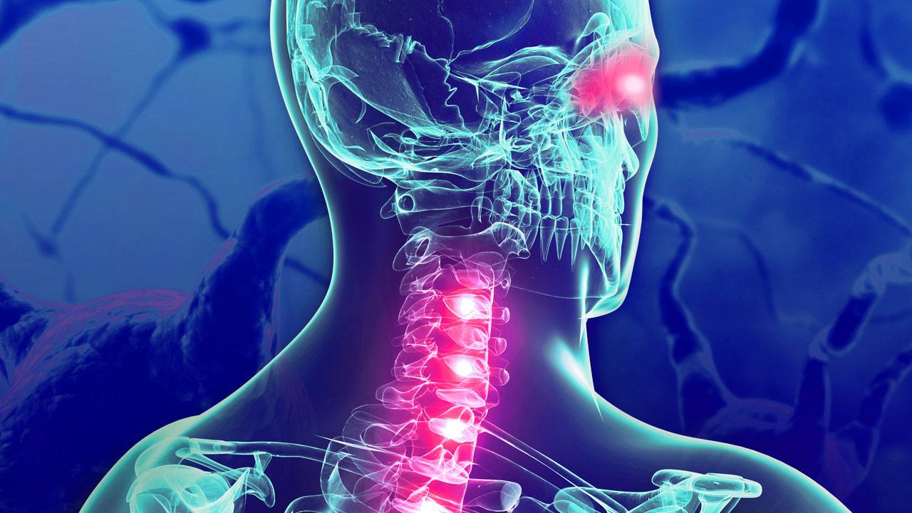
Neuromyelitis Optica (NMO), also known as Devic’s disease or Neuromyelitis Optica Spectrum Disorder (NMOSD), is a rare, severe autoimmune inflammatory disorder of the central nervous system. It is characterized by immune-mediated demyelination and axonal damage predominantly targeting the optic nerves and spinal cord, leading to episodes of optic neuritis (inflammation of the optic nerve) and transverse myelitis (inflammation of the spinal cord).
NMO is now recognized as distinct from multiple sclerosis (MS), with which it was historically confused. The key differentiating factor is that in most cases of NMO, the immune system produces antibodies (called NMO-IgG or anti-AQP4 antibodies) that attack aquaporin-4, a protein that serves as a water channel in cells supporting nerve cells in the central nervous system. This specific autoimmune mechanism distinguishes NMO from other demyelinating diseases.
Affected Body Parts/Organs
NMO primarily affects:
Optic Nerves: The nerves that transmit visual information from the retina to the brain, resulting in vision problems ranging from pain with eye movement to severe vision loss or blindness.
Spinal Cord: Particularly the long segments of the thoracic and cervical spinal cord, leading to motor weakness or paralysis, sensory disturbances, and bladder/bowel dysfunction.
Area Postrema: A region in the brain stem that controls vomiting, which can lead to intractable nausea, vomiting, and hiccups when affected.
Brain Stem: Can lead to symptoms such as double vision, facial numbness, hearing loss, vertigo, or difficulty swallowing when involved.
Diencephalon: Including the hypothalamus and thalamus, potentially causing sleep disorders, endocrine abnormalities, or autonomic dysfunction.
Cerebrum: Less commonly affected, but lesions can occur in the cerebral hemispheres, particularly in children and in specific regions like the periventricular white matter.
Prevalence and Significance
NMO is considered a rare disease with the following epidemiological characteristics:
- Global Prevalence: Varies from 0.5 to 10 per 100,000 population, depending on geographic region and ethnicity.
- Gender Distribution: Strong female predominance with a female-to-male ratio ranging from 3:1 to 9:1.
- Age of Onset: Typically occurs in adults with a median age of onset around 35-45 years, but can affect children and older adults.
- Ethnic Variation: Higher prevalence in East Asian and African ancestry populations compared to Caucasian populations.
The significance of NMO lies in its:
Severity: Without treatment, NMO can cause rapid, severe, and permanent damage to vision and spinal cord function.
Disability Impact: Up to 60% of patients become functionally blind in at least one eye or require ambulatory assistance within 5-7 years of disease onset if untreated.
Mortality Risk: The 5-year mortality rate was historically as high as 25-30% before modern treatments, primarily due to respiratory failure from cervical spinal cord involvement.
Diagnostic Challenges: Misdiagnosis as MS is common and potentially harmful, as some MS treatments can exacerbate NMO.
Treatment Advancements: Recent years have seen significant progress with the FDA approval of three targeted therapies specifically for NMOSD (eculizumab, inebilizumab, and satralizumab), improving prognosis considerably.
Research Significance: The discovery of the AQP4 autoantibody has transformed understanding of autoimmune CNS disorders and led to a paradigm shift in classification, diagnosis, and treatment of demyelinating diseases.
NMO represents an important model of antibody-mediated CNS autoimmunity and has led to improved understanding of the mechanisms underlying other neurological autoimmune conditions.
2. History & Discoveries
First Identification
The earliest documented cases of what would later be recognized as neuromyelitis optica date back to the early 19th century:
- In 1804, Antoine Portal described a patient with visual loss and spinal cord inflammation.
- In 1844, Giovanni Battista Pescetto reported a case of a 42-year-old woman with simultaneous blindness and paralysis.
However, the formal recognition of NMO as a distinct clinical entity is attributed to:
- Eugène Devic (1858-1930), a French neurologist, who presented a case of a 45-year-old woman with concurrent optic neuritis and myelitis at the Congress of French Medicine in Lyon in 1894.
- Fernand Gault, Devic’s student, who that same year published his doctoral thesis titled “On Neuromyelitis Optica,” reviewing 16 similar cases from the literature. This is why the condition was historically known as “Devic’s disease.”
Major Discoveries and Breakthroughs
The understanding of NMO has evolved dramatically over more than a century:
Early Classification (1894-1950s):
- Initially described as a monophasic, severe syndrome with simultaneous optic neuritis and myelitis.
- Often considered a variant or severe form of multiple sclerosis.
Recognition of Relapsing Course (1950s-1990s):
- Growing recognition that many patients experienced relapses, challenging the initial monophasic definition.
- Clinical criteria expanded to include cases with sequential rather than simultaneous involvement of optic nerves and spinal cord.
Distinct Imaging and Pathological Features (1990s):
- Identification of characteristic longitudinally extensive transverse myelitis (LETM) spanning three or more vertebral segments on MRI.
- Recognition of distinct pathological features including perivascular complement deposition and eosinophilic infiltration.
Serum Biomarker Discovery (2004-2005):
- The groundbreaking discovery of NMO-IgG (later identified as anti-AQP4 antibodies) by Vanda Lennon, Brian Weinshenker, and colleagues at the Mayo Clinic in 2004.
- In 2005, the target antigen of NMO-IgG was identified as aquaporin-4 (AQP4), the most abundant water channel in the central nervous system.
Revised Diagnostic Criteria (2006-2015):
- The 2006 diagnostic criteria incorporated the NMO-IgG serostatus.
- The 2015 International Panel for NMO Diagnosis (IPND) introduced the broader term “NMOSD” and stratified diagnostic criteria by AQP4 antibody status.
Recognition of MOG Antibody Disease (2010s):
- Identification of a subset of patients with NMO-like symptoms who have antibodies against myelin oligodendrocyte glycoprotein (MOG) rather than AQP4.
- Growing consensus that MOG antibody-associated disease represents a distinct entity with different treatment considerations.
FDA Approval of Targeted Therapies (2019-2021):
- Eculizumab (Soliris), a complement inhibitor, approved in 2019.
- Inebilizumab (Uplizna), a CD19 B-cell depleting agent, approved in 2020.
- Satralizumab (Enspryng), an IL-6 receptor antagonist, approved in 2020.
Evolution of Medical Understanding
The conceptualization of NMO has undergone several paradigm shifts:
From Monophasic Syndrome to Relapsing Disease (1894-1990s):
- Initially described as a single, devastating attack affecting optic nerves and spinal cord simultaneously.
- Gradually recognized that many patients (80-90%) experience a relapsing course.
From MS Variant to Distinct Disease (1990s-2004):
- Long considered a severe variant or subtype of multiple sclerosis.
- Clinical, radiological, and pathological differences led to questions about this classification.
- The discovery of NMO-IgG definitively established NMO as a distinct disease entity.
From Clinical to Serological Definition (2004-2015):
- Shifted from purely clinical criteria to incorporation of serological testing.
- Recognition that anti-AQP4 antibody positivity is more specific than clinical features alone.
From NMO to NMOSD (2015-present):
- Expansion of the clinical spectrum to include patients with involvement of other CNS regions beyond optic nerves and spinal cord.
- Recognition of “limited” forms with either isolated optic neuritis or transverse myelitis.
- The 2015 IPND criteria formalized the concept of NMOSD as a broader spectrum.
From Single Entity to Antibody-Defined Spectrum (2010s-present):
- Recognition of different antibody-associated diseases with overlapping clinical features.
- Distinction between AQP4-IgG positive NMOSD, MOG antibody-associated disease, and double-seronegative cases.
From Empiric to Targeted Treatment (2019-present):
- Evolution from general immunosuppression to targeted therapies based on disease mechanisms.
- Development of treatments specifically targeting the complement cascade, B-cells, and IL-6 signaling.
This historical progression from a clinically defined syndrome to a mechanistically understood autoimmune channelopathy exemplifies how advances in neuroimmunology can transform disease classification and treatment paradigms. The story of NMO illustrates the importance of biomarkers in neurological disease and the potential for targeted therapeutic approaches based on specific pathogenic mechanisms.
3. Symptoms
Early Symptoms
Neuromyelitis optica typically presents with acute attacks affecting the optic nerves, spinal cord, or both. Early symptoms can vary depending on which part of the nervous system is initially affected:
Optic Neuritis (30-50% of initial presentations):
- Rapid onset of vision loss (can progress over hours to days)
- Eye pain, especially with eye movement
- Altered color perception (particularly red desaturation)
- Visual field defects, often central
- Relative afferent pupillary defect
- May affect one or both eyes simultaneously (bilateral involvement more common than in MS)
Transverse Myelitis (30-40% of initial presentations):
- Sudden onset of weakness or paralysis in legs or arms
- Sensory disturbances (numbness, tingling, burning sensations)
- Sharp, radicular pain along spine or bands around the torso
- Bladder and bowel dysfunction (urinary retention, incontinence)
- Sexual dysfunction
- Lhermitte’s sign (electric shock-like sensation down spine upon neck flexion)
Area Postrema Syndrome (10-15% of initial presentations):
- Intractable nausea and vomiting
- Persistent hiccups lasting more than 48 hours
- Vertigo and dizziness
- May occur in isolation or precede other symptoms by weeks or months
Other Early Presentations:
- Brainstem syndromes with double vision, facial numbness, vertigo, or hearing changes
- Narcolepsy or excessive daytime sleepiness (with hypothalamic involvement)
- Seizures or encephalopathy (more common in children)
Advanced Stage Symptoms
As the disease progresses, especially without appropriate treatment, symptoms can worsen significantly:
Severe Visual Impairment:
- Complete blindness in one or both eyes
- Optic atrophy visible on examination
- Visual field constriction
- Persistent visual disturbances even between attacks
Advanced Spinal Cord Dysfunction:
- Spastic paraplegia or tetraplegia
- Tonic spasms and painful muscle cramps
- Severe sensory deficits
- Permanent bladder and bowel dysfunction requiring catheterization
- Respiratory compromise (with high cervical lesions)
- Autonomic dysreflexia
- Pressure ulcers and complications of immobility
Chronic Pain Syndromes:
- Neuropathic pain resistant to conventional analgesics
- Painful tonic spasms
- Trigeminal neuralgia
- Radicular pain
Cognitive and Psychiatric Symptoms:
- Cognitive impairment (more subtle than in MS but can affect executive function)
- Depression and anxiety
- Fatigue
- Emotional lability
- Sleep disorders
Systemic Complications:
- Venous thromboembolism
- Urinary tract infections
- Respiratory complications
- Osteoporosis and fractures secondary to immobility
Common vs. Rare Symptoms
Common Symptoms (>50% of patients):
- Unilateral or bilateral optic neuritis
- Longitudinally extensive transverse myelitis
- Bladder dysfunction
- Neuropathic pain
- Sensory disturbances
- Motor weakness
- Fatigue
Less Common Symptoms (10-50% of patients):
- Area postrema syndrome (intractable vomiting, hiccups)
- Brainstem syndromes
- Lhermitte’s sign
- Tonic spasms
- Pruritus (itching)
Rare Symptoms (<10% of patients):
- Narcolepsy and hypersomnia
- Syndrome of inappropriate antidiuretic hormone secretion (SIADH)
- Seizures (more common in children)
- Posterior reversible encephalopathy syndrome (PRES)
- Hearing loss
- Cardiac dysrhythmias (with medullary involvement)
Symptom Progression Over Time
NMO typically follows one of two disease courses:
Relapsing Course (80-90% of patients):
- Characterized by discrete attacks followed by periods of stability
- Partial or no recovery between attacks leading to stepwise accumulation of disability
- Higher relapse frequency in the first two years after onset
- Relapse frequency typically 0.5-1.0 per year without treatment
- Higher relapse rates in AQP4-IgG positive patients compared to MOG-IgG positive patients
Monophasic Course (10-20% of patients):
- Single attack affecting both optic nerves and spinal cord, without subsequent relapses
- More common in seronegative or MOG-IgG positive patients
- Generally better prognosis if treated promptly
- Increasingly rare with improved diagnosis and recognition of relapsing forms
Progression Pattern:
- Acute Phase: Rapid symptom development over hours to days, reaching maximum severity within 1-2 weeks
- Plateau Phase: Period of stable symptoms lasting days to weeks
- Recovery Phase: Gradual improvement over weeks to months, often incomplete
- Interictal Phase: Period between attacks with stable symptoms
- Chronic Phase: After multiple attacks, accumulation of fixed deficits
Unlike multiple sclerosis, NMO rarely shows a secondary progressive course with gradual worsening in the absence of discrete attacks. However, disability accumulates through incomplete recovery from repeated relapses rather than through progressive neurodegeneration.
Prognostic Factors for Symptom Progression:
- More frequent and severe relapses in AQP4-IgG positive patients
- Worse recovery from optic neuritis in AQP4-IgG positive vs. MOG-IgG positive cases
- Higher disability associated with:
- Older age at onset
- Male gender
- Higher attack frequency
- Longer delay to treatment
- Presence of coexisting autoimmune diseases
Importantly, early recognition and appropriate immunosuppressive treatment can significantly alter the natural history of NMO, reducing relapse frequency and accumulation of disability. The introduction of targeted therapies has further improved outcomes, highlighting the critical importance of prompt diagnosis and treatment initiation.
4. Causes
Biological Mechanisms
Neuromyelitis optica is fundamentally an autoimmune astrocytopathy, where the immune system attacks a specific target in the central nervous system. Several biological mechanisms contribute to disease pathogenesis:
Autoantibody Production: The primary biological cause of NMO in approximately 70-80% of cases is the production of autoantibodies (NMO-IgG) that target aquaporin-4 (AQP4), a water channel protein highly expressed on astrocytes in the central nervous system. In another 10-20% of cases with NMO-like presentations, antibodies target myelin oligodendrocyte glycoprotein (MOG) instead.
Key Immunological Processes:
Loss of Immune Tolerance: The immune system fails to recognize AQP4 as “self,” leading to autoantibody production. The exact mechanism of this breach in tolerance remains unclear.
Antibody-Mediated Pathology: AQP4-IgG are predominantly of the IgG1 subclass, which efficiently activates complement and binds to Fc receptors on inflammatory cells.
Blood-Brain Barrier Disruption: Antibodies must cross the blood-brain barrier (BBB) to reach their target. Conditions that compromise BBB integrity (inflammation, infection) may facilitate this process.
Complement Activation: When AQP4-IgG binds to AQP4, it activates the complement cascade, leading to the formation of membrane attack complexes and subsequent cell lysis.
Recruitment of Inflammatory Cells: The process attracts neutrophils, eosinophils, and macrophages, which contribute to tissue damage.
Secondary Demyelination: The primary attack on astrocytes leads to secondary damage to myelin and oligodendrocytes, rather than the direct myelin attack seen in multiple sclerosis.
Astrocyte Dysfunction and Loss: Damage to astrocytes disrupts multiple functions including maintaining the BBB, regulating extracellular fluid homeostasis, and supporting neuronal function.
Regional Susceptibility: The predilection for the optic nerves and spinal cord relates to:
- High expression of AQP4 in these regions
- Anatomical characteristics (narrow spaces with limited compensation for swelling)
- Regional differences in blood-brain barrier properties
Cellular and Humoral Immunity: Both B-cell and T-cell mediated processes contribute:
- B-cells produce pathogenic antibodies
- T-cells, particularly IL-6 producing T-helper cells, support B-cell survival and antibody production
- Reduced regulatory T-cell function may contribute to loss of tolerance
Genetic and Hereditary Factors
NMO is not typically considered a hereditary disorder, but genetic factors play a role in susceptibility:
HLA Associations:
- DRB1*03:01 in Caucasians
- DPB1*05:01 in Southern Han Chinese
- DRB1*04:05 in Japanese
- DRB108:01 and DRB108:03 in Brazilian populations
These HLA associations differ from those seen in multiple sclerosis, supporting the distinct nature of NMO.
Non-HLA Genetic Factors:
- Polymorphisms in genes encoding cytokines (IL-6, IL-17)
- Variations in complement component genes
- Fc gamma receptor polymorphisms affecting antibody affinity
- CYP2C9 polymorphisms influencing drug metabolism
Familial Cases:
- Rare familial cases have been reported (less than 3% of all NMO cases)
- Most commonly seen in identical twins or first-degree relatives
- Suggest a complex interplay between genetic predisposition and environmental triggers
Genetic Architecture:
- Complex, polygenic susceptibility rather than simple Mendelian inheritance
- Lower concordance in identical twins compared to strongly genetic autoimmune diseases
- Environmental factors likely more important than genetic factors
Environmental Factors and Triggers
While the fundamental cause of NMO is autoimmune, various environmental factors can trigger disease onset or relapses:
Infectious Triggers:
- Viral infections (influenza, varicella zoster, HIV)
- Tuberculosis
- Non-specific upper respiratory or gastrointestinal infections
- Molecular mimicry between microbial antigens and self-proteins may initiate autoimmunity
Vaccination:
- Case reports of NMO onset or relapses following vaccinations
- Includes influenza, HPV, and yellow fever vaccines
- Likely represents an activation of pre-existing autoimmune tendencies rather than direct causation
Hormonal Influences:
- Disease onset or relapses during pregnancy or postpartum period
- Changes in estrogen and progesterone levels may influence immune system function
- Oral contraceptive use has been associated with reduced relapse rates in some studies
Vitamin D Deficiency:
- Associated with increased NMO risk and possibly more severe disease course
- May explain some geographic and racial variations in prevalence
- Similar to other autoimmune conditions where vitamin D has immunomodulatory effects
Ultraviolet Radiation Exposure:
- Inverse correlation between UV radiation and NMO prevalence in some studies
- Potentially mediated through vitamin D production
- May partially explain geographic variations
Occupational or Chemical Exposures:
- Limited evidence, but reports of clusters among individuals exposed to specific solvents or heavy metals
- Silica exposure has been associated with various autoimmune diseases including NMO
- Smoking may increase risk and worsen disease outcomes
Gut Microbiome:
- Emerging evidence for altered gut microbiota in NMO patients
- Potential role in regulating immune tolerance
- Therapeutic implications still under investigation
Physical or Emotional Stress:
- Reported as a trigger for relapses by many patients
- May act through stress hormones that modulate immune function
- Difficult to quantify objectively in clinical studies
The multifactorial nature of NMO causation helps explain its complex epidemiological patterns and the variation in disease expression among individuals. Current evidence suggests that genetic susceptibility combined with environmental triggers leads to loss of immune tolerance to AQP4 or MOG, initiating the autoimmune process that characterizes this disorder.
5. Risk Factors
Demographic Risk Factors
Gender:
- Strong female predominance with a female-to-male ratio of 3:1 to 9:1
- The gender disparity is most pronounced in AQP4-IgG positive NMOSD (up to 9:1)
- Less pronounced in MOG-IgG associated disease (approximately 1.2:1)
- Suggests hormonal influences on disease susceptibility
Age:
- Can occur at any age, from early childhood to elderly patients
- Peak onset between 35-45 years for AQP4-IgG positive disease
- Earlier onset (median age 30-35) for MOG-IgG associated disease
- Childhood onset (before age 18) in approximately 3-5% of cases
- Bimodal age distribution in some populations with peaks in the 30s and 60s
Ethnicity and Race:
- Higher prevalence in non-Caucasian populations:
- Significantly higher rates in East Asians (particularly Japanese, Chinese, Korean)
- Higher prevalence in African and Afro-Caribbean populations
- Lower rates in Northern European populations
- In the United States, African Americans have a 3-4 fold higher risk than Caucasians
- These variations persist even after migration, suggesting genetic rather than purely environmental factors
Geographic Distribution:
- Higher prevalence near the equator, unlike multiple sclerosis which shows the opposite pattern
- High prevalence in countries like:
- Japan (3.9 per 100,000)
- Southern India (2.6 per 100,000)
- Caribbean islands
- Parts of Africa
- Lower prevalence in Northern Europe and North America (0.7-1.0 per 100,000)
- Clusters reported in specific regions, suggesting possible environmental triggers
Genetic and Immunological Risk Factors
HLA Associations:
- Different HLA associations across ethnic groups:
- DRB1*03:01 in Caucasians
- DPB1*05:01 in Southern Han Chinese
- DRB1*04:05 in Japanese populations
- These differ from MS-associated HLA alleles, supporting distinct genetic predisposition
Coexisting Autoimmune Conditions:
- 20-30% of NMO patients have another autoimmune disease
- Most commonly:
- Sjögren’s syndrome
- Systemic lupus erythematosus (SLE)
- Myasthenia gravis
- Autoimmune thyroid diseases
- Rheumatoid arthritis
- Having one autoimmune condition increases risk for developing NMO
Family History:
- First-degree relatives have slightly increased risk of NMO
- Family history of other autoimmune diseases also increases risk
- Lower familial aggregation than in many other autoimmune diseases
Immune System Variations:
- Altered B-cell homeostasis
- Abnormal cytokine profiles (especially IL-6, IL-17)
- Decreased regulatory T-cell function
- Complement system polymorphisms
Environmental and Occupational Risk Factors
Infectious Exposures:
- Certain infectious agents may trigger disease onset through molecular mimicry:
- Mycobacterium tuberculosis
- Helicobacter pylori
- Various viruses (varicella zoster, influenza, HIV)
- Higher prevalence in countries with higher infectious disease burden
Vitamin D Status:
- Low vitamin D levels associated with increased risk and potentially more severe disease
- May partially explain geographical and seasonal variations in disease onset
Ultraviolet Radiation Exposure:
- Inverse relationship between UV exposure and NMO risk
- Contrary to the pattern seen in MS, suggesting different pathogenic mechanisms
Smoking:
- Associated with increased risk and potentially worse outcomes
- Effect less pronounced than in MS
Chemical Exposures:
- Limited evidence for specific occupational exposures
- Silica exposure (in mining, construction, manufacturing) associated with increased risk
- Some heavy metals and organic solvents implicated in case reports
Hormonal Factors:
- Pregnancy and postpartum period as vulnerable times
- Oral contraceptive use may modify risk
- Menopause associated with changes in disease activity in some women
Impact of Pre-existing Conditions
Other Autoimmune Diseases:
- Pre-existing autoimmune conditions significantly increase NMO risk
- May share genetic susceptibility factors
- Immunosuppressive treatment for the primary condition may mask or delay NMO diagnosis
Infectious Diseases:
- Chronic infections may create a pro-inflammatory environment
- HIV infection has been associated with NMOSD, possibly due to immune dysregulation
- Tuberculosis reported as both a triggering and complicating factor
Metabolic Disorders:
- Obesity associated with increased risk and worse outcomes
- May act through proinflammatory adipokines and generally increased inflammation
- Diabetes has not been clearly established as a risk factor
Malignancy:
- Paraneoplastic NMOSD reported in association with:
- Breast cancer
- Lung cancer
- Thymoma
- Lymphoma
- May involve cross-reactivity between tumor antigens and self-proteins
Prior CNS Injury:
- Some evidence that prior CNS trauma or inflammation may increase BBB permeability
- May allow circulating antibodies access to the CNS
- Could explain focal susceptibility in some patients
Medication Use:
- Certain medications may trigger or unmask NMO:
- TNF-alpha inhibitors used for rheumatologic conditions
- Interferon-beta used for presumed MS
- Some antibiotics with immunomodulatory properties
Understanding these risk factors has important implications for disease prevention, early diagnosis, and management strategies. While many risk factors are non-modifiable (gender, ethnicity, genetic predisposition), others may represent opportunities for intervention or risk stratification. The complex interplay between demographic, genetic, and environmental factors underscores the multifactorial nature of NMO pathogenesis.
6. Complications
Neurological Complications
Visual System Complications:
- Permanent visual impairment (20/200 or worse) in 60-70% of untreated patients
- Complete blindness in one or both eyes in up to 30% of patients
- Optic atrophy and disc pallor
- Visual field defects persisting after acute inflammation resolves
- Reduced color vision and contrast sensitivity
- Retinal nerve fiber layer thinning visible on optical coherence tomography
- Nystagmus and internuclear ophthalmoplegia (with brainstem involvement)
Spinal Cord Complications:
- Permanent paraplegia or quadriplegia in 30-40% of untreated patients
- Spasticity and muscle spasms requiring ongoing management
- Sensory deficits including loss of pain and temperature sensation
- Neuropathic pain syndromes affecting quality of life
- Pressure ulcers and skin breakdown secondary to immobility
- Heterotopic ossification in paralyzed limbs
- Contractures and muscle atrophy
Bladder, Bowel, and Sexual Dysfunction:
- Neurogenic bladder requiring intermittent catheterization (50-80% of patients)
- Recurrent urinary tract infections
- Urinary incontinence or retention
- Bladder stones and renal complications from chronic urinary stasis
- Neurogenic bowel dysfunction (constipation, incontinence)
- Sexual dysfunction (erectile dysfunction, decreased libido, anorgasmia)
Brainstem and Cerebral Complications:
- Intractable hiccups and vomiting from area postrema involvement
- Respiratory failure with high cervical or medullary lesions
- Sleep disorders, including narcolepsy with hypothalamic involvement
- Cognitive impairment affecting executive function and memory
- Seizures (more common in pediatric cases)
- Encephalopathy and altered consciousness
- Dysautonomia (blood pressure fluctuations, temperature dysregulation)
Systemic Complications
Respiratory Complications:
- Respiratory muscle weakness or paralysis with high cervical lesions
- Aspiration pneumonia due to swallowing difficulties
- Sleep-disordered breathing and sleep apnea
- Reduced vital capacity and ineffective cough
- Respiratory failure requiring ventilatory support
- Ventilator-associated complications in severe cases
Cardiovascular Complications:
- Autonomic dysreflexia with dangerously high blood pressure (with lesions above T6)
- Orthostatic hypotension
- Deep vein thrombosis and pulmonary embolism due to immobility
- Cardiac arrhythmias (with medullary involvement)
Musculoskeletal Complications:
- Osteoporosis due to immobility and sometimes steroid treatment
- Pathological fractures
- Scoliosis and spinal deformities
- Joint contractures
- Heterotopic ossification
- Muscle atrophy
Genitourinary Complications:
- Recurrent urinary tract infections
- Pyelonephritis and urosepsis
- Renal calculi
- Hydronephrosis
- Chronic kidney disease from recurrent infections or urinary retention
Gastrointestinal Complications:
- Dysphagia and risk of aspiration
- Constipation sometimes alternating with fecal incontinence
- Fecal impaction
- Paralytic ileus
- Malnutrition and weight loss
Treatment-Related Complications
Corticosteroid-Related:
- Osteoporosis and avascular necrosis
- Weight gain and cushingoid features
- Diabetes mellitus
- Hypertension
- Cataracts
- Increased infection risk
- Psychiatric effects (mood changes, insomnia)
Immunosuppressive Therapy-Related:
- Increased susceptibility to infections
- Bone marrow suppression
- Liver or kidney toxicity
- Malignancy risk with long-term use
- Infertility concerns
- Specific complications related to individual agents:
- Progressive multifocal leukoencephalopathy (with rituximab, rarely)
- Meningococcal infections (with eculizumab)
- Serum sickness reactions
- Infusion reactions
Plasma Exchange Complications:
- Catheter-related infections and thrombosis
- Hypotension during procedures
- Citrate toxicity and electrolyte disturbances
- Bleeding complications
- Transfusion reactions
Long-term Impact on Health and Function
Disability Progression:
- Approximately 60% of untreated patients require a wheelchair and/or are functionally blind within 5 years
- Stepwise accumulation of disability with each attack
- EDSS (Expanded Disability Status Scale) scores typically 4.0-6.0 within 5 years without treatment
- Earlier disability milestones compared to MS
Impact on Life Expectancy:
- 5-year mortality rates of 25-30% in historical untreated cohorts
- Mortality decreased to below 10% with current treatments
- Most common causes of death:
- Respiratory failure
- Urosepsis and complications of urinary tract infections
- Complications of immobility
- Treatment-related complications
Factors Affecting Prognosis:
- Antibody status (AQP4-IgG typically worse than MOG-IgG)
- Age at onset (worse prognosis with older age)
- Gender (slightly worse outcomes in males)
- Ethnicity (more severe in Afro-Caribbean populations)
- Time to treatment initiation
- Number of attacks in first two years
- Location of initial lesions (worse with brainstem involvement)
Quality of Life Impact:
- High rates of depression and anxiety (40-60%)
- Social isolation
- Employment difficulties (50-80% unemployment rates)
- Economic burden from medical costs and lost productivity
- Caregiver burden and strain on family relationships
- Pain as a major contributor to reduced quality of life
Relative Impact Compared to Other Neurological Disorders:
- More severe visual outcomes than MS
- More extensive spinal cord damage than MS
- Higher risk of respiratory failure than MS
- More rapid disability accumulation than MS
- Greater treatment responsiveness than traditionally thought
- Better cognitive preservation than MS
With the advent of newer targeted therapies and earlier diagnosis, these complications have become less common and less severe. However, patients diagnosed late or who have accumulated damage from early attacks before diagnosis may still face significant long-term complications. This underscores the critical importance of early recognition, prompt treatment, and comprehensive supportive care to minimize the impact of this potentially devastating disorder.
7. Diagnosis & Testing
Diagnostic Criteria and Approach
The diagnosis of neuromyelitis optica spectrum disorder (NMOSD) relies on a combination of clinical presentation, serological testing, and neuroimaging findings. The current standard is the 2015 International Panel for NMO Diagnosis (IPND) criteria, which stratifies diagnostic requirements based on AQP4-IgG antibody status:
For AQP4-IgG Positive Patients:
- At least one core clinical characteristic:
- Optic neuritis
- Acute myelitis
- Area postrema syndrome
- Acute brainstem syndrome
- Symptomatic narcolepsy or acute diencephalic syndrome with NMOSD-typical diencephalic MRI lesions
- Symptomatic cerebral syndrome with NMOSD-typical brain lesions
- Exclusion of alternative diagnoses
For AQP4-IgG Negative or Unknown Status Patients:
- At least two core clinical characteristics occurring as a result of one or more clinical attacks and meeting all of the following requirements:
- At least one core clinical characteristic must be optic neuritis, acute myelitis with longitudinally extensive transverse myelitis (LETM), or area postrema syndrome
- Dissemination in space (two or more different core clinical characteristics)
- Fulfillment of additional MRI requirements:
- For optic neuritis: Brain MRI showing normal findings or only nonspecific white matter lesions, OR optic nerve MRI with T2-hyperintense lesion or T1-weighted gadolinium-enhancing lesion extending over >1/2 optic nerve length or involving optic chiasm
- For acute myelitis: MRI showing LETM (≥3 contiguous segments) OR ≥3 contiguous segments of focal spinal cord atrophy in patients with history compatible with acute myelitis
- For area postrema syndrome: Dorsal medulla/area postrema lesions
- For acute brainstem syndrome: Periependymal brainstem lesions
- Exclusion of alternative diagnoses
Laboratory Testing
Serological Testing:
Anti-AQP4 Antibody Testing:
- Gold standard biomarker for NMOSD
- Multiple testing methodologies available:
- Cell-based assays (highest sensitivity 76-99% and specificity 98-100%)
- Immunofluorescence
- ELISA (lower sensitivity)
- Flow cytometry
- Results may be affected by recent plasma exchange or immunosuppressive therapy
- Testing should be performed before initiating immunosuppression when possible
- Titers may correlate with disease activity in some patients
Anti-MOG Antibody Testing:
- Present in 10-40% of AQP4-negative cases with NMO-like phenotype
- Only cell-based assays recommended due to conformational epitope recognition
- More common in:
- Bilateral simultaneous optic neuritis
- Optic neuritis with papillitis
- Transverse myelitis with lower spinal cord involvement
- Monophasic course
- Children
Other Serum Testing:
- Complete blood count
- Comprehensive metabolic panel
- Erythrocyte sedimentation rate and C-reactive protein
- Autoimmune panel (ANA, ENA, RF) to detect coexisting autoimmune diseases
- Thyroid function tests
- Vitamin B12 levels
- Infectious disease screening (syphilis, HIV, HTLV-1) as part of differential diagnosis
- Paraneoplastic antibody panel in appropriate cases
Cerebrospinal Fluid (CSF) Analysis:
CSF Cell Count and Protein:
- Pleocytosis (>5 cells/mm³) in 35-75% of acute attacks
- Sometimes with neutrophilic predominance (unlike MS)
- Protein elevation in 45-75% of cases
- Severe attacks can show very high cell counts (>100 cells/mm³)
Oligoclonal Bands:
- Present in only 15-30% of NMO patients (vs. 85-95% in MS)
- Absence helpful in distinguishing from MS
- May be transiently present during acute attacks
Other CSF Markers:
- Elevated glial fibrillary acidic protein (GFAP) during acute attacks
- Elevated IL-6 levels
- Detectable AQP4-IgG in CSF in some cases (lower sensitivity than serum)
- Cytokine profiles different from MS
Imaging Studies
Magnetic Resonance Imaging (MRI):
Spinal Cord MRI:
- Longitudinally extensive transverse myelitis (LETM) spanning ≥3 vertebral segments
- Central gray matter involvement with “H” or “butterfly” pattern on axial images
- Bright spotty lesions representing microcystic change
- Cord swelling in acute phase
- Cord atrophy in chronic phase
- Preferential cervical and upper thoracic involvement
- Gadolinium enhancement common in acute lesions
Brain MRI:
- Normal in up to 60% of patients (especially early in disease)
- When present, lesions typically located in:
- Area postrema/dorsal medulla
- Periventricular areas (especially third ventricle)
- Corpus callosum (can mimic MS but typically involves the central portion rather than Dawson’s fingers)
- Corticospinal tracts
- Brainstem
- Lesions often have distinctive characteristics:
- Periependymal distribution
- Cloud-like enhancement
- Large, tumefactive appearance
- Less ovoid than MS lesions
Orbital MRI:
- More extensive and posterior involvement of optic nerves than in MS
- Often bilateral
- May involve the optic chiasm
- Long segment involvement
- More severe gadolinium enhancement
Other Imaging Modalities:
Optical Coherence Tomography (OCT):
- Measures retinal nerve fiber layer thickness and macular volume
- More severe thinning compared to MS optic neuritis
- Helpful for monitoring disease progression and recovery
- Can detect subclinical damage
Visual Evoked Potentials (VEP):
- Demonstrates prolonged latencies and reduced amplitudes
- May detect subclinical optic nerve involvement
- Less specific but more widely available than OCT
PET Scanning:
- Research tool, not routine in clinical practice
- Can demonstrate altered glucose metabolism
- May show abnormalities in normal-appearing white matter
Differential Diagnosis
The differential diagnosis of NMOSD includes:
Multiple Sclerosis:
- Most important differential diagnosis
- Distinguished by:
- AQP4-IgG positivity (specific for NMOSD)
- LETM (rare in MS)
- More severe optic neuritis, often bilateral
- Different brain MRI pattern
- Lower frequency of oligoclonal bands
- Different response to MS treatments (some worsen NMOSD)
MOG Antibody-Associated Disease:
- Now considered distinct from both MS and AQP4+ NMOSD
- Features include:
- More frequent involvement of lower spinal cord
- Better recovery from attacks
- More frequent bilateral optic neuritis
- Often monophasic in adults (more frequently relapsing in children)
- Optic nerve head swelling more common
Acute Disseminated Encephalomyelitis (ADEM):
- More common in children
- Typically monophasic
- More diffuse brain involvement
- Often follows viral illness or vaccination
Sarcoidosis:
- Can cause myelopathy and optic neuropathy
- Distinguished by:
- Systemic involvement
- Hilar lymphadenopathy
- Elevated ACE levels
- Different MRI appearance (often meningeal enhancement)
Systemic Lupus Erythematosus with CNS involvement:
- May coexist with NMOSD
- Look for systemic lupus features
- Positive ANA, anti-dsDNA antibodies
Sjögren’s Syndrome with CNS involvement:
- May coexist with NMOSD
- Sicca symptoms (dry eyes, dry mouth)
- Positive anti-SSA/SSB antibodies
Paraneoplastic Syndromes:
- Associated anti-neuronal antibodies
- Underlying malignancy
- Older age of onset typically
Infectious Causes:
- HIV myelopathy
- HTLV-1 associated myelopathy
- Syphilis
- Tuberculosis
- Viral myelitis
Vascular Disorders:
- Spinal dural arteriovenous fistula
- Anterior spinal artery infarction
- Usually monophasic
Nutritional Deficiencies:
- Vitamin B12 deficiency
- Copper deficiency myelopathy
Early Detection and Diagnostic Challenges
Challenges in Diagnosis:
- Average diagnostic delay of 1-5 years from symptom onset
- Misdiagnosis as MS in 30-50% of cases before AQP4 testing
- Seronegative cases particularly challenging
- Limited access to specialized testing in some regions
- Atypical presentations, especially in children
- Coexistence with other autoimmune diseases complicating the picture
Strategies for Earlier Detection:
AQP4 antibody testing in all patients with:
- Severe or bilateral optic neuritis
- Recurrent optic neuritis
- Longitudinally extensive transverse myelitis
- Intractable vomiting or hiccups without clear cause
- Optic neuritis or myelitis in non-Caucasian patients
- Poor response to MS therapies
Education of primary care physicians, ophthalmologists, and neurologists about red flags for NMOSD
Repeat antibody testing if initial results negative but clinical suspicion high
Use of most sensitive assay methodologies (cell-based assays)
Consider testing for both AQP4 and MOG antibodies in appropriate clinical scenarios
Accurate and timely diagnosis is critical for initiating appropriate treatment, preventing relapses, and avoiding harmful therapies. The diagnostic approach continues to evolve as understanding of the NMOSD spectrum expands and testing methodologies improve.
8. Treatment Options
Acute Attack Management
The immediate treatment of acute NMO attacks aims to minimize inflammation and tissue damage, thereby reducing the severity of residual deficits.
High-Dose Corticosteroids:
- First-line therapy for acute attacks
- Intravenous methylprednisolone 1000mg daily for 3-5 days
- Mechanism: Reduces inflammation, stabilizes blood-brain barrier, induces T-cell apoptosis
- Typically followed by oral prednisone taper over 2-8 weeks
- Side effects: Hyperglycemia, hypertension, insomnia, mood disturbances, gastric irritation
Plasma Exchange (PLEX):
- Indicated for patients with severe attacks or inadequate response to steroids
- Typically 5-7 exchanges over 10-14 days
- Mechanism: Removes circulating antibodies, complement factors, and cytokines
- More effective when initiated early (within 5 days of symptom onset)
- Side effects: Hypotension, citrate toxicity, catheter-related complications, infection risk
Intravenous Immunoglobulin (IVIG):
- Alternative for patients who cannot undergo PLEX
- Typical dose: 2g/kg divided over 3-5 days
- Mechanism: Blocks Fc receptors, neutralizes complement, modulates cytokines
- Less evidence for efficacy compared to PLEX
- Side effects: Headache, aseptic meningitis, thrombotic events, renal dysfunction
Escalation Protocol:
- Many centers use a stepwise approach:
- IV methylprednisolone for 5 days
- If inadequate improvement by day 5, initiate PLEX
- Consider IVIG if PLEX is contraindicated or unavailable
- In severe cases (particularly respiratory compromise), consider early combination of steroids and PLEX
Supportive Care During Acute Attacks:
- Respiratory support if high cervical lesions present
- Bladder management (catheterization as needed)
- DVT prophylaxis
- Pressure ulcer prevention
- Early rehabilitation involvement
- Psychological support
Preventive Treatment
Long-term immunosuppressive therapy is essential to prevent relapses and accumulation of disability.
FDA-Approved Targeted Therapies:
Eculizumab (Soliris):
- Humanized monoclonal antibody targeting C5 complement protein
- Dosing: 900mg weekly for first 4 weeks, then 1200mg every 2 weeks
- Efficacy: 94% reduction in relapse risk in PREVENT trial
- Mechanism: Inhibits terminal complement activation
- Side effects: Increased risk of meningococcal infections (vaccination required), headache, upper respiratory infections
- Considerations: Very expensive, requires ongoing infusions
Inebilizumab (Uplizna):
- Humanized monoclonal antibody targeting CD19+ B cells
- Dosing: 300mg IV on days 1 and 15, then every 6 months
- Efficacy: 77% reduction in relapse risk in N-MOmentum trial
- Mechanism: Depletes B cells (including plasmablasts and some plasma cells)
- Side effects: Infusion reactions, increased infection risk, hypogammaglobulinemia
- Considerations: Broader B-cell depletion than rituximab
Satralizumab (Enspryng):
- Humanized monoclonal antibody targeting IL-6 receptor
- Dosing: 120mg SC at weeks 0, 2, 4, then every 4 weeks
- Efficacy: 74% reduction in relapse risk in AQP4+ patients in SAkuraStar trial
- Mechanism: Blocks IL-6 signaling, reducing inflammation and antibody production
- Side effects: Injection site reactions, upper respiratory infections, headache
- Considerations: Subcutaneous administration allows self-injection
Off-Label Immunosuppressive Therapies:
Rituximab:
- Most widely used therapy before FDA-approved options
- Anti-CD20 monoclonal antibody depleting B cells
- Dosing: Various protocols including 375mg/m² weekly for 4 weeks, or 1000mg x 2 doses 2 weeks apart
- Maintenance: Typically every 6 months, often guided by CD19/CD20 cell counts
- Efficacy: 72-80% reduction in relapse rates in observational studies
- Side effects: Infusion reactions, hypogammaglobulinemia, increased infection risk
Mycophenolate Mofetil:
- Inhibits inosine monophosphate dehydrogenase, reducing B and T cell proliferation
- Dosing: 1000-3000mg daily in divided doses
- Efficacy: 60-75% reduction in relapse rates
- Side effects: Gastrointestinal upset, bone marrow suppression, increased infection risk
Azathioprine:
- Purine synthesis inhibitor affecting lymphocyte proliferation
- Dosing: 2-3mg/kg daily
- Efficacy: 50-60% reduction in relapse rates
- Side effects: Liver toxicity, bone marrow suppression, increased malignancy risk with long-term use
- Monitoring: Regular blood counts, liver function tests, TPMT testing before initiation
Other Options:
- Tocilizumab (IL-6 receptor antagonist, similar to satralizumab)
- Methotrexate (folate antagonist)
- Cyclophosphamide (alkylating agent, typically for refractory cases)
- Combination therapies (limited evidence)
Treatment Selection Considerations:
- AQP4 antibody status (seronegative patients may respond differently)
- Attack severity and frequency
- Comorbidities and contraindications
- Pregnancy plans (most immunosuppressants contraindicated)
- Cost and insurance coverage
- Patient preference regarding route of administration
- Monitoring requirements
- Regional availability
Monitoring on Treatment:
- Regular clinical assessment (typically every 3-6 months)
- MRI surveillance (typically annually or with new symptoms)
- AQP4-IgG titers (utility for monitoring controversial)
- Medication-specific monitoring (e.g., CD19/CD20 counts for rituximab)
- Screening for long-term medication complications
Symptomatic Management
Management of residual symptoms is essential for improving quality of life.
Visual Impairment Management:
- Low vision aids and rehabilitation
- Orientation and mobility training
- Screen reading software and adaptive technology
- Registration with visual impairment services
- Management of visual phenomena (e.g., phosphenes, photopsias)
Spasticity Management:
- Physical therapy with stretching programs
- Oral medications (baclofen, tizanidine, dantrolene)
- Botulinum toxin injections for focal spasticity
- Intrathecal baclofen pump for severe cases
- Surgical interventions rarely needed
Pain Management:
- Neuropathic pain agents (gabapentin, pregabalin)
- Antidepressants (amitriptyline, duloxetine)
- Membrane stabilizers (carbamazepine, lamotrigine) for paroxysmal symptoms
- Topical agents (lidocaine, capsaicin)
- Pain psychology approaches (cognitive behavioral therapy)
- Specialized pain programs for refractory cases
Bladder and Bowel Management:
- Intermittent catheterization for urinary retention
- Anticholinergics for detrusor overactivity
- Botulinum toxin bladder injections
- Bowel programs combining dietary modification, medications, and timed evacuation
- Urological and colorectal specialist involvement
Fatigue Management:
- Energy conservation techniques
- Structured exercise programs
- Sleep hygiene
- Medications (modafinil, amantadine) in selected cases
- Address contributing factors (depression, sleep disorders, medication side effects)
Cognitive and Mood Disorders:
- Neuropsychological assessment and cognitive rehabilitation
- Treatment of depression and anxiety
- Psychotherapy and counseling
- Social support and peer connection
Mobility and Activities of Daily Living:
- Physical and occupational therapy
- Appropriate mobility aids
- Home modifications
- Vocational rehabilitation
- Driving assessment and adaptive equipment
Rehabilitative Approaches
A multidisciplinary approach to rehabilitation is crucial for maximizing functional recovery.
Physical Therapy:
- Strength and conditioning exercises
- Gait training
- Balance and coordination therapy
- Aquatic therapy
- Management of spasticity
- Education on energy conservation
Occupational Therapy:
- Activities of daily living assessment and training
- Upper limb rehabilitation
- Cognitive strategies
- Environmental modifications
- Assistive technology assessment
Speech and Language Therapy:
- Swallowing assessment and strategies
- Communication aids for those with brainstem involvement
- Cognitive communication strategies
Neuro-ophthalmological Rehabilitation:
- Visual field defect compensation strategies
- Ocular motor exercises
- Prism therapy for diplopia
- Adaptation strategies for vision loss
Psychological Support:
- Adjustment to diagnosis
- Coping strategies for chronic illness
- Management of anxiety and depression
- Family support and education
Vocational Rehabilitation:
- Workplace accommodations
- Job modification or retraining
- Disability benefits guidance
- Return to work planning
Emerging Treatments and Clinical Trials
Several novel therapeutic approaches are under investigation:
Complement Pathway Targeted Therapies:
- Ravulizumab (longer-acting C5 inhibitor)
- Pegcetacoplan (C3 inhibitor)
- Oral complement inhibitors in development
B-Cell Directed Therapies:
- Next-generation anti-CD20 antibodies
- Bruton’s tyrosine kinase (BTK) inhibitors
- Proteasome inhibitors targeting plasma cells
- BAFF and APRIL pathway inhibitors
Tolerization Approaches:
- AQP4 peptide tolerization therapy
- Regulatory T-cell enhancement
- Mesenchymal stem cell therapy
Neuroprotective Strategies:
- Remyelination-promoting compounds
- Growth factors
- Erythropoietin (shown to have neuroprotective properties)
Novel Delivery Systems:
- Extended-release formulations
- Blood-brain barrier penetrating therapies
- Intrathecal delivery systems
Combination Therapy Approaches:
- Acute attack therapy combined with early preventive treatment
- Sequential immunotherapy protocols
- Combination of mechanisms (e.g., complement inhibition plus B-cell depletion)
Biomarker-Guided Treatment:
- Personalized therapy based on cytokine profiles
- AQP4-IgG subclass and titer-guided treatment intensity
- Genetic modifier-informed therapy selection
The treatment landscape for NMO has transformed dramatically in recent years, from a condition with limited options to one with multiple FDA-approved targeted therapies. This has significantly improved prognosis, with reduced relapse rates, disability accumulation, and mortality. The individualization of treatment approaches based on patient characteristics, preferences, and biomarkers represents the frontier of NMO management.
9. Prevention & Precautionary Measures
Primary Prevention
Unlike some neurological disorders, there are currently no proven strategies for primary prevention of neuromyelitis optica, as the initial trigger for the autoimmune process remains unknown. However, some measures may theoretically reduce risk based on our understanding of disease mechanisms:
Vitamin D Optimization:
- Low vitamin D levels are associated with increased risk and severity of many autoimmune diseases, including NMOSD
- Maintaining adequate vitamin D levels (typically >30 ng/mL) may be reasonable
- Particularly relevant for high-risk populations (e.g., those with family history of autoimmunity)
- Sources include sunlight exposure, diet, and supplements
Management of Coexisting Autoimmune Conditions:
- Proper control of other autoimmune diseases may theoretically reduce risk
- Particularly important for conditions like Sjögren’s syndrome and SLE that are associated with NMOSD
- Regular follow-up with rheumatologists or immunologists for those with autoimmune comorbidities
Smoking Cessation:
- Smoking is associated with increased risk and potentially worse outcomes in multiple autoimmune diseases
- May influence blood-brain barrier integrity and immune system function
- Additional health benefits beyond potential NMO risk reduction
Infection Prevention:
- Some infections may trigger autoimmunity through molecular mimicry or bystander activation
- Basic hygiene practices and appropriate vaccinations
- Prompt treatment of significant infections
Genetic Counseling:
- While NMOSD is not typically inherited in a Mendelian pattern, familial cases do occur
- Relatives of NMO patients have slightly increased risk
- Genetic counseling may be appropriate for families with multiple affected members
Relapse Prevention
Preventing relapses is critical for individuals already diagnosed with NMOSD:
Adherence to Immunosuppressive Therapy:
- Consistent use of prescribed preventive medications
- Regular monitoring as recommended for specific agents
- Communication with healthcare providers about side effects or concerns
- Avoid abrupt discontinuation of treatment
Trigger Avoidance:
- Identify and manage personal triggers when possible
- Common reported triggers include:
- Infections (particularly upper respiratory)
- Physical and emotional stress
- Excessive fatigue
- Hormonal fluctuations
- Surgery or trauma
- Certain medications (TNF inhibitors, interferons)
Infection Prevention and Management:
- Prompt treatment of infections
- Influenza vaccination (generally safe in NMOSD)
- Pneumococcal vaccination for those on immunosuppression
- Meningococcal vaccination mandatory for patients on eculizumab
- Good hygiene practices, especially during seasons of high infection risk
Pregnancy Planning and Management:
- Pre-conception consultation with neurologist and high-risk obstetrician
- Planned transition to pregnancy-compatible medications when possible
- Increased monitoring during pregnancy and postpartum period
- Awareness of increased relapse risk in postpartum period
- Prompt resumption of preventive therapy after delivery
Avoidance of Contraindicated Treatments:
- Certain MS therapies can worsen NMOSD:
- Interferon-beta
- Natalizumab
- Fingolimod
- Alemtuzumab
- Clear communication with all healthcare providers about diagnosis
Regular Monitoring:
- Clinical follow-up every 3-6 months
- MRI surveillance (typically annually or with new symptoms)
- Prompt reporting of new or worsening symptoms
- Regular screening for medication side effects
Lifestyle Modifications
While no specific lifestyle interventions have been proven to change NMOSD outcomes, general health-promoting measures may be beneficial:
Balanced Diet:
- Mediterranean-style diet with anti-inflammatory properties
- Adequate intake of omega-3 fatty acids
- Limited processed foods and refined sugars
- Appropriate caloric intake to maintain healthy weight
- Consider vitamin D-rich foods if levels are low
Regular Physical Activity:
- Adapted to individual ability level
- May improve overall health and reduce comorbidities
- Can help manage fatigue and depression
- Improved cardiovascular health may support recovery
- Water-based exercises often well-tolerated with spasticity
Stress Management:
- Stress reduction techniques (meditation, mindfulness, yoga)
- Psychological support and counseling
- Adequate sleep hygiene
- Work-life balance adjustments as needed
- Support groups and peer connections
Cognitive Health:
- Mental stimulation and cognitive exercise
- Social engagement
- Cognitive rehabilitation when needed
- Management of mood disorders
Environmental Factors:
- Maintaining comfortable ambient temperature (heat can worsen symptoms)
- Home modifications for safety and accessibility
- Ergonomic workstations to reduce fatigue
- Adaptive equipment as needed
Monitoring and Early Intervention
Early detection of relapses or complications can significantly improve outcomes:
Education on Warning Signs:
- New or changing neurological symptoms
- Changes in vision (blurring, pain, color desaturation)
- Sensory changes (numbness, tingling, pain)
- Motor weakness
- Changes in bladder or bowel function
- Intractable hiccups or vomiting
Regular Medical Follow-up:
- Scheduled neurologist visits
- Coordinated care with other specialists
- Regular visual assessments with ophthalmology or neuro-ophthalmology
- Urological follow-up for bladder management
- Mental health screening
Imaging Surveillance:
- Baseline MRI of brain and spinal cord after diagnosis
- Periodic surveillance MRIs (frequency individualized)
- OCT for monitoring optic nerve damage
- Functional measures of vision and mobility
Blood Monitoring:
- Complete blood count and liver function with immunosuppressive therapy
- Specific monitoring based on medication (e.g., CD19/CD20 counts with rituximab)
- Consider periodic AQP4-IgG testing, though utility for monitoring is debated
- Screening for comorbidities (e.g., diabetes, hypertension)
Emergency Planning:
- Written relapse action plan
- Contact information for medical team
- Pre-authorization for emergency treatments when possible
- Medical alert identification
- Education of family members on warning signs
Special Considerations
Vaccination Guidance:
- Generally avoid live vaccines while on immunosuppression
- Annual inactivated influenza vaccine recommended
- Pneumococcal vaccination advised
- COVID-19 vaccination recommended but may have reduced effectiveness
- Timing vaccination between B-cell depleting therapy doses when possible
- Meningococcal vaccination mandatory before starting eculizumab
Travel Considerations:
- Pre-travel consultation with neurologist
- Medication supply and storage plans
- Medical evacuation insurance
- Location of medical facilities at destination
- Letter explaining medical condition and medications
- Considerations for temperature sensitivity
Surgery and Procedure Planning:
- Pre-procedure consultation with neurologist
- Medication adjustments as needed
- Stress-dose steroids sometimes recommended
- Careful anesthesia planning
- Post-procedure monitoring for relapses
While there is no way to completely prevent the development of NMOSD or guarantee against relapses, a combination of appropriate immunosuppressive therapy, trigger avoidance, healthy lifestyle practices, and vigilant monitoring can significantly reduce risk and improve outcomes. The individualization of preventive strategies based on patient-specific factors, comorbidities, and preferences is essential for optimal management.
10. Global & Regional Statistics
Global Prevalence and Incidence
Overall Prevalence:
- Worldwide prevalence estimates range from 0.5 to 10 per 100,000 population
- Global number of cases estimated at 100,000-200,000 individuals
- Significant regional and ethnic variations
- Challenges in accurate estimation due to:
- Changing diagnostic criteria over time
- Variable access to AQP4-IgG testing
- Potential misdiagnosis as MS
- Limited epidemiological studies in many regions
Incidence Rates:
- Annual incidence typically reported between 0.05 and 0.4 per 100,000 person-years
- Higher incidence in females (0.07-0.73 per 100,000) than males (0.02-0.26 per 100,000)
- Some evidence for increasing incidence over time, likely due to improved recognition and diagnosis
Age and Gender Distribution:
- Strong female predominance with female-to-male ratios of:
- 3:1 in Caucasian populations
- Up to 9:1 in East Asian populations
- 7:1 in African American populations
- Mean age of onset typically between 35-45 years
- Pediatric onset in approximately 3-5% of cases
- Some reports of bimodal age distribution with peaks in young adulthood and middle age
Regional Variations
East Asia:
- Highest prevalence rates globally
- Japan: 3.9 per 100,000
- South Korea: 2.6-4.1 per 100,000
- Southern China: 1.6-3.1 per 100,000
- High AQP4-IgG seropositivity rates (70-90%)
- NMOSD comprises approximately 15-40% of demyelinating disease cases in these regions (compared to <5% in Western countries)
South and Southeast Asia:
- India: Prevalence of 2.6-2.7 per 100,000
- Thailand: 1.9 per 100,000
- Malaysia: 1.2 per 100,000
- Regional variations within countries, with higher rates in southern India
- Limited data from many countries in the region
North America:
- United States: 0.7-1.0 per 100,000 overall
- Significant racial disparities:
- African Americans: 2.5-4.6 per 100,000
- Hispanic Americans: 1.8-3.1 per 100,000
- Caucasian Americans: 0.7-1.1 per 100,000
- Asian Americans: 1.9-2.4 per 100,000
- Canada: 1.0 per 100,000, with higher rates in Indigenous populations
Europe:
- Generally lower rates than other regions
- Denmark: 1.1 per 100,000
- United Kingdom: 0.7-0.8 per 100,000
- Spain: 0.89 per 100,000
- Italy: 1.2 per 100,000
- Higher rates in populations with African or Asian ancestry
- NMOSD accounts for approximately 1-2% of demyelinating disease cases
Latin America:
- Brazil: 1.1-1.6 per 100,000
- Mexico: 1.3 per 100,000
- Caribbean: Higher rates reported, particularly in Afro-Caribbean populations
- Limited data from many countries in the region
- Often intermediate prevalence between European and African populations
Africa:
- Very limited epidemiological data
- Some studies suggest higher prevalence than in Caucasian populations
- Significant diagnostic challenges due to limited healthcare resources
- Likely substantial underdiagnosis and underreporting
Oceania:
- Australia: 0.7 per 100,000
- New Zealand: 0.7 per 100,000
- Higher rates in Indigenous and Polynesian populations
- Good diagnostic capabilities but relatively small population base
Middle East:
- Iran: 1.1 per 100,000
- Variable rates across the region
- Limited population-based studies
- Evidence of different clinical presentations compared to Western cohorts
Mortality and Survival Rates
Mortality Rates:
- 5-year mortality in historical (pre-2000) untreated cohorts: 25-30%
- Current 5-year mortality with modern treatments: 7-10%
- 10-year mortality rates: 20-25% in older studies, 10-15% with current treatments
- Leading causes of death:
- Respiratory complications (particularly with high cervical lesions)
- Infections (particularly urosepsis)
- Treatment-related complications
- Comorbidities
Survival Rates:
- 5-year survival: 90-93% with current treatments
- 10-year survival: 85-90% with current treatments
- Factors associated with poorer survival:
- Older age at onset
- Male sex
- High attack frequency
- Respiratory involvement
- Delayed diagnosis and treatment
- Limited access to healthcare
Disability Accumulation:
- EDSS 6.0 (requires unilateral assistance to walk) reached by:
- 50-60% of untreated patients within 5 years
- 25-30% of treated patients within 5 years
- Blindness in at least one eye:
- 60-70% of untreated patients within 5 years
- 25-35% of treated patients within 5 years
Temporal Trends
Diagnostic Rates:
- Increasing diagnosis rates worldwide since AQP4-IgG discovery in 2004
- Shortened time to diagnosis (from 3-5 years historically to 1-2 years currently)
- Improved recognition of atypical presentations
- Earlier treatment initiation
Treatment Availability:
- Dramatic improvements in treatment options since 2019
- Significant regional disparities in access to newer therapies
- Cost barriers for targeted therapies in many regions
- Rituximab remains most widely used globally due to cost and availability
Outcomes:
- Improving mortality and disability outcomes over time
- Greatest improvements in regions with:
- Early diagnosis
- Access to immunosuppressive therapy
- Multidisciplinary care
- Persistent disparities in outcomes based on socioeconomic factors
Economic Impact
Healthcare Costs:
- Annual direct medical costs per patient: $50,000-$100,000 USD (United States)
- Significantly lower in other healthcare systems but still substantial
- Main cost drivers:
- Medication costs (particularly newer targeted therapies)
- Hospitalization for attacks
- Rehabilitation services
- Long-term care for severely disabled patients
Indirect Costs:
- Lost productivity: 50-70% employment reduction
- Caregiver burden and lost caregiver productivity
- Disability payments and social support services
- Estimated lifetime economic burden of $1.1-1.9 million per patient
Regional Economic Disparities:
- Limited access to newer therapies in low and middle-income countries
- Catastrophic healthcare expenditures for families in regions without universal healthcare
- Productivity losses disproportionately affect developing economies
- Limited specialized care availability in many regions
The global landscape of NMOSD shows significant disparities in prevalence, diagnosis, treatment access, and outcomes. These disparities are driven by a complex interplay of genetic factors, environmental influences, healthcare system differences, and socioeconomic factors. Continued efforts to improve epidemiological data collection, diagnostic capacity, and treatment access worldwide are essential to addressing these disparities and improving outcomes for all patients with NMOSD.
11. Recent Research & Future Prospects
Latest Treatment Advances
The therapeutic landscape for NMOSD has transformed dramatically in recent years, with several groundbreaking advancements:
FDA-Approved Targeted Therapies (2019-2020):
Eculizumab (Soliris): First FDA-approved therapy for AQP4-IgG positive NMOSD (June 2019)
- Terminal complement inhibitor targeting C5
- PREVENT trial showed 94% reduction in attack risk
- Revolutionized treatment paradigm by targeting a specific disease mechanism
Inebilizumab (Uplizna): Approved June 2020
- Anti-CD19 B-cell depleting monoclonal antibody
- N-MOmentum trial demonstrated 77% reduction in relapse risk
- Broader B-cell depletion than traditional anti-CD20 therapies
Satralizumab (Enspryng): Approved August 2020
- IL-6 receptor antagonist
- SAkuraStar and SAkuraSky trials showed approximately 74% reduction in relapse risk in AQP4+ patients
- First self-administered subcutaneous option
Emerging Treatment Approaches:
Next-Generation Complement Inhibitors:
- Ravulizumab (Ultomiris): Long-acting C5 inhibitor requiring less frequent dosing than eculizumab
- Pegcetacoplan: C3 inhibitor potentially offering more complete complement blockade
- Small molecule oral complement inhibitors in development
Enhanced B-Cell Targeted Therapies:
- Obinutuzumab: Type II anti-CD20 antibody with enhanced antibody-dependent cellular cytotoxicity
- Ofatumumab: Fully human anti-CD20 antibody with subcutaneous administration
- BTK inhibitors: Target B-cell receptor signaling and potentially affect plasma cells
- Plasma cell-directed therapies (proteasome inhibitors, anti-CD38)
Novel Immunomodulatory Approaches:
- JAK inhibitors: Target multiple cytokine pathways simultaneously
- S1P receptor modulators: Sequester lymphocytes in lymphoid tissues
- Selective T-cell costimulation modifiers
- Combination therapies targeting multiple pathways
Individualized Treatment Algorithms:
- Biomarker-guided therapy selection
- Serological monitoring to guide treatment intensity
- Risk stratification tools for treatment decisions
- Sequential therapy approaches
Ongoing Research Initiatives
Major Research Programs and Collaborations:
Guthy-Jackson Charitable Foundation Initiatives:
- International NMO biorepository and database
- Collaborative international research network
- Patient registry with longitudinal data collection
- Focus on biomarkers and precision medicine approaches
CIRCLES Study (Collaborative International Research in Clinical and Longitudinal Experience Studies):
- Multicenter, longitudinal observational study
- Collection of clinical data, imaging, and biospecimens
- Over 1,500 participants enrolled across multiple countries
- Platform for biomarker discovery and natural history characterization
NMOSD Clinical Consortium:
- Standardization of outcomes measures
- Development of clinical trial platforms
- Comparative effectiveness research
- Patient-reported outcome validation
Pharmaceutical Industry Initiatives:
- Continued development of targeted therapies
- Post-marketing surveillance studies
- Expanded indication studies (pediatric populations, seronegative disease)
- Biomarker development programs
Key Research Areas:
Pathophysiology and Mechanisms:
- Detailed characterization of AQP4-IgG epitopes and effector functions
- Understanding mechanisms of BBB disruption
- Role of T cells in disease pathogenesis
- Environmental triggers of autoimmunity
Biomarker Development:
- Predictive biomarkers for attack risk
- Treatment response biomarkers
- Prognostic biomarkers for disability outcomes
- Imaging markers of disease activity and progression
Imaging Advances:
- Advanced MRI techniques (DTI, MTR, MR spectroscopy)
- PET imaging of inflammation and neurodegeneration
- Optical coherence tomography protocols
- AI-assisted lesion detection and classification
Clinical Trial Design:
- Novel endpoints beyond relapse rate
- Patient-reported outcome measures
- Adaptive trial designs
- Biomarker-driven studies
Future Therapeutic Directions
Potential Paradigm-Shifting Approaches:
Antigen-Specific Immunotherapy:
- AQP4 peptide tolerization therapy
- Engineered AQP4-specific regulatory T cells
- Chimeric antigen receptor (CAR) regulatory T cells
- Nanoparticle-based antigen presentation for tolerance induction
Neuroprotection and Repair:
- Remyelination-promoting compounds
- Neurotrophic factors to support damaged axons
- Cell-based therapies (neural precursor cells, oligodendrocyte progenitors)
- Combined approaches addressing both inflammation and repair
Gene Therapy and Editing:
- Viral vector delivery of anti-inflammatory genes
- CRISPR-based approaches to modify autoreactive B cells
- RNA therapeutics to modulate disease-associated genes
- Gene therapy for complement regulation
Aquaporumab Technology:
- Engineered non-pathogenic antibodies that competitively bind AQP4
- Block binding of pathogenic AQP4-IgG without triggering complement
- Potentially fewer side effects than current approaches
- Combining binding specificity with effector function engineering
Central Nervous System Delivery Systems:
- Blood-brain barrier penetrating technologies
- Nanoparticle drug delivery
- Intrathecal administration routes
- Exosome-based delivery systems
Preventative Approaches Under Investigation:
Risk Factor Modification:
- Vitamin D supplementation trials
- Microbiome modulation
- Early intervention in high-risk individuals
- Lifestyle intervention studies
Early Disease Modification:
- Treatment of AQP4-IgG positive individuals without clinical disease
- Early aggressive therapy to prevent disability accumulation
- “Hit hard, hit early” paradigms being evaluated
- Biomarker-guided preventive strategies
Combination Prevention Strategies:
- Multi-target approaches addressing different pathways
- Sequential therapy protocols
- Pulsed vs. continuous treatment strategies
- Personalized prevention based on risk profiles
Challenges and Future Directions
Current Challenges in NMOSD Research and Treatment:
Disease Heterogeneity:
- Better understanding of seronegative disease needed
- Distinguishing MOG antibody disease as a separate entity
- Variable treatment responses even within AQP4+ patients
- Challenges in predicting individual disease courses
Treatment Optimization:
- Determining optimal treatment duration
- Strategies for treatment de-escalation or discontinuation
- Management of treatment failures
- Sequential therapy protocols
- Cost-effectiveness of newer therapies
Pediatric NMOSD:
- Limited data on safety and efficacy of targeted therapies
- Developmental considerations in treatment selection
- Long-term effects of immunosuppression
- Different phenotypic features requiring specialized approaches
Global Access Disparities:
- High cost of newer therapies limiting access
- Diagnostic capability variations worldwide
- Need for cost-effective treatment protocols
- Knowledge transfer to regions with limited resources
Future Research Priorities:
Precision Medicine Approaches:
- Biomarker-guided treatment selection
- Genetic and immunological profiling
- Predictive models for individual prognosis
- Tailored monitoring strategies
Understanding Disease Initiation:
- Triggers of autoimmunity
- Factors determining CNS localization
- Role of peripherally-produced vs. CNS-produced antibodies
- Gut-brain axis influences
Long-term Outcomes:
- Effects of sustained remission on disability
- Cognitive outcomes with modern treatments
- Quality of life with targeted therapies
- Aging with NMOSD
Convergence with Other Autoimmune CNS Disorders:
- Common pathways with other autoantibody-mediated CNS diseases
- Shared therapeutic approaches
- Overlap syndromes and their management
- Lessons from related disorders
The field of NMOSD research and treatment is evolving rapidly, with unprecedented progress in the past decade. The transition from a condition with limited treatment options and poor prognosis to one with multiple targeted therapies and improving outcomes represents one of the success stories of translational neuroimmunology. Future advances are likely to further refine our understanding of disease mechanisms, optimize treatment selection, address remaining unmet needs, and potentially move toward disease prevention and cure.
12. Interesting Facts & Lesser-Known Insights
Unique Biological Features
Water Channel Autoimmunity:
- NMOSD is the first confirmed autoimmune disease targeting a water channel protein
- AQP4 is the most abundant water channel in the CNS, regulating water homeostasis
- AQP4 exists in different conformational arrays (M1 and M23 isoforms), with the M23 isoform forming orthogonal arrays that are preferentially targeted by antibodies
- This unique target explains many distinctive features of the disease, including lesion locations in areas of high AQP4 expression
Astrocyte Footprint:
- Unlike most demyelinating diseases that primarily target myelin, NMOSD is fundamentally an astrocytopathy
- The disease produces a unique “astrocyte footprint” pattern where astrocyte damage precedes secondary demyelination
- This pattern is visible in pathological specimens and helps distinguish NMOSD from multiple sclerosis
- The astrocyte-specific nature of the disease has prompted rethinking of astrocyte roles in other neurological disorders
Complement-Mediated Pathology:
- NMOSD lesions show a distinctive pattern of complement deposition not seen in most other CNS inflammatory conditions
- This complement signature guided the development of eculizumab, the first FDA-approved therapy
- The success of complement inhibition in NMOSD has influenced approaches to other neurological diseases
- The role of complement in NMOSD highlights the interface between adaptive and innate immunity
Regional Vulnerability Patterns:
- Regions with high AQP4 expression and limited anatomical compensation for swelling are most vulnerable
- The area postrema lacks a blood-brain barrier, potentially explaining why it’s often affected early
- The alignment of AQP4 molecules in orthogonal arrays is particularly dense in optic nerves and spinal cord
- This explains the classical optico-spinal presentation of the disease
Historical and Evolutionary Perspectives
Changing Classification:
- NMOSD has undergone multiple identity shifts over 120+ years:
- Initially described as a unique syndrome (Devic’s disease)
- Later considered a severe variant of MS
- Then recognized as a distinct entity with the discovery of AQP4-IgG
- Now understood as part of a spectrum of autoantibody-mediated CNS disorders
- This evolution mirrors the broader progression from syndromic to mechanistic disease classification in neurology
Diagnostic Technology Evolution:
- Early diagnosis relied on autopsy findings of necrotic lesions in spinal cord and optic nerves
- Progressed to crude detection of the fragile X chromosome in the 1900s (interestingly, unrelated to the disease mechanism)
- MRI revolutionized diagnosis with recognition of LETM pattern
- AQP4-IgG discovery in 2004 transformed classification
- Cell-based assays now provide ~99% specificity for diagnosis
Cross-cultural Recognition:
- Cases resembling NMOSD appear in historical medical texts from multiple cultures:
- Traditional Chinese medicine described cases of sudden blindness with paralysis
- Ancient Indian Ayurvedic texts contain descriptions of similar syndromes
- Historical European medical literature documented cases dating back to the 1700s
- Traditional treatments often involved anti-inflammatory approaches, presaging modern therapy
Evolutionary Considerations:
- AQP4 is highly conserved across species, suggesting essential biological functions
- The autoimmune target is virtually identical across human populations
- Some researchers speculate that the female predominance may relate to evolutionary pressure on immune responses to protect offspring during pregnancy
- Genetic risk factors show interesting patterns across populations, suggesting potential evolutionary selection pressures
Surprising Clinical Observations
Pregnancy Paradox:
- NMOSD often worsens in the postpartum period rather than during pregnancy itself
- This contrasts with many other autoimmune diseases that improve during pregnancy
- The postpartum relapse risk remains elevated for up to 12 months after delivery
- This pattern has implications for family planning and treatment strategies for women of childbearing age
Pain Prominence:
- Pain is a much more prominent feature in NMOSD than in MS
- Tonic painful spasms (Uhthoff phenomenon) are particularly characteristic
- Many patients report pain as their most disabling symptom, more so than weakness or visual loss
- The mechanisms involve both inflammatory and neuropathic components
- This has led to greater emphasis on pain management in treatment protocols
Hiccup Connection:
- Intractable hiccups and vomiting can be the presenting symptom of NMOSD
- This unusual presentation often leads to gastroenterology referrals before neurological evaluation
- It results from involvement of the area postrema in the dorsal medulla
- Recognition of this association has improved early diagnosis
Narcolepsy Link:
- Some patients develop narcolepsy-like symptoms due to hypothalamic involvement
- The discovery of this connection has led to greater understanding of the role of aquaporin-4 in sleep regulation
- This presentation is more common in certain ethnic groups, particularly East Asian populations
- It responds differently to conventional narcolepsy treatments
Misconceptions vs. Medical Facts
Myth: NMOSD is just a severe form of multiple sclerosis. Fact: NMOSD and MS are distinct diseases with different pathological mechanisms, treatment responses, and prognoses. NMOSD is an antibody-mediated astrocytopathy, while MS is primarily a T-cell mediated condition affecting myelin.
Myth: NMOSD always affects both the optic nerves and spinal cord simultaneously. Fact: While the classical Devic’s description involved simultaneous optic neuritis and myelitis, modern understanding recognizes that attacks can occur sequentially, and some patients may never experience both manifestations.
Myth: All NMOSD patients are positive for AQP4 antibodies. Fact: Approximately 70-80% of patients meeting clinical criteria for NMOSD have detectable AQP4-IgG. The remainder include those with MOG antibodies (now considered a distinct entity) and truly seronegative cases.
Myth: NMOSD always leads to blindness and paralysis. Fact: With modern treatments, particularly when started early, many patients maintain good functional vision and mobility. The prognosis has improved dramatically in the past decade.
Myth: The same treatments used for MS are effective for NMOSD. Fact: Several MS treatments (interferons, natalizumab, fingolimod) can actually worsen NMOSD. The therapeutic approach is fundamentally different, emphasizing B-cell depletion, complement inhibition, and IL-6 blockade.
Myth: NMOSD primarily affects young adults, similar to MS. Fact: While NMOSD can occur at any age, the average age of onset is later than MS (late 30s to early 40s vs. late 20s to early 30s), and the age range is broader.
Myth: NMOSD is a progressive neurological disease similar to secondary progressive MS. Fact: NMOSD typically follows a relapsing pattern with discrete attacks and stable periods between attacks. Progressive neurological deterioration without distinct relapses is uncommon in NMOSD, unlike MS where many patients develop secondary progression.
Practical Insights for Patients and Clinicians
Diagnostic Red Flags:
- Severe optic neuritis with poor recovery
- Bilateral simultaneous optic neuritis
- Longitudinally extensive transverse myelitis (≥3 vertebral segments)
- Intractable hiccups or vomiting
- Area postrema or brainstem syndromes
- Poor response to MS therapies or worsening after interferons
- Non-Caucasian ethnicity, especially with demyelinating disease
- Coexisting autoimmune conditions
Treatment Decision Factors:
- Serostatus (AQP4+ vs. MOG+ vs. double negative) strongly influences treatment choice
- Attack severity and location affect treatment urgency and intensity
- Comorbidities significantly impact treatment selection
- Pregnancy plans require careful consideration in treatment decisions
- Age and gender influence both disease course and treatment response
- Access to healthcare resources and monitoring affects treatment feasibility
Living with NMOSD:
- Heat sensitivity is common and can temporarily worsen symptoms (Uhthoff’s phenomenon)
- Pain management often requires multimodal approaches
- Energy conservation techniques help manage fatigue
- Psychological support is crucial for adjustment to diagnosis
- Patient support organizations provide valuable resources and community
- Many patients benefit from service dogs trained to assist with specific limitations
Emerging Clinical Pearls:
- Novel biomarkers like glial fibrillary acidic protein (GFAP) in CSF may help distinguish NMOSD from MS
- OCT showing more severe retinal damage patterns can suggest NMOSD over MS
- Early plasma exchange (within 5 days of symptom onset) improves outcomes
- Rituximab dosing guided by CD19/CD20 cell counts rather than fixed intervals may optimize treatment
- Combination therapy approaches showing promise in refractory cases
- Careful monitoring of covert relapses with subclinical imaging activity
Research Participation Opportunities:
- Patient registries collecting natural history data
- Biospecimen repositories advancing biomarker research
- Clinical trials for novel therapies
- Patient-reported outcome measure development
- Telehealth and remote monitoring studies
NMOSD represents a paradigm shift in our understanding of autoimmune neurological disorders, moving from syndromic classification to mechanism-based definitions and targeted therapies. The dramatic improvements in diagnosis and treatment over the past two decades exemplify how basic science discoveries can transform patient care. As research continues to advance, the prospects for patients with this once devastating disorder continue to improve, offering hope for even better outcomes in the future.