
⚠️ Disclaimer: The information provided in this article is for educational purposes only and does not constitute medical advice. RevisionTown does not provide diagnosis, treatment, or medical recommendations. Always consult a qualified healthcare professional regarding any medical condition, symptoms, or concerns.
Read More – 🏥 Medical Disclaimer
Comprehensive Report on Mitochondrial Disease
1. Overview
What is Mitochondrial Disease?
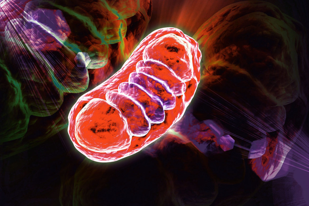
Mitochondrial disease (mito disease) refers to a group of disorders caused by dysfunction of the mitochondria, specialized structures within cells that generate approximately 90% of the energy needed by the body to sustain life and support growth. Specifically, mitochondria convert the energy from food molecules into adenosine triphosphate (ATP), the cellular energy currency that powers virtually all cellular functions.
Mitochondrial diseases result from failures in the mitochondria’s function, particularly in the oxidative phosphorylation process (OXPHOS) – the biochemical pathway in which nutrients are converted to energy. When mitochondria fail to produce sufficient energy, cells begin to malfunction or die, leading to a wide range of symptoms and multi-organ system effects.
These disorders are characterized by their extraordinary diversity in presentation, severity, age of onset, progression, and inheritance patterns. Mitochondrial diseases can present at any age from birth to late adulthood and can affect virtually any organ system, making them challenging to diagnose and treat.
Detailed Definition
At a molecular level, mitochondrial diseases involve defects in:
Mitochondrial DNA (mtDNA) – Mutations in the small circular genome contained within mitochondria themselves, which encodes 37 genes essential for mitochondrial function.
Nuclear DNA (nDNA) – Mutations in the nuclear genome that affect proteins involved in mitochondrial function, structure, or maintenance. The nuclear genome contains over 1,500 genes that encode proteins required for mitochondrial function.
Intergenomic communication – Disruptions in the coordination between nuclear and mitochondrial genomes.
The dysfunction can occur in several ways:
- Defects in the electron transport chain (ETC) complexes that generate ATP
- Abnormalities in mitochondrial dynamics (fusion, fission, movement, and quality control)
- Disturbances in mitochondrial protein import
- Deficits in mitochondrial translation
- Impaired mitochondrial DNA maintenance and replication
Affected Body Parts/Organs
Mitochondrial diseases can theoretically affect any organ system due to the ubiquitous presence of mitochondria in nearly all human cells (red blood cells being a notable exception). However, tissues and organs with high energy demands are particularly vulnerable:
Commonly Affected Systems:
Neurological System
- Brain (encephalopathy, seizures, stroke-like episodes)
- Peripheral nerves (neuropathy)
- Muscle (myopathy, weakness, exercise intolerance)
Cardiac System
- Heart muscle (cardiomyopathy)
- Conduction defects (heart block, arrhythmias)
Ophthalmological System
- Retina (retinopathy, vision loss)
- Eye muscles (ophthalmoplegia, ptosis)
- Optic nerve (optic atrophy)
Auditory System
- Inner ear (sensorineural hearing loss)
Endocrine System
- Pancreas (diabetes)
- Thyroid (hypothyroidism)
- Adrenal glands (adrenal insufficiency)
- Growth hormone deficiency
Gastrointestinal System
- Liver (hepatopathy)
- Digestive tract (dysmotility, pseudo-obstruction)
- Pancreas (exocrine pancreatic insufficiency)
Renal System
- Kidneys (tubulopathy, Fanconi syndrome)
Hematological System
- Bone marrow (sideroblastic anemia, pancytopenia)
Dermatological System
- Skin (rashes, abnormal pigmentation)
The pattern of organ involvement varies widely between different mitochondrial disease types and even between patients with the same genetic defect, creating significant diagnostic challenges.
Prevalence and Significance
Prevalence:
Mitochondrial diseases are among the most common groups of inherited metabolic disorders:
- The prevalence of mitochondrial disease caused by pathogenic mitochondrial DNA mutations is approximately 1 in 5,000 individuals.
- When including nuclear DNA mutations affecting mitochondrial function, the prevalence increases to approximately 1 in 4,300.
- The prevalence of carrier status for pathogenic mitochondrial DNA mutations in the general population may be as high as 1 in 200.
- Specific syndromes have varying prevalence; for instance, MELAS (Mitochondrial Encephalomyopathy, Lactic Acidosis, and Stroke-like episodes) affects approximately 1 in 4,000 individuals.
Significance:
Mitochondrial diseases have profound significance across multiple dimensions:
Medical Significance:
- Mitochondrial dysfunction is implicated not only in primary mitochondrial diseases but also in many common conditions including Parkinson’s disease, Alzheimer’s disease, heart disease, and certain cancers.
- These disorders represent some of the most challenging conditions to diagnose and manage due to their heterogeneity and multi-system involvement.
Scientific Significance:
- Mitochondrial diseases provide unique insights into bioenergetics, genetics, and cellular metabolism.
- Mitochondrial DNA inheritance follows maternal patterns, offering a distinct perspective on human genetics different from the traditional Mendelian inheritance of nuclear genes.
Public Health Significance:
- Mitochondrial diseases often lead to significant disability, reduced quality of life, and shortened lifespan.
- The chronic, progressive nature of many mitochondrial diseases creates substantial healthcare utilization and socioeconomic impact.
- As awareness increases, the recognized prevalence is likely to grow, suggesting previously undiagnosed cases are common.
Economic Impact:
- The average annual medical cost per mitochondrial disease patient is estimated at $24,000-$100,000 in the United States, depending on disease severity.
- Indirect costs including lost productivity, caregiver burden, and disability services substantially increase the overall societal impact.
Research Significance:
- Mitochondrial diseases represent a frontier in the development of gene therapies and other cutting-edge treatments.
- Understanding mitochondrial dysfunction offers potential insights into aging processes and age-related diseases.
The significance of mitochondrial disease has grown substantially in recent decades as improvements in diagnostic technologies have revealed their true prevalence and their connections to more common diseases, aging, and neurodegeneration.
2. History & Discoveries
First Identification and Early Understanding
The journey to understanding mitochondrial diseases spans over a century, beginning with the discovery of mitochondria themselves and gradually evolving toward recognition of their role in human disease:
Early Mitochondrial Biology:
1857-1890s: The structures later named mitochondria were first observed by Rudolf Albert von Kölliker, Richard Altmann, and Carl Benda using early microscopes.
1898: Carl Benda coined the term “mitochondria” from the Greek words “mitos” (thread) and “chondros” (granule), describing their appearance during cell division.
1912-1913: B.F. Kingsbury and Otto Warburg established mitochondria’s role in cellular respiration.
1940s: Mitochondria were isolated and their structure began to be elucidated, recognizing the inner and outer membranes.
1953: The presence of DNA in mitochondria was first reported by Nass and Nass, though its significance wasn’t immediately understood.
First Recognition of Mitochondrial Disease:
1962: Rolf Luft and colleagues at the Karolinska Institute in Stockholm, Sweden, described the first clinically documented case of mitochondrial disease in a 35-year-old woman with severe hypermetabolism not caused by thyroid dysfunction. This condition, later named Luft’s disease, was characterized by excessive heat production and profuse sweating even at rest.
1963: Electron microscopy revealed abnormal mitochondria in the patient’s skeletal muscle, establishing the connection between clinical symptoms and mitochondrial dysfunction.
Key Historical Figures
Several pioneering scientists made fundamental contributions to our understanding of mitochondrial disease:
Rolf Luft (1914-2007): Swedish endocrinologist who, along with his colleagues, documented the first case of mitochondrial disease in 1962.
Margaret M. and Joanne E. Nass: In 1963, provided the first evidence that mitochondria contain their own DNA, a critical discovery for understanding the genetics of mitochondrial diseases.
Lars Ernster (1920-1998): Biochemist who made significant contributions to understanding oxidative phosphorylation defects in mitochondrial diseases.
Douglas C. Wallace: Revolutionary figure who in 1988 reported the first maternally inherited mitochondrial DNA mutation causing human disease (Leber’s Hereditary Optic Neuropathy or LHON). His work established the field of human mitochondrial genetics.
Anita Harding (1952-1995): Neurologist who made substantial contributions to understanding mitochondrial disorders, particularly those affecting the nervous system.
Salvatore DiMauro: Neurologist who advanced the understanding of mitochondrial encephalomyopathies and developed a classification system for mitochondrial diseases.
Gottfried Schatz (1936-2015): Biochemist who elucidated many aspects of mitochondrial protein import and biogenesis.
Major Discoveries and Breakthroughs
The field of mitochondrial medicine has been marked by several transformative discoveries:
1963-1965: Identification of mitochondrial DNA (mtDNA) as a separate genetic system from nuclear DNA.
1977: Human mitochondrial DNA was completely sequenced, becoming one of the first genomes fully sequenced.
1988: First identification of a pathogenic mtDNA mutation causing human disease by Douglas Wallace’s team, who discovered a point mutation in the mtDNA gene ND4 causing Leber’s Hereditary Optic Neuropathy (LHON).
1990: Discovery of large-scale mtDNA deletions causing Kearns-Sayre syndrome and chronic progressive external ophthalmoplegia (CPEO).
1990: Identification of the A3243G mutation in tRNALeu causing MELAS syndrome (Mitochondrial Encephalomyopathy, Lactic Acidosis, and Stroke-like episodes).
1990s: Recognition of nuclear gene mutations affecting mitochondrial function, expanding the genetic basis of mitochondrial diseases beyond mtDNA itself.
1995: First identification of mutations in nuclear genes encoding mitochondrial proteins causing mitochondrial disease (Flavoprotein subunit of succinate dehydrogenase in Leigh syndrome).
1999: Discovery of polymerase gamma (POLG) mutations, revealing a major cause of mitochondrial disorders through defects in mtDNA maintenance.
2000s: Recognition of mitochondrial dynamics (fusion, fission) abnormalities in disease, with the identification of mutations in fusion proteins (OPA1, MFN2) and fission proteins (DNM1L).
2010: Development of the “Mito Exome,” a targeted next-generation sequencing approach for diagnosing mitochondrial disorders.
2015: First clinical application of mitochondrial replacement therapy to prevent transmission of mtDNA disorders.
2018: FDA approval of the first treatment specifically indicated for a condition caused by mitochondrial DNA mutations (idebenone for LHON).
Evolution of Medical Understanding
The conceptualization of mitochondrial diseases has evolved dramatically:
1960s-1970s: Early Recognition Phase
- Initial focus on rare, severe cases with obvious metabolic derangements
- Limited diagnostic tools restricted identification to the most severe phenotypes
- Mitochondrial diseases considered extremely rare conditions
1980s: Genetic Revolution
- Discovery of mtDNA mutations fundamentally changed understanding of inheritance
- Recognition of maternal inheritance pattern for mtDNA disorders
- Beginning appreciation of the heteroplasmy concept (mixed populations of normal and mutant mtDNA)
1990s: Expansion Phase
- Recognition of the broad spectrum of clinical presentations
- Development of mitochondrial disease classification systems
- Growing appreciation of nuclear-mitochondrial genomic interactions
- Establishment of specialized mitochondrial disease centers
2000s: Systems Biology Approach
- Understanding mitochondria as dynamic organelles that undergo fusion, fission, and quality control
- Recognition of mitochondrial dysfunction in common diseases (Parkinson’s, Alzheimer’s)
- Appreciation of mitochondria’s role beyond energy production (calcium homeostasis, apoptosis, reactive oxygen species signaling)
- Development of standardized diagnostic criteria
2010s-Present: Precision Medicine Era
- Next-generation sequencing revolutionizing diagnosis
- Growing recognition of mitochondrial dysfunction in aging
- Development of targeted therapies based on specific mechanisms
- Emergence of genetic therapies and mitochondrial replacement techniques
- Appreciation of mitochondrial diseases as part of a continuum of mitochondrial dysfunction disorders
This evolution has transformed mitochondrial medicine from a niche subspecialty focused on rare disorders to a field with implications for understanding and treating many common diseases, aging, and neurodegeneration. Contemporary understanding views mitochondrial diseases not as discrete entities but as a spectrum of disorders with overlapping phenotypes, complex genetics, and variable tissue specificity.
3. Symptoms
Early Symptoms
Mitochondrial diseases can manifest with a wide range of symptoms that vary significantly depending on which cells are affected, the severity of mitochondrial dysfunction, and the patient’s age. Early symptoms are often subtle and nonspecific, contributing to diagnostic delays:
Common Early Neurological Symptoms:
- Developmental delays in infants and children
- Loss of previously acquired skills (developmental regression)
- Exercise intolerance and easy fatigability
- Muscle weakness that may fluctuate
- Coordination problems (ataxia)
- Migraine-like headaches
- Seizures, particularly those that are difficult to control
- Learning disabilities or cognitive delays
Common Early Sensory Symptoms:
- Vision problems (blurred vision, visual field defects)
- Hearing difficulties or progressive hearing loss
- Balance problems
- Reduced pain or temperature sensation
Common Early Systemic Symptoms:
- Failure to thrive in infants
- Short stature
- Unexplained vomiting or constipation
- Temperature regulation problems
- Elevated lactate levels in blood or cerebrospinal fluid
- Excessive fatigue after routine activities
Early-Onset Presentation Patterns:
Neonatal Presentation:
- Poor feeding and weight gain
- Hypotonia (“floppy baby”)
- Respiratory difficulties
- Early seizures
- Liver dysfunction
Infantile Presentation:
- Developmental milestone delays
- Poor growth
- Feeding difficulties
- Hypotonia progressing to spasticity
- Early evidence of sensory impairments
Childhood Presentation:
- Exercise intolerance
- Academic difficulties
- Intermittent symptoms during times of stress or illness
- Subtle sensory impairments
- Movement disorders
Adolescent Presentation:
- Exercise intolerance
- Unexplained fatigue
- Muscle cramps with exercise
- Ptosis (drooping eyelids)
- Gastrointestinal dysmotility
Advanced-Stage Symptoms
As mitochondrial disease progresses, symptoms typically become more severe and multi-systemic:
Advanced Neurological Manifestations:
- Progressive external ophthalmoplegia (inability to move the eyes)
- Ptosis (drooping eyelids) that worsens over time
- Stroke-like episodes (in MELAS and similar syndromes)
- Dementia or cognitive decline
- Severe seizure disorders
- Movement disorders (dystonia, chorea, parkinsonism)
- Peripheral neuropathy with loss of sensation or neuropathic pain
- Autonomic dysfunction affecting blood pressure, heart rate, and temperature regulation
Advanced Muscular Manifestations:
- Progressive muscle weakness
- Muscle wasting (atrophy)
- Exercise-induced muscle breakdown (rhabdomyolysis)
- Respiratory muscle weakness requiring ventilatory support
Advanced Multi-System Manifestations:
- Cardiomyopathy (heart muscle disease)
- Heart conduction defects requiring pacemaker
- Severe gastrointestinal dysmotility requiring feeding tubes
- Pancreatic insufficiency
- Liver failure
- Kidney dysfunction
- Endocrine abnormalities (diabetes, growth hormone deficiency, adrenal insufficiency)
- Complete vision or hearing loss
Advanced Disease Patterns:
Encephalomyopathic Pattern:
- Progressive cognitive decline
- Seizures
- Stroke-like episodes
- Movement disorders
Myopathic Pattern:
- Progressive muscle weakness
- Respiratory compromise
- Swallowing difficulties
- Cardiac involvement
Multi-organ Failure Pattern:
- Progressive involvement of multiple organ systems
- Requires multidisciplinary care
- May lead to life-threatening complications
Common vs. Rare Symptoms
Common Symptoms (Present in >50% of patients):
- Fatigue and exercise intolerance
- Muscle weakness
- Neurological symptoms (seizures, developmental delays, cognitive issues)
- Gastrointestinal problems (constipation, dysmotility, reflux)
- Ptosis and external ophthalmoplegia
- Hearing loss
Less Common Symptoms (Present in 20-50% of patients):
- Diabetes mellitus
- Cardiomyopathy
- Migraine-like headaches
- Retinal degeneration
- Growth failure
- Liver dysfunction
- Peripheral neuropathy
- Movement disorders
Rare Symptoms (Present in <20% of patients):
- Stroke-like episodes
- Renal tubulopathy
- Exocrine pancreatic failure
- Bone marrow failure
- Cyclic vomiting
- Retinal pigmentary changes
- Corneal clouding
- Hypertrophic cardiomyopathy
- Wolff-Parkinson-White syndrome
- Severe lactic acidosis crisis
Symptom Progression Over Time
The progression of mitochondrial disease varies considerably based on the specific genetic defect, heteroplasmy level (for mtDNA mutations), age of onset, and involved organs. However, some general patterns can be described:
Progressive Patterns:
Steady Progressive Decline:
- Gradual worsening of symptoms over years
- Relentless progression without significant remissions
- Common in severe nuclear gene defects
Step-wise Progression:
- Periods of stability punctuated by episodes of acute decompensation
- Often triggered by physiological stressors (illness, surgery, etc.)
- After each episode, baseline function may be lower
- Common in many mtDNA disorders like MELAS
Relapsing-Remitting Course:
- Symptoms fluctuate with periods of improvement and deterioration
- May mimic multiple sclerosis in some cases
- Less common but seen in some mitochondrial cytopathies
Delayed-Onset Progressive:
- Normal early development followed by symptom onset and progression
- May begin after a symptom-free period of decades
- Seen in some mtDNA disorders like LHON
Timeline of Progression:
Neonatal/Infantile Onset: Often rapid progression with significant disability or mortality within first years of life
Childhood Onset: Variable progression; may have periods of stability interspersed with decline
Adolescent/Adult Onset: Typically slower progression, sometimes over decades
Factors Affecting Progression:
Heteroplasmy Level: Higher proportions of mutant mtDNA generally correlate with more severe disease and faster progression
Tissue Distribution: Patterns of mutant mtDNA distribution across tissues affects which symptoms progress
Environmental Stressors: Infections, medications, surgeries, and physiological stress can accelerate progression
Genetic Modifiers: Nuclear genetic background can modify the expression and progression of mtDNA disorders
Nutritional Status: Adequate nutrition and avoidance of fasting appears to slow progression in some patients
Treatment Interventions: Symptomatic treatments and supportive care may alter the natural history of certain manifestations
Disease-Specific Progression Examples:
Leigh Syndrome: Often rapid progression with developmental regression, brainstem dysfunction, and respiratory failure, frequently fatal in childhood
MELAS: Step-wise progression with stroke-like episodes causing cumulative neurological damage
MERRF (Myoclonic Epilepsy with Ragged Red Fibers): Progressive myoclonus, seizures, ataxia, and muscle weakness over decades
Kearns-Sayre Syndrome: Progressive external ophthalmoplegia, pigmentary retinopathy, and cardiac conduction abnormalities typically beginning before age 20
LHON: Rapid loss of central vision typically over weeks to months, often with stabilization after the acute phase
Understanding the diversity of symptom presentation and progression patterns is crucial for clinical recognition, appropriate monitoring, and management planning in mitochondrial diseases.
4. Causes
Biological Causes
Mitochondrial diseases result from dysfunction of the mitochondria, primarily affecting their ability to produce adequate ATP through oxidative phosphorylation. At the molecular level, this dysfunction stems from several interconnected mechanisms:
Genetic Mutations:
Mitochondrial DNA (mtDNA) Mutations:
- Point Mutations: Single nucleotide changes in mtDNA genes
- Examples: m.3243A>G (MELAS), m.8344A>G (MERRF), m.11778G>A (LHON)
- Large-scale Deletions/Duplications: Removal or duplication of significant mtDNA segments
- Examples: Common deletion causing Kearns-Sayre syndrome or CPEO
- mtDNA Depletion: Reduction in mtDNA copy number
- Examples: TK2 or DGUOK deficiency causing mtDNA depletion syndrome
- Point Mutations: Single nucleotide changes in mtDNA genes
Nuclear DNA (nDNA) Mutations:
- OXPHOS Subunit Genes: Mutations in nuclear-encoded components of respiratory chain complexes
- Examples: NDUFV1 (Complex I), SDHA (Complex II), UQCRQ (Complex III)
- Assembly Factor Genes: Mutations in proteins required for respiratory chain complex assembly
- Examples: SURF1 (Complex IV), BCS1L (Complex III)
- mtDNA Maintenance Genes: Mutations affecting replication, repair, or maintenance of mtDNA
- Examples: POLG, TWNK, MPV17
- Mitochondrial Translation Genes: Mutations affecting protein synthesis within mitochondria
- Examples: GFM1, TSFM, MRPS16
- Mitochondrial Import Genes: Mutations affecting protein transport into mitochondria
- Examples: TIMM8A, DNAJC19
- Mitochondrial Dynamics Genes: Mutations affecting fusion, fission, or movement of mitochondria
- Examples: OPA1, MFN2, DNM1L
- Mitochondrial Quality Control Genes: Mutations affecting removal of damaged mitochondria
- Examples: PINK1, PRKN (parkin)
- Cofactor Metabolism Genes: Mutations affecting synthesis of essential mitochondrial cofactors
- Examples: CoQ10 biosynthesis genes (COQ2, PDSS1)
- OXPHOS Subunit Genes: Mutations in nuclear-encoded components of respiratory chain complexes
Biochemical Mechanisms of Dysfunction:
Respiratory Chain Deficiency:
- Reduced activity of one or more OXPHOS complexes
- Decreased ATP production
- Increased production of reactive oxygen species (ROS)
Coenzyme Q10 (CoQ10) Deficiency:
- Reduced electron carrier essential for complexes I, II, and III
- Impaired ATP synthesis and increased ROS production
Pyruvate Dehydrogenase Complex Deficiency:
- Impaired conversion of pyruvate to acetyl-CoA
- Reduced substrate entry into the Krebs cycle
Krebs Cycle Defects:
- Impaired generation of reducing equivalents (NADH, FADH2)
- Metabolic intermediate accumulation
mtDNA Maintenance Defects:
- Qualitative (mutations) or quantitative (depletion) mtDNA abnormalities
- Secondary respiratory chain deficiency
Mitochondrial Protein Import Defects:
- Failure to properly localize nuclear-encoded proteins to mitochondria
- Disruption of mitochondrial proteostasis
Mitochondrial Dynamics Disruption:
- Abnormal fusion/fission balance
- Impaired mitochondrial quality control
- Disrupted mitochondrial distribution in cells
Genetic and Hereditary Factors
Mitochondrial diseases exhibit unique and complex inheritance patterns due to the dual genomic control of mitochondrial function:
mtDNA Inheritance Patterns:
Maternal Inheritance:
- mtDNA is exclusively transmitted from mother to all her children
- Affected mothers can transmit disease to all offspring, but only daughters can further transmit to next generation
- Examples: MELAS, MERRF, LHON
Heteroplasmy and Threshold Effect:
- Cells contain multiple copies of mtDNA (polyplasmy)
- Mutations may affect some but not all mtDNA copies (heteroplasmy)
- Symptoms appear when the proportion of mutant mtDNA exceeds a tissue-specific threshold
- This creates variable expressivity even within the same family
Mitotic Segregation:
- Random distribution of mitochondria during cell division
- Can lead to changing mutation load in different tissues over time
- Explains why some symptoms may appear or worsen with age
Bottleneck Effect:
- Dramatic reduction in mtDNA copy number during oogenesis
- Results in unpredictable transmission of heteroplasmy levels to offspring
- Explains why a mildly affected mother can have severely affected children or vice versa
Nuclear DNA Inheritance Patterns:
Autosomal Recessive:
- Most common inheritance pattern for nuclear gene defects affecting mitochondria
- Requires two mutated copies of the gene
- Parents are typically asymptomatic carriers
- Examples: SURF1 mutations in Leigh syndrome, TK2 mutations in mtDNA depletion
Autosomal Dominant:
- Less common but significant pattern
- Single mutated copy sufficient to cause disease
- Examples: OPA1 mutations in dominant optic atrophy, some POLG mutations
X-linked:
- Rare for mitochondrial disorders
- Males typically more severely affected
- Examples: PDHA1 mutations (pyruvate dehydrogenase deficiency), NDUFA1 mutations
De Novo Mutations:
- New mutations not present in parents
- Common in severe, early-onset mitochondrial disorders
- Examples: De novo SDHA mutations, de novo mtDNA deletions
Complex Inheritance Phenomena:
Digenic/Polygenic Inheritance:
- Mutations in multiple genes contribute to phenotype
- Examples: Combined effects of mtDNA variants and nuclear modifiers
Genetic Modifiers:
- Nuclear genetic background influences expression of mtDNA mutations
- Explains phenotypic variability in patients with identical mtDNA mutations
Epigenetic Factors:
- Methylation and other epigenetic modifications affect expression of mitochondrial genes
- May contribute to tissue-specific manifestations
Environmental Causes and Triggers
While primary mitochondrial diseases are genetic in origin, environmental factors can trigger onset, exacerbate symptoms, or contribute to secondary mitochondrial dysfunction:
Pharmacological Triggers:
Mitochondrial Toxins:
- Certain antibiotics (aminoglycosides, linezolid)
- Antiretroviral drugs (particularly older NRTIs like AZT)
- Valproic acid (particularly in POLG-related disorders)
- Statins (rarely cause mitochondrial myopathy)
- Chemotherapeutics (doxorubicin, cisplatin)
Metabolic Stressors:
- Alcohol (chronic exposure)
- Environmental toxins (rotenone, cyanide, carbon monoxide)
- Heavy metals (mercury, lead)
- Pesticides (paraquat, organophosphates)
Physiological Stressors:
Infection and Fever:
- Increased metabolic demand during infections
- Fever increases energy requirements
- Can trigger decompensation in previously stable patients
Prolonged Fasting:
- Depletes glycogen stores
- Forces greater reliance on fatty acid oxidation and ketogenesis
- Can trigger metabolic crises in certain mitochondrial disorders
Surgery and Anesthesia:
- Surgical stress increases metabolic demands
- Certain anesthetics may impair mitochondrial function
- Perioperative fasting compounds metabolic stress
Extreme Physical Exertion:
- Exceeds ATP production capacity
- Can trigger rhabdomyolysis in susceptible individuals
- May cause prolonged post-exertional malaise
Other Environmental Factors:
Aging:
- Progressive accumulation of mtDNA mutations
- Decreased mitochondrial function with age
- May unmask subclinical mitochondrial defects
Oxidative Stress:
- Environmental pollutants increasing ROS production
- UV radiation damage
- Cigarette smoke exposure
Nutritional Factors:
- Deficiencies in essential cofactors (vitamins B2, B3, folate, B12)
- Copper, iron, and zinc deficiencies affecting respiratory chain components
- Protein malnutrition impairing mitochondrial protein synthesis
Acquired Mitochondrial Dysfunction:
- Autoimmune conditions (anti-mitochondrial antibodies)
- Paraneoplastic phenomena
- Secondary to other metabolic disorders
Understanding these causal factors is essential for diagnosis, genetic counseling, prevention of symptom triggers, and development of targeted therapies. The complex interplay between genetic predisposition and environmental factors explains much of the clinical heterogeneity observed in mitochondrial disease patients.
5. Risk Factors
Demographic Risk Factors
Mitochondrial diseases can affect individuals of any age, gender, or ethnic background, but certain demographic factors are associated with different risks:
Age-Related Risk Factors:
Neonatal and Infantile Period:
- Highest risk for severe, multi-system mitochondrial disorders
- Nuclear gene mutations more commonly manifest in early life
- Leigh syndrome, mtDNA depletion syndromes, and POLG-related disorders often present in this age group
- Mortality is highest for infantile-onset mitochondrial disease
Childhood:
- Period when many moderate-severity disorders become apparent
- School performance issues may first highlight cognitive effects
- Exercise intolerance often becomes more noticeable
- Progressive disorders may show developmental plateaus or regression
Adolescence and Young Adulthood:
- Peak onset period for certain mtDNA disorders (e.g., LHON typically 15-35 years)
- Increased metabolic demands of puberty may unmask latent disease
- Stress of academic demands or athletic participation may reveal exercise intolerance
Middle Age:
- Late-onset presentations of milder mtDNA disorders
- Accumulated mtDNA damage may reach threshold levels
- Progressive hearing loss, diabetes, and myopathy often manifest
- CPEO (Chronic Progressive External Ophthalmoplegia) often presents in this age range
Elderly:
- Age-related accumulation of somatic mtDNA mutations
- Interaction between mitochondrial defects and age-related diseases
- Sarcopenia (age-related muscle loss) may be accelerated in subclinical mitochondrial dysfunction
Gender-Related Risk Factors:
Female-Specific Factors:
- Maternal transmission of mtDNA mutations
- Pregnancy can exacerbate symptoms or trigger disease onset
- Some studies suggest slightly higher penetrance of certain mtDNA mutations in females
- X-linked mitochondrial disorders show carrier status in females
Male-Specific Factors:
- More severely affected by X-linked mitochondrial disorders
- LHON shows male predominance (80% of cases are male)
- Some studies suggest different threshold effects in male tissues
Ethnic and Ancestral Risk Factors:
Population-Specific mtDNA Haplogroups:
- Certain mtDNA backgrounds modify disease expression
- Haplogroup J increases risk for LHON if carrying primary LHON mutations
- Some haplogroups may be protective for certain phenotypes
Founder Effects:
- Higher prevalence of specific mutations in certain populations
- Examples:
- A3243G mutation (MELAS) more common in certain Japanese populations
- High prevalence of LHON in Northeast England
- Increased Leigh syndrome in French Canadian populations (LRPPRC mutations)
- Higher MNGIE prevalence in Middle Eastern populations due to consanguinity
Consanguinity:
- Increases risk of autosomal recessive mitochondrial disorders
- Particularly important in populations with high rates of consanguineous marriage
Genetic Risk Factors
Genetic risk factors represent the primary determinants of mitochondrial disease risk:
Primary Genetic Risk Factors:
Carrying Pathogenic mtDNA Mutations:
- Maternal family history of mitochondrial disease
- Being offspring of a woman carrying heteroplasmic mtDNA mutations
- Risk correlates with heteroplasmy level in the mother’s oocytes
Carrier Status for Nuclear Mitochondrial Genes:
- Heterozygous mutations in autosomal recessive mitochondrial disease genes
- Risk for having affected children when both parents are carriers
- Carrier frequency for some mitochondrial disease genes approaches 1:100
Inherited Nuclear Genetic Variants:
- Autosomal dominant mutations with reduced penetrance
- X-linked carrier status
- Digenic inheritance requiring multiple genetic “hits”
Genetic Modifiers:
Nuclear Background Effects:
- Nuclear genes that modify mtDNA mutation expression
- Polymorphisms affecting mitochondrial function
- Variants in antioxidant pathways
mtDNA Haplogroup:
- Background mtDNA variants that can increase or decrease disease risk
- Influence penetrance and expression of pathogenic mutations
Epigenetic Factors:
- Methylation patterns affecting expression of mitochondrial genes
- Inherited epigenetic modifications
Risk Quantification:
Heteroplasmy Levels:
- Higher levels generally correlate with greater disease risk
- Tissue-specific thresholds vary (typically 60-90% mutant mtDNA required for symptoms)
- Blood heteroplasmy poorly predicts risk in some disorders
Recurrence Risks:
- mtDNA point mutations: Highly variable maternal transmission risk (0-100%)
- mtDNA deletions: Generally low recurrence risk (<5%) except in mtDNA maintenance disorders
- Autosomal recessive: 25% risk for each pregnancy of carrier parents
- Autosomal dominant: 50% risk for affected parent’s offspring
Environmental and Lifestyle Risk Factors
While primarily genetic, mitochondrial disease risk and progression are influenced by environmental and lifestyle factors:
Exposure-Related Risks:
Medication Exposures:
- Aminoglycoside antibiotics can trigger hearing loss in those with susceptible mtDNA mutations
- Valproic acid can precipitate liver failure in POLG-related disorders
- Statins may increase myopathy risk in subclinical mitochondrial disease
- Certain anesthetics may pose higher risks
Environmental Toxins:
- Occupational exposure to mitochondrial toxins (pesticides, heavy metals)
- Industrial chemicals affecting mitochondrial function
- Cigarette smoke (damages mtDNA and respiratory chain)
Nutritional Factors:
- Deficiencies in cofactors needed for mitochondrial function
- Vitamin B deficiencies (particularly riboflavin, niacin, folate)
- Inadequate protein intake
- Micronutrient deficiencies (iron, copper, zinc, magnesium)
Lifestyle-Related Risks:
Physical Activity Patterns:
- Extremes of exercise (complete inactivity or excessive exertion)
- Abrupt increases in physical demands
- Inadequate recovery between physical activities
Dietary Patterns:
- Prolonged fasting or irregular meal patterns
- Very low carbohydrate diets in certain mitochondrial disorders
- Excessive alcohol consumption
- High oxidative stress diets (high in processed foods, low in antioxidants)
Sleep and Stress:
- Chronic sleep deprivation impacts mitochondrial function
- Psychological stress increases oxidative stress
- Circadian rhythm disruption affects mitochondrial dynamics
Medical Condition Risks:
Acute Physiological Stressors:
- Infections and febrile illnesses
- Surgery and anesthesia
- Trauma and critical illness
- Pregnancy and delivery
Chronic Diseases:
- Autoimmune conditions
- Chronic inflammatory disorders
- Endocrine disorders (especially diabetes)
- Neurodegenerative diseases
Impact of Pre-existing Conditions
Several pre-existing conditions can significantly influence mitochondrial disease risk, manifestation, and progression:
Endocrine Conditions:
Diabetes Mellitus:
- Both contributor to and result of mitochondrial dysfunction
- Complicates metabolic management
- Increases risk of multisystem complications
- May accelerate progression of certain mitochondrial disorders
Thyroid Disorders:
- Hypothyroidism increases mitochondrial disease symptom burden
- Hyperthyroidism increases metabolic demands
- Thyroid function affects overall energy metabolism
Neurological Conditions:
Epilepsy:
- Seizures increase metabolic demands
- Anti-epileptic medications may impact mitochondrial function
- Status epilepticus can trigger decompensation
Migraine:
- Shares pathophysiological features with mitochondrial disorders
- May indicate subclinical mitochondrial dysfunction
- Complicates symptom management
Cardiovascular Conditions:
Pre-existing Cardiomyopathy:
- Compounds cardiac manifestations of mitochondrial disease
- Limits physiological reserve
- Complicates medication choices
Conduction Abnormalities:
- Increases risk of serious arrhythmias
- May require earlier pacemaker intervention
Other Relevant Conditions:
Chronic Kidney Disease:
- Alters drug clearance for mitochondrial medications
- Increases metabolic stress
- Complicates management of mitochondrial tubulopathies
Chronic Liver Disease:
- Impairs detoxification
- Reduces metabolic reserve
- May accelerate hepatic manifestations of mitochondrial disease
Autoimmune Disorders:
- Inflammatory stress affects mitochondrial function
- Immunosuppressive treatments may impact energy metabolism
- May share underlying pathophysiological mechanisms
Understanding these risk factors is crucial for:
- Identifying high-risk individuals who may benefit from genetic testing
- Genetic counseling for family planning
- Developing prevention strategies for symptom triggers
- Creating personalized management approaches
- Research into modifiable factors that could alter disease course
The combination of genetic predisposition and environmental factors creates a complex risk landscape that explains the remarkable clinical heterogeneity seen in mitochondrial disorders. Clinicians must consider this entire spectrum of risk factors when evaluating patients and developing management strategies.
6. Complications
Direct Complications of Mitochondrial Disease
Mitochondrial diseases can lead to various complications across multiple organ systems, reflecting the ubiquitous need for mitochondrial energy production throughout the body:
Neurological Complications:
Seizure Disorders:
- Prevalence: 35-60% of patients
- Types: Focal, generalized, myoclonic, status epilepticus
- Challenges: Often drug-resistant, may worsen with certain anti-epileptics
- Impact: Cognitive impacts, risk of sudden unexpected death in epilepsy (SUDEP)
Stroke-like Episodes:
- Prevalence: Common in MELAS (80%), less frequent in other syndromes
- Characteristics: Focal neurological deficits not confined to vascular territories
- Pathophysiology: Metabolic dysfunction, impaired autoregulation, neuronal hyperexcitability
- Consequences: Cumulative brain injury, cognitive decline, focal deficits
Cognitive Impairment and Dementia:
- Prevalence: 30-70% depending on specific syndrome
- Pattern: May be static or progressive
- Features: Executive dysfunction, memory impairment, processing speed deficits
- Impact: Educational challenges, loss of independence, behavioral changes
Movement Disorders:
- Types: Dystonia, chorea, ataxia, parkinsonism
- Prevalence: 30-50% in pediatric cases, variable in adults
- Associated syndromes: Leigh syndrome, POLG-related disorders
- Impact: Mobility limitations, pain, functional impairment
Cardiac Complications:
Cardiomyopathy:
- Types: Hypertrophic, dilated, or non-compaction
- Prevalence: 20-40% of patients
- Risk factors: Specific mutations (particularly in tRNA genes), infantile onset
- Progression: May be rapidly progressive or slowly evolving
- Consequences: Heart failure, sudden cardiac death
Conduction Defects:
- Types: Atrioventricular block, bundle branch block, long QT syndrome
- Prevalence: Up to 30% in certain syndromes (Kearns-Sayre syndrome)
- Management needs: Pacemaker or defibrillator implantation
- Risks: Sudden cardiac death if undetected
Endocrine Complications:
Diabetes Mellitus:
- Prevalence: 5-25% (higher in MIDD – Maternally Inherited Diabetes and Deafness)
- Characteristics: Usually non-autoimmune, mixed insulin secretion/resistance defect
- Management challenges: Increased risk of lactic acidosis with metformin
- Associated features: Often occurs with hearing loss in A3243G mutation
Growth Hormone Deficiency:
- Prevalence: 10-35% of pediatric patients
- Impact: Short stature, altered body composition
- Treatment considerations: Efficacy and safety of growth hormone therapy
Adrenal Insufficiency:
- Prevalence: Rare overall, higher in specific syndromes
- Risks: Adrenal crisis during stress
- Screening needs: Periodic evaluation in high-risk patients
Ophthalmological Complications:
Optic Atrophy:
- Prevalence: Near 100% in LHON, common in other syndromes
- Progression: May be acute or insidious
- Functional impact: Central vision loss, legal blindness
- Recovery potential: Variable based on genetic cause
Retinopathy:
- Types: Pigmentary retinopathy, macular degeneration
- Associated syndromes: Kearns-Sayre syndrome, NARP
- Impact: Night blindness, peripheral vision loss, central vision impairment
Progressive External Ophthalmoplegia (PEO):
- Features: Restricted eye movements, ptosis
- Functional impact: Visual field limitation, compensatory head posturing
- Management needs: Ptosis surgery considerations
Gastrointestinal Complications:
Dysmotility:
- Manifestations: Gastroparesis, intestinal pseudo-obstruction, constipation
- Prevalence: 20-60% of patients
- Impact: Malnutrition, bacterial overgrowth, abdominal pain
- Management challenges: Limited effective treatments
Liver Disease:
- Types: Hepatic steatosis, fibrosis, acute liver failure
- Risk factors: POLG mutations, infantile onset, mtDNA depletion
- Triggers: Valproic acid, infections, fasting
- Outcomes: Progressive liver failure may require transplantation
Exocrine Pancreatic Insufficiency:
- Prevalence: Uncommon overall, higher in certain syndromes (Pearson)
- Impact: Malabsorption, malnutrition, failure to thrive
- Management: Enzyme replacement therapy
Renal Complications:
Tubulopathies:
- Types: Fanconi syndrome, renal tubular acidosis
- Associated syndromes: More common in tRNA mutations, cytochrome c oxidase deficiency
- Impact: Electrolyte disturbances, growth impairment, nephrocalcinosis
Glomerular Disease:
- Less common than tubular dysfunction
- Manifestations: Proteinuria, progressive renal insufficiency
- Management: Standard nephroprotective strategies
Long-term Impact on Health
The chronic and progressive nature of many mitochondrial diseases leads to cumulative health impacts:
Physical Health Impacts:
Multisystem Deterioration:
- Progressive involvement of previously unaffected organ systems
- Accumulating physical disabilities
- Increasing medical complexity
Exercise Intolerance and Deconditioning:
- Worsening exercise capacity
- Muscle atrophy from disuse
- Cardiorespiratory deconditioning
- Secondary complications of immobility
Nutritional Challenges:
- Progressive dysphagia and aspiration risk
- Malnutrition from GI dysmotility
- Increased metabolic demands with decreased intake
- Eventual dependence on enteral feeding
Increased Susceptibility:
- Greater vulnerability to infections
- Reduced physiological reserve during stress
- Prolonged recovery from illness
- Accelerated features of aging
Functional and Quality of Life Impacts:
Progressive Loss of Independence:
- Mobility limitations requiring assistive devices or wheelchairs
- Self-care difficulties
- Increasing caregiver dependence
- Home and environmental modification needs
Educational and Vocational Impacts:
- Learning challenges affecting educational attainment
- Cognitive limitations affecting vocational options
- Fatigue limiting work capacity
- Early retirement due to disability
Psychosocial Consequences:
- Social isolation due to symptoms and limitations
- Relationship challenges
- Depression and anxiety
- Financial strain from medical costs and lost income
Healthcare Burden:
- Frequent hospitalizations
- Multiple specialist involvement
- Complex medication regimens
- Invasive interventions (feeding tubes, ventilatory support)
Disability and Fatality Rates
Mitochondrial diseases vary dramatically in their impact on disability and mortality:
Disability Patterns:
Spectrum of Disability:
- Range from minimal impairment to profound multi-system disability
- Approximately 50% of patients experience significant disability
- 30-40% require mobility assistance
- 10-25% become dependent on feeding tubes
- 5-15% require ventilatory support
Disability Progression:
- Childhood-onset typically associated with developmental impacts
- Adult-onset often affects established skills and independence
- Step-wise loss of function common after physiological stressors
- Variable plateau periods between deteriorations
Functional Outcomes by Disease Type:
- Leigh syndrome: 75-90% develop significant motor disabilities
- MELAS: Progressive neurological disability in 60-80%
- MERRF: Progressive ataxia and myoclonus impacting function in 70-90%
- LHON: Visual disability but often preserved general function
- CPEO: Progressive limitation in activities requiring full visual fields
Mortality Patterns:
Mortality Rates by Age of Onset:
- Neonatal-onset: 50-90% mortality within first years
- Infantile-onset: 30-50% 5-year mortality
- Childhood-onset: 10-30% 10-year mortality
- Adult-onset: Highly variable, often with near-normal life expectancy in milder forms
Syndrome-Specific Mortality:
- Leigh syndrome: Historically 50% mortality within 2 years of onset
- Alpers-Huttenlocher syndrome: Often fatal within months to years of onset
- Pearson syndrome: High early mortality, survivors may develop Kearns-Sayre phenotype
- MELAS: Reduced life expectancy, with median survival 30-40 years from diagnosis
- LHON: Generally normal life expectancy despite visual disability
Common Causes of Death:
- Respiratory complications (central hypoventilation, aspiration pneumonia)
- Cardiac events (arrhythmias, heart failure)
- Status epilepticus
- Complications of liver failure
- Sepsis during metabolic decompensations
Prognostic Factors:
- Age of onset (earlier onset generally worse prognosis)
- Specific genetic defect
- Multi-system vs. isolated organ involvement
- Seizure burden
- Cardiac involvement
- Timely supportive interventions
Long-term Survival Trends:
Improved Survival Over Time:
- Advances in supportive care have improved outcomes
- Earlier diagnosis allowing preventive strategies
- Better management of complications
- Multidisciplinary care approaches
Quality of Extended Survival:
- Increased survival often accompanied by significant disability
- Palliative and supportive care needs
- Long-term mechanical ventilation considerations
- Extended transitions from pediatric to adult care
Predictors of Better Outcomes:
- Access to specialized mitochondrial disease centers
- Comprehensive symptom management
- Aggressive prevention of metabolic decompensation
- Early intervention for complications
- Strong social and family support
Understanding these complications is essential for anticipatory care, appropriate monitoring, timely intervention, and realistic prognostic discussions with patients and families. The unpredictable and variable nature of mitochondrial disease progression necessitates individualized care plans with regular reassessment and adaptation.
7. Diagnosis & Testing
Clinical Evaluation and Diagnostic Approach
Diagnosing mitochondrial disease typically follows a stepwise approach, moving from clinical suspicion based on symptoms and signs to confirmatory molecular testing:
Initial Clinical Evaluation:
Detailed Medical History:
- Symptom onset, progression, and pattern
- Family history with multi-generational pedigree
- Maternal lineage analysis for suspected mtDNA disorders
- Developmental milestones and regression
- Exacerbating factors and environmental triggers
- Response to previous treatments
Comprehensive Physical Examination:
- Neurological assessment (strength, reflexes, coordination, gait)
- Ophthalmological evaluation (ptosis, ophthalmoplegia, retinopathy)
- Cardiac examination
- Growth parameters and dysmorphic features
- Hearing assessment
- Skin and hair examination
Clinical Diagnostic Criteria:
- Bernier Criteria: Classifies diagnostic likelihood based on clinical, metabolic, and histological findings
- Modified Walker Criteria: Points-based system for probable, possible, and definite mitochondrial disease
- Mitochondrial Disease Criteria (MDC): System incorporating clinical features and laboratory findings
- Nijmegen Criteria: Specifically for children, with age-related considerations
Clinical Red Flags Suggesting Mitochondrial Disease:
Symptom Patterns:
- Multi-system involvement
- Progressive course
- Fluctuating or exercise-related symptoms
- Maternal inheritance pattern
- Symptom combinations without unifying alternative diagnosis
Specific Clinical Constellations:
- Stroke-like episodes + diabetes + hearing loss (suggesting MELAS)
- Progressive external ophthalmoplegia + retinopathy + heart block (suggesting Kearns-Sayre)
- Myoclonus + ataxia + ragged-red fibers (suggesting MERRF)
- Subacute visual loss in young adult (suggesting LHON)
- Leigh syndrome neuroimaging pattern with regression
Laboratory Testing
Laboratory investigation is crucial for supporting the diagnosis and characterizing the specific type of mitochondrial disease:
First-Tier Biochemical Testing:
Blood Tests:
- Lactate and pyruvate: Elevated in 50-60% of patients, particularly during decompensation
- Creatine kinase (CK): Elevated in myopathic presentations
- Comprehensive metabolic panel: Assessing liver function, renal function, glucose
- Complete blood count: May show anemia or pancytopenia in certain syndromes
- Amino acids profile: May show elevated alanine
- Thyroid function tests: Evaluating endocrine involvement
Urine Tests:
- Organic acids: May show elevated TCA cycle intermediates
- Amino acids: Generalized aminoaciduria in renal tubulopathy
- Myoglobin: Evidence of rhabdomyolysis in acute episodes
Cerebrospinal Fluid (CSF) Analysis:
- Lactate: Often elevated even when blood lactate is normal
- Protein: May be elevated
- Glucose: Usually normal (distinguishing from other neurometabolic disorders)
Specialized Biochemical Testing:
Fibroblast Enzyme Analysis:
- Measurement of respiratory chain complex activities
- Oxygen consumption studies
- ATP synthesis capacity
Muscle Biochemistry:
- Spectrophotometric analysis of respiratory chain complexes
- Coenzyme Q10 levels
- Pyruvate dehydrogenase complex activity
Metabolic Stress Testing:
- Exercise testing with serial lactate measurements
- Glucose and fatty acid oxidation studies
- Nuclear magnetic resonance (NMR) spectroscopy
Tissue Biopsies and Histopathology
Tissue sampling provides valuable diagnostic information, particularly in cases without clear genetic findings:
Skeletal Muscle Biopsy:
Histological Features:
- Ragged-red fibers: Accumulation of abnormal mitochondria (modified Gomori trichrome stain)
- Cytochrome c oxidase (COX) negative fibers: Indicating complex IV deficiency
- Succinate dehydrogenase (SDH) hyperreactive fibers: Signs of mitochondrial proliferation
- Abnormal mitochondrial size and distribution
Electron Microscopy Findings:
- Abnormal mitochondrial ultrastructure
- Paracrystalline inclusions
- Abnormal cristae organization
- Mitochondrial proliferation
Immunohistochemistry:
- Respiratory chain complex subunit expression
- Mitochondrial content markers
Liver Biopsy:
- Micro- or macrovesicular steatosis
- Cholestasis
- Iron accumulation
- Reduced COX staining
- Particularly valuable in suspected mtDNA depletion syndromes
Cardiac Muscle Biopsy:
- Rarely performed but may show similar features to skeletal muscle
- Useful in isolated cardiomyopathy of suspected mitochondrial origin
Skin Biopsy:
- Source for fibroblast culture
- Less invasive alternative to muscle biopsy for some analyses
Genetic Testing
Genetic testing has revolutionized mitochondrial disease diagnosis and largely replaced invasive biopsies as the primary diagnostic approach:
mtDNA Analysis:
Targeted Mutation Testing:
- Specific common point mutations (A3243G, A8344G, T8993G/C)
- Best for patients with classical syndromes like MELAS, MERRF, LHON
mtDNA Sequencing:
- Next-generation sequencing of entire mitochondrial genome
- Detecting both common and rare variants
- Heteroplasmy quantification
mtDNA Deletion/Duplication Analysis:
- Long-range PCR or array CGH
- Southern blot analysis
- Detection of single large-scale deletions or multiple deletions
mtDNA Copy Number Analysis:
- Quantitative PCR
- Critical for diagnosing mtDNA depletion syndromes
Nuclear DNA Analysis:
Targeted Gene Panels:
- Analysis of 100-400 genes associated with mitochondrial function
- Useful when clinical presentation suggests specific gene categories
Whole Exome Sequencing (WES):
- Analysis of all protein-coding regions
- High diagnostic yield for rare presentations
- Can identify novel disease-causing genes
Whole Genome Sequencing (WGS):
- Comprehensive analysis including non-coding regions
- Highest diagnostic yield but most costly
- Can detect structural variants missed by exome sequencing
RNA Sequencing:
- Evaluates consequences of splice variants
- May identify gene expression abnormalities
- Emerging approach for cases with negative DNA testing
Special Considerations in Genetic Testing:
Tissue Selection:
- Blood DNA may miss mutations present in affected tissues
- Muscle, urine sediment, or buccal cells often preferred for mtDNA analysis
- Multiple tissue testing may be required for heteroplasmic mutations
Heteroplasmy Assessment:
- Quantification of mutant vs. wild-type mtDNA ratio
- Critical for predicting disease severity and progression
- May require deep sequencing techniques
Segregation Studies:
- Testing family members to confirm variant pathogenicity
- Tracking transmission patterns
Functional and Imaging Studies
Functional testing and imaging provide critical information about organ involvement and disease severity:
Functional Studies:
Cardiac Evaluation:
- Electrocardiogram (ECG): Conduction abnormalities
- Echocardiogram: Cardiomyopathy assessment
- Holter monitoring: Arrhythmia detection
Neurophysiological Testing:
- Electroencephalogram (EEG): Seizure activity
- Nerve conduction studies/EMG: Neuropathy and myopathy patterns
- Visual evoked potentials: Optic pathway function
- Brainstem auditory evoked responses: Hearing pathway assessment
Pulmonary Function Testing:
- Spirometry: Respiratory muscle weakness
- Sleep studies: Central or obstructive apnea
Exercise Testing:
- Cycle ergometry with lactate monitoring
- 31P-MR spectroscopy during exercise
- 6-minute walk test for functional capacity
Imaging Studies:
Brain MRI:
- Stroke-like lesions not conforming to vascular territories (MELAS)
- Symmetric basal ganglia lesions (Leigh syndrome)
- Cerebral atrophy
- White matter abnormalities
- Calcifications
Magnetic Resonance Spectroscopy (MRS):
- Elevated lactate peak in affected brain regions
- Reduced N-acetylaspartate (neuronal loss)
Muscle Imaging:
- MRI patterns of selective muscle involvement
- T1 changes indicating fatty replacement
- STIR sequence showing edema/inflammation
Cardiac MRI:
- Detailed assessment of cardiomyopathy
- Tissue characterization
- Functional evaluation
PET Scanning:
- Fluorodeoxyglucose (FDG) uptake patterns
- Regional metabolic defects
Diagnostic Challenges and Considerations
Several factors complicate the diagnostic process for mitochondrial diseases:
Diagnostic Limitations:
Phenotypic Heterogeneity:
- Same genetic defect causing different clinical presentations
- Different mutations causing similar clinical syndromes
- Challenges in genotype-phenotype correlation
Genetic Complexity:
- Dual genomic control (nuclear and mitochondrial)
- Heteroplasmy affecting expression
- Tissue-specific manifestations
- Secondary mitochondrial dysfunction in other disorders
Technical Limitations:
- Some genes remain difficult to analyze
- Detecting low-level heteroplasmy requires specialized techniques
- Variant interpretation challenges with novel mutations
Diagnostic Algorithms:
Modern diagnostic approaches typically follow a tiered strategy:
Initial Evaluation:
- Clinical assessment using standardized criteria
- Basic metabolic and biochemical screening
- Family history and pedigree analysis
First-tier Testing:
- Targeted genetic testing if syndrome-specific features present
- mtDNA sequencing and deletion analysis
- Nuclear gene panel testing based on clinical presentation
Second-tier Testing:
- Whole exome or genome sequencing
- Tissue biopsy for biochemical and histopathological studies
- Functional studies to assess pathogenicity of variants
Special Situations:
- Prenatal diagnosis options
- Pre-implantation genetic diagnosis
- Family variant testing
- Novel gene discovery approaches
Diagnostic Efficacy:
Current diagnostic yield varies by approach:
- Clinical criteria alone: 5-10% definitive diagnosis
- Biochemical testing: 30-60% supportive evidence
- Targeted genetic testing: 10-25% for specific syndromes
- Comprehensive mtDNA analysis: 15-20% overall
- Nuclear gene panels: 25-40%
- Whole exome sequencing: 35-55%
- Combined genetic approaches: Up to 60-70%
Emerging Diagnostic Technologies:
Next-Generation Sequencing Advances:
- Long-read sequencing for complex structural variants
- Single-cell sequencing for heteroplasmy mapping
- Nanopore technology for real-time sequencing
Multi-omics Approaches:
- Integrated genomics, transcriptomics, proteomics
- Metabolomics profiling
- Systems biology analyses of mitochondrial function
Functional Assays:
- Patient-derived cellular models
- High-throughput functional screening
- CRISPR-based functional validation
The diagnosis of mitochondrial disease remains challenging despite advances in technology. A systematic, step-wise approach combining clinical assessment, biochemical testing, and state-of-the-art genetic analysis offers the best diagnostic yield. The diagnostic pathway continues to evolve with rapid technological advances, increasing understanding of the genetic basis of mitochondrial diseases, and improved accessibility of comprehensive genetic testing.
8. Treatment Options
Current Treatment Approaches
Treatment for mitochondrial diseases remains largely supportive and symptomatic, although targeted approaches are emerging. Management typically involves a multidisciplinary team addressing the diverse manifestations of these complex disorders:
Supportive and Symptomatic Therapy:
Neurological Symptom Management:
- Seizure control: Levetiracetam, lamotrigine, and benzodiazepines are generally preferred; valproate is contraindicated in POLG-related disorders
- Headache management: Migraine prophylaxis and treatment, avoiding triggers
- Movement disorder treatment: Targeted medication for dystonia, chorea, or spasticity
- Pain management: Multimodal approach for neuropathic and myopathic pain
Cardiac Management:
- Cardiomyopathy treatment: Standard heart failure medications (ACE inhibitors, beta-blockers)
- Rhythm management: Pacemaker or defibrillator implantation for conduction defects
- Monitoring: Regular cardiac evaluations to detect progression
Ophthalmological Support:
- Visual aids: For vision loss
- Ptosis management: Crutches, lid tape, or surgical correction
- Regular monitoring: For retinal disease progression
Gastrointestinal Symptom Management:
- Dysmotility treatment: Prokinetics, dietary modifications
- Nutritional support: Enteral feeding when necessary
- Liver function monitoring: Regular assessment
Endocrine Management:
- Diabetes treatment: Insulin therapy often required
- Hormone replacement: For deficiencies (thyroid, growth hormone, etc.)
- Regular screening: For developing endocrinopathies
Physical and Occupational Therapy:
- Maintaining function and preventing contractures
- Adaptive equipment and assistive technology
- Energy conservation techniques
Respiratory Support:
- Non-invasive ventilation for sleep-disordered breathing
- Secretion management
- Invasive ventilation decisions based on disease trajectory and goals of care
Metabolic and Nutritional Therapies
Approaches targeting metabolic dysfunction are a cornerstone of mitochondrial disease management:
Nutritional Interventions:
Dietary Modifications:
- Avoiding fasting: Regular meal timing, including nighttime snacks in children
- Balanced macronutrient intake: Protein, carbohydrate, and fat ratios individualized to specific disorders
- Modified ketogenic diets: In selected cases, particularly pyruvate dehydrogenase deficiency
- Low glycemic index diet: For those with insulin resistance or diabetes
Tube Feeding:
- Nasogastric or gastrostomy feeding for inadequate oral intake
- Continuous nighttime feeding to prevent catabolism
- Specialized formulas for specific metabolic needs
Supplementation Approaches:
Cofactor Supplementation:
Coenzyme Q10 (CoQ10/Ubiquinone):
- Dosage: 5-30 mg/kg/day in children; 100-600 mg/day in adults
- Evidence: Strong for primary CoQ10 deficiencies; moderate for secondary deficiencies
- Mechanism: Electron carrier in respiratory chain, antioxidant
Riboflavin (Vitamin B2):
- Dosage: 100-400 mg/day
- Evidence: Strong for riboflavin-responsive disorders (ACAD9, ETF, ETFDH defects)
- Mechanism: Precursor for FAD, essential for complexes I and II
L-Carnitine:
- Dosage: 50-100 mg/kg/day
- Evidence: Beneficial in secondary carnitine deficiency
- Mechanism: Essential for fatty acid transport into mitochondria
Thiamine (Vitamin B1):
- Dosage: 100-600 mg/day
- Evidence: Effective for thiamine-responsive PDH deficiency
- Mechanism: Cofactor for pyruvate dehydrogenase complex
Antioxidant Therapy:
- Vitamin C and E:
- Dosage: Vitamin C 200-2000 mg/day; Vitamin E 400-800 IU/day
- Mechanism: Free radical scavenging
- Alpha-lipoic acid:
- Dosage: 200-600 mg/day
- Mechanism: Recycling other antioxidants, metal chelation
- N-acetylcysteine:
- Dosage: 600-1800 mg/day
- Mechanism: Glutathione precursor, antioxidant
- Vitamin C and E:
Metabolic Modifiers:
Arginine and Citrulline:
- Dosage: Arginine 150-300 mg/kg/day (acute); 100-150 mg/kg/day (maintenance)
- Evidence: May reduce stroke-like episode frequency in MELAS
- Mechanism: Nitric oxide precursor, improves vasodilation
Creatine monohydrate:
- Dosage: 5-10 g/day
- Mechanism: Alternative energy buffer in muscle and brain
Folinic acid:
- Dosage: 1-5 mg/kg/day
- Particular benefit in Kearns-Sayre syndrome
Pharmaceutical Interventions
Several medications have been investigated specifically for mitochondrial diseases:
Approved Medications:
Idebenone:
- Synthetic analog of CoQ10 with improved bioavailability
- Approved in Europe for Leber’s Hereditary Optic Neuropathy (LHON)
- Dosage: 900 mg/day
- Evidence: Moderately effective in visual recovery, particularly in recent-onset cases
- Mechanism: Bypasses complex I, antioxidant properties
EPI-743 (Vatiquinone):
- Synthetic para-benzoquinone
- Received FDA approval in 2023 for Leigh syndrome
- Dosage: Based on clinical trials
- Mechanism: Modulates cellular redox balance, targets NQO1 enzyme
Repurposed Medications:
Dichloroacetate (DCA):
- Activates pyruvate dehydrogenase by inhibiting PDH kinase
- Reduces lactate levels
- Mixed results in clinical trials
- Use limited by peripheral neuropathy side effects
L-cysteine:
- Precursor for glutathione synthesis
- May improve mitochondrial function in some conditions
- Evidence primarily from small studies
Bezafibrate:
- PPAR agonist that increases mitochondrial biogenesis
- Preliminary evidence in muscle biopsies
- Clinical trials ongoing
Experimental Therapies in Clinical Trials:
Nicotinamide Riboside and NAD+ Precursors:
- Enhances mitochondrial biogenesis and function
- Multiple clinical trials underway
- Promising results in cellular and animal models
Mitochondrial-targeted Antioxidants:
- MitoQ, SkQ1, and similar compounds
- Selectively concentrate in mitochondria
- Enhanced antioxidant efficacy
Novel Metabolic Modulators:
- KL1333: NAD+ modulator in Phase 2 trials
- Omaveloxolone: Nrf2 activator showing promise in Friedrich’s ataxia
- Rapamycin: mTOR inhibitor with potential benefits in specific mitochondrial disorders
Surgical and Invasive Interventions
Surgical approaches address specific complications or manifestations:
Pacemaker/Defibrillator Implantation:
- Essential for cardiac conduction defects
- Prophylactic in high-risk patients (e.g., Kearns-Sayre syndrome)
Gastrostomy Tube Placement:
- For nutritional support
- Reducing aspiration risk
Cochlear Implantation:
- For sensorineural hearing loss
- Variable efficacy in mitochondrial deafness
Ptosis Surgery:
- Frontalis sling or levator resection
- Functional and cosmetic improvement
Deep Brain Stimulation:
- For medication-refractory dystonia
- Case series show benefit in selected patients
Genetic and Cellular Therapies
Cutting-edge approaches targeting the genetic basis of mitochondrial disease represent the frontier of treatment:
Mitochondrial Replacement Therapy (MRT):
- Prevents transmission of mtDNA mutations from mother to child
- Techniques include maternal spindle transfer or pronuclear transfer
- Creates embryo with mother’s nuclear DNA and donor’s mitochondria
- Limited clinical application to date
- Legal in UK and some other countries; regulatory restrictions elsewhere
Gene Therapy Approaches:
Gene Replacement:
- Delivering functional copies of defective nuclear genes
- Adeno-associated virus (AAV) vectors showing promise
- Challenges include delivery to affected tissues and expression levels
Allotopic Expression:
- Relocating mtDNA-encoded genes to the nucleus
- Engineering nuclear-encoded proteins to be imported into mitochondria
- Technical challenges in protein import and assembly
mtDNA Editing:
- CRISPR-based approaches being adapted for mitochondrial genome
- Alternative nucleases (TALENs, ZFNs) showing preliminary success
- Challenges in delivery and specificity
Heteroplasmy Shifting:
- Mitochondrially targeted nucleases to selectively eliminate mutant mtDNA
- Allows wild-type mtDNA to repopulate cells
- Promising results in animal models and cell lines
Stem Cell and Regenerative Approaches:
Induced Pluripotent Stem Cells (iPSCs):
- Patient-derived cells corrected and differentiated to affected cell types
- Potential for transplantation of gene-corrected cells
- In early research stages
Mitochondrial Augmentation Therapy:
- Transfer of healthy mitochondria into affected cells
- Preliminary clinical application in some settings
- Mechanism and durability uncertain
Mesenchymal Stem Cell Therapy:
- Potential immunomodulatory and regenerative effects
- Limited evidence in mitochondrial disease specifically
Emerging Clinical Trials and Research Directions
The field of mitochondrial disease therapeutics is rapidly evolving:
Active Clinical Trial Areas:
Small Molecule Approaches:
- NAD+ precursors and boosters
- Mitochondrial biogenesis activators
- Redox modulators and antioxidants
- Metabolic pathway modulators
Biological Therapies:
- AAV-mediated gene therapy for specific mutations
- RNA-based therapies for splice defects
- Antisense oligonucleotides for specific mutations
Novel Delivery Systems:
- Mitochondrial-targeted peptides
- Nanoparticle delivery
- Extracellular vesicle approaches
Clinical Trial Challenges:
Disease Heterogeneity:
- Varied clinical presentations
- Different genetic causes
- Challenge in defining homogeneous trial populations
Outcome Measures:
- Lack of validated biomarkers correlating with clinical outcomes
- Variable natural history complicating endpoint selection
- Need for disease-specific outcome measures
Rare Disease Challenges:
- Small patient populations
- Geographic dispersion
- Funding limitations
Research Consortia and Networks:
- North American Mitochondrial Disease Consortium (NAMDC)
- Mitochondrial Clinical Research Network (MCRN)
- European Network for Mitochondrial Diseases (ENMD)
- International Mito Patients (IMP)
- Various disease-specific research foundations
The treatment landscape for mitochondrial diseases has evolved from purely supportive care to include targeted metabolic approaches and emerging genetic therapies. While curative treatments remain elusive for most forms of mitochondrial disease, symptomatic management has improved quality of life and survival. The next decade promises significant advances as gene therapy, mitochondrial replacement, and novel pharmacological approaches move from research to clinical application.
9. Prevention & Precautionary Measures
Genetic Counseling and Family Planning
Given the genetic basis of mitochondrial diseases, prevention largely centers on genetic counseling and reproductive options:
Genetic Counseling Process:
Risk Assessment:
- Identifying inheritance pattern (maternal, autosomal recessive, dominant, X-linked)
- Calculating recurrence risk for future pregnancies
- Assessment of extended family members at risk
Education:
- Explaining genetic basis of specific mitochondrial disease
- Discussing heteroplasmy and the bottleneck effect for mtDNA mutations
- Exploring variability in disease expression
- Providing information on reproductive options
Psychosocial Support:
- Addressing guilt feelings in parents
- Decision-making support regarding family planning
- Connecting with support groups and resources
Reproductive Options:
For Nuclear Gene (nDNA) Mutations:
- Preimplantation Genetic Testing (PGT):
- IVF with testing of embryos before implantation
- Selection of embryos without disease-causing mutations
- High success rates for single-gene disorders
- Limitations: Cost, need for IVF, availability
- Prenatal Diagnosis:
- Chorionic villus sampling (11-13 weeks)
- Amniocentesis (15-20 weeks)
- Analysis of fetal cells for specific mutations
- Limitations: Performed after pregnancy is established, ethical considerations regarding termination
- Gamete Donation:
- Use of donor eggs or sperm to avoid transmission of mutations
- Limitations: Loss of genetic connection to one parent
- Preimplantation Genetic Testing (PGT):
For Mitochondrial DNA (mtDNA) Mutations:
- Preimplantation Genetic Testing for Mitochondrial Disease (PGT-M):
- Assessment of mtDNA mutation load in embryos
- Selection of embryos with mutation load below disease threshold
- Limitations: Unpredictable heteroplasmy changes during development
- Prenatal Diagnosis:
- More complex interpretation than for nuclear genes
- Heteroplasmy levels in sampled tissue may not reflect all tissues
- Limited predictive value for some mutations
- Mitochondrial Donation/Replacement:
- Maternal spindle transfer: Transfer of nuclear material from affected mother’s egg to enucleated donor egg
- Pronuclear transfer: Transfer of pronuclei from affected zygote to enucleated donor zygote
- Results in offspring with mother’s nuclear DNA and donor’s mtDNA
- Limitations: Regulatory restrictions, limited availability, ethical considerations
- Oocyte Donation:
- Use of donor eggs to prevent mtDNA transmission
- Most established and accessible option
- Limitation: Loss of maternal genetic contribution
- Preimplantation Genetic Testing for Mitochondrial Disease (PGT-M):
Adoption and Foster Parenting:
- Alternative family-building options
- Avoids genetic transmission concerns
- Important option to present during counseling
Cascade Genetic Testing:
- Identifying At-Risk Relatives:
- Constructing detailed family pedigree
- Identifying relatives who should consider testing
- Testing Strategy:
- Testing for known familial mutation
- For mtDNA mutations, testing maternal relatives
- For nuclear mutations, testing based on inheritance pattern
- Presymptomatic Testing:
- Testing asymptomatic at-risk individuals
- Special considerations for testing minors
- Psychological support during testing process
Preventive Medical Management
For individuals with diagnosed mitochondrial disease or at genetic risk, preventive measures aim to avoid disease onset or slow progression:
Monitoring and Surveillance:
Regular Medical Evaluations:
- Baseline and periodic assessment of multiple organ systems
- Standardized assessment protocols based on specific disease
- Frequency determined by disease severity and progression rate
System-Specific Monitoring:
- Cardiac: Annual echocardiogram, ECG, consideration of Holter monitoring
- Neurological: Regular neurological examinations, consideration of MRI
- Ophthalmological: Annual comprehensive eye examination
- Audiological: Regular hearing assessments
- Endocrine: Glucose tolerance testing, thyroid function studies
- Renal and hepatic function assessment
Developmental Monitoring:
- Regular developmental assessments in children
- Educational supports and interventions as needed
- Neuropsychological testing when indicated
Preventive Interventions:
Stress Management:
- Avoiding physiological stressors that may trigger decompensation
- Careful management during intercurrent illness
- Preoperative and perioperative precautions
- Fever management protocols
Exercise and Physical Activity:
- Individualized exercise prescription
- Supervised, gradual conditioning programs
- Avoiding extremes (both inactivity and overexertion)
- Regular physical therapy assessment
Nutritional Planning:
- Avoiding fasting
- Regular meal spacing
- Maintaining adequate hydration
- Nutritionist involvement in care
Supplementation Strategies:
- Prophylactic use of coenzyme Q10, riboflavin, and other cofactors
- Consideration of antioxidants
- Targeted supplementation based on specific deficiency
Immunizations:
- Adherence to standard vaccination schedules
- Annual influenza vaccination
- Pneumococcal vaccination
- Aggressive management of infectious exposures
Preventive Medications:
For Specific Syndromes:
- Arginine/citrulline for MELAS (may prevent stroke-like episodes)
- Idebenone for LHON (may prevent second eye involvement)
- CoQ10 for CoQ10 deficiencies (prevents progression)
Emergency Protocols:
- Individualized emergency letters
- Glucose management protocols
- Metabolic crisis prevention strategies
- Medication lists including contraindicated drugs
Lifestyle Modifications and Environmental Precautions
Lifestyle factors can significantly impact disease course and quality of life:
Energy Management Strategies:
Activity Pacing:
- Balancing activity with rest periods
- Energy conservation techniques
- Prioritizing important activities
- Scheduling around energy patterns
Environmental Modifications:
- Temperature control (avoiding extremes)
- Assistive devices to reduce energy expenditure
- Home adaptations for accessibility
- Energy-efficient household organization
Sleep Hygiene:
- Optimizing sleep quality and quantity
- Regular sleep schedule
- Addressing sleep disorders (sleep apnea, etc.)
- Appropriate nighttime positioning and support
Dietary Considerations:
Meal Planning:
- Regular meal timing (typically every 3-4 hours)
- Overnight feeding consideration for children
- Balanced nutrient composition
- Adequate caloric intake
Specific Dietary Approaches:
- Low glycemic index diet if insulin resistant
- Modified fat intake based on fatty acid oxidation capacity
- Ketogenic diet for pyruvate dehydrogenase deficiency
- Avoidance of fasting mimicking diets
Hydration:
- Maintaining optimal hydration status
- Electrolyte considerations
- Increased needs during illness or hot weather
Environmental Exposure Management:
Medication Precautions:
- Avoiding known mitochondrial toxins
- Caution with:
- Valproic acid (particularly in POLG mutations)
- Aminoglycoside antibiotics (with mtDNA mutations)
- Statins (risk of myopathy)
- Certain anesthetics
- Medication review by mitochondrial specialist
Occupational Considerations:
- Energy demands of occupation
- Workplace accommodations
- Avoidance of extreme physical demands
- Limiting exposure to environmental toxins
Environmental Toxin Avoidance:
- Limiting exposure to pesticides
- Air quality considerations
- Water quality assessment
- Household chemical evaluation
Special Considerations for High-Risk Periods
Certain high-risk periods require additional precautionary measures:
Illness Management:
Acute Illness Protocol:
- Early medical intervention for infections
- Liberal use of antipyretics for fever
- Increased caloric and fluid intake
- Consideration of IV fluids for inadequate intake
- Avoidance of fasting during illness
- Low threshold for hospitalization
Hospital Management Guidelines:
- Glucose-containing IV fluids
- Avoidance of prolonged NPO status
- Careful medication selection
- Consideration of stress-dose steroids if adrenal insufficiency
- Monitoring for neurological deterioration
Surgical Considerations:
Preoperative Planning:
- Mitochondrial disease specialist involvement
- Anesthesia consultation
- Minimizing fasting duration
- Consideration of dextrose-containing IV fluids
Anesthetic Considerations:
- Individualized anesthetic plan
- Caution with propofol (particularly for prolonged sedation)
- Temperature management
- Vigilant monitoring for malignant hyperthermia-like reactions
Postoperative Care:
- Early nutritional resumption
- Pain management (avoiding respiratory depression)
- Monitoring for metabolic decompensation
- Physical therapy to prevent deconditioning
Pregnancy Management:
Preconception Planning:
- Baseline evaluation of cardiac, respiratory, and metabolic status
- Medication review and adjustment
- Supplementation optimization
- Multidisciplinary planning
Prenatal Care:
- High-risk obstetrical management
- Regular cardiac monitoring
- Increased caloric requirements
- Glucose tolerance monitoring
- Early anesthesia consultation
Labor and Delivery Planning:
- Consideration of planned delivery
- Energy conservation during labor
- Appropriate pain management
- Avoidance of prolonged fasting
- Careful fluid management
Postpartum Considerations:
- Close monitoring for decompensation
- Lactation support and nutritional management
- Sleep protection strategies
- Depression screening
- Contraception counseling
While primary prevention through genetic counseling and reproductive options remains the most effective approach for mitochondrial diseases, secondary prevention through lifestyle modifications, monitoring, and targeted interventions can significantly impact quality of life and disease progression. A proactive approach to high-risk periods is particularly important in preventing metabolic decompensation and maintaining stability.
10. Global & Regional Statistics
Prevalence and Incidence
Mitochondrial diseases represent one of the most common groups of inherited metabolic disorders, although precise epidemiological data is challenging to obtain due to their clinical heterogeneity, varied genetic causes, and evolving diagnostic capabilities:
Global Prevalence Estimates:
Overall Mitochondrial Disease Prevalence:
- Comprehensive mitochondrial disease (including both mtDNA and nDNA causes): approximately 1 in 4,300 individuals
- Pathogenic mtDNA mutations: approximately 1 in 5,000 individuals
- Carrier frequency for pathogenic mtDNA mutations: approximately 1 in 200 individuals in the general population
- Mitochondrial disorders due to nuclear gene defects: approximately 1 in 5,000 to 1 in 10,000
Specific Syndrome Prevalence:
- LHON (Leber’s Hereditary Optic Neuropathy): 1 in 25,000 to 1 in 50,000
- MELAS (Mitochondrial Encephalomyopathy, Lactic Acidosis, and Stroke-like episodes): 1 in 4,000 to 1 in 15,000
- MERRF (Myoclonic Epilepsy with Ragged Red Fibers): 1 in 50,000 to 1 in 100,000
- Leigh syndrome: 1 in 40,000 live births
- CPEO (Chronic Progressive External Ophthalmoplegia): 1 in 30,000
Carrier Frequency for Common Mutations:
- m.3243A>G (MELAS): approximately 1 in 400
- m.11778G>A (LHON): approximately 1 in 1,000 in European populations
- Large-scale mtDNA deletions: 1 in 5,000 to 1 in 10,000
Incidence Data:
Annual Incidence Estimates:
- Overall mitochondrial disease: 1-2 per 10,000 births
- Clinically affected individuals: 1 per 5,000 to 1 per 10,000 per year
- Leigh syndrome: 1 per 32,000 to 1 per 40,000 live births
Age-Specific Incidence:
- Childhood-onset: Higher incidence of nuclear gene defects
- Adult-onset: Higher incidence of mtDNA mutations
- LHON: Peak incidence in young adults (15-35 years)
Incidence Trends:
- Increasing diagnosed incidence over past decades due to improved recognition and diagnostic capabilities
- Shift from phenotype-based to genetics-based diagnosis
Regional Variations
The prevalence and types of mitochondrial disorders show significant geographic and ethnic variations:
North America:
United States:
- Estimated prevalence: 1 in 4,000-5,000
- Higher recognition in areas with specialized mitochondrial centers
- Ethnic variations reflecting immigration patterns
- Estimated 20,000-40,000 diagnosed cases
Canada:
- Similar overall prevalence to US
- French-Canadian founder mutations (LRPPRC causing Leigh syndrome)
- Specialized centers in major cities
- Universal healthcare facilitates diagnosis
Europe:
United Kingdom:
- Well-documented prevalence: 1 in 4,300
- Northeast England LHON cluster (1 in 9,000 affected)
- Advanced mitochondrial replacement therapy regulation
- National mitochondrial disease service
Finland:
- Higher prevalence of certain nuclear gene defects due to founder effect
- GRACILE syndrome specific to Finnish population
- Relatively homogeneous population with distinct mtDNA haplogroups
Mediterranean Region:
- Higher prevalence of certain mtDNA haplogroups affecting disease expression
- Founder mutations in specific communities
- Varying diagnostic capabilities between countries
Asia:
Japan:
- Well-studied epidemiology: 1 in 5,000-10,000
- Higher prevalence of certain mtDNA mutations
- Especially good data on A3243G (MELAS) prevalence
- Advanced diagnostic capabilities and research
China:
- Limited nationwide epidemiological data
- Higher prevalence in certain ethnic regions
- Rapidly expanding diagnostic capabilities
- Growing specialized treatment centers
South Asia:
- Limited epidemiological data
- Higher rates of consanguinity increasing recessive disorders
- Significant under-diagnosis
- Primary mtDNA mutations similar to global patterns
Other Regions:
Australia/New Zealand:
- Prevalence similar to European estimates
- Specialized centers in major cities
- Indigenous populations potentially under-represented in data
Latin America:
- Limited prevalence studies
- Higher prevalence of certain mutations in isolated populations
- Variable diagnostic capabilities by country and region
Africa:
- Significantly limited epidemiological data
- Unique mtDNA haplogroups with potential disease implications
- Major gaps in diagnostic capabilities
- Likely substantial under-diagnosis
Factors Influencing Regional Variations:
Genetic Factors:
- Population-specific founder mutations
- mtDNA haplogroup distribution
- Consanguinity rates affecting recessive disorders
- Genetic isolation in certain communities
Healthcare System Factors:
- Availability of genetic testing
- Specialist recognition and expertise
- Healthcare access
- Diagnostic coding and registry systems
Research Factors:
- Concentration of research in certain regions
- Variations in case definition and diagnostic criteria
- Surveillance systems and patient registries
- Research funding disparities
Mortality and Survival Rates
Mortality and survival in mitochondrial disease vary dramatically based on specific diagnosis, age of onset, and affected organ systems:
Overall Mortality Data:
Life Expectancy Impacts:
- Severe, infantile-onset: Often reduced to months or few years
- Childhood-onset: Variable, often reduced by decades
- Adult-onset: Often near-normal life expectancy with disease-specific exceptions
- Late-onset (>50 years): Minimal impact on life expectancy
Mortality Rate Estimates:
- Pediatric mitochondrial disease: 5-year mortality of 5-50% (syndrome-dependent)
- Adult mitochondrial disease: Annual mortality of 1-3%
- MELAS: Median age of death approximately 45 years
- MERRF: Median age of death approximately 50-55 years
- LHON: Generally normal life expectancy despite visual disability
Syndrome-Specific Survival Data:
Leigh Syndrome:
- 5-year survival rate: 50-60%
- Median age at death: 2.4 years (range 0-21 years)
- Survival determinants: age of onset, brainstem involvement, respiratory failure
MELAS Syndrome:
- Survival typically into adulthood
- Estimated median survival: 45-50 years
- Major causes of death: cardiac complications, status epilepticus, respiratory failure
Pearson Syndrome:
- Early mortality high: 50% by age 4
- Survivors often develop Kearns-Sayre syndrome
- Progressive course with multisystem deterioration
Alpers-Huttenlocher Syndrome:
- Typically fatal in childhood
- Median survival: 1-12 years from diagnosis
- Accelerated course with valproic acid exposure
LHON:
- Normal life expectancy despite visual disability
- Rare cases with additional neurological features may have reduced survival
Causes of Death:
Primary Causes:
- Respiratory failure (central or due to muscle weakness)
- Cardiac complications (arrhythmias, cardiomyopathy)
- Status epilepticus and neurological crises
- Infections and sepsis during metabolic decompensation
Age-Related Patterns:
- Infants: Metabolic crises, respiratory failure
- Children: Status epilepticus, respiratory failure
- Adults: Cardiac events, complications of multi-organ dysfunction
Preventable Contributing Factors:
- Aspiration pneumonia
- Medication toxicity
- Metabolic decompensation during illness
- Delayed treatment of complications
Survival Trends Over Time:
Improving Trends:
- Earlier diagnosis allowing preventive management
- Improved supportive care
- Better management of complications
- Multidisciplinary care approaches
Factors Associated with Improved Survival:
- Access to specialized mitochondrial disease centers
- Implementation of emergency protocols
- Symptom-specific interventions
- Avoidance of known toxins and triggers
Data Limitations:
- Limited long-term natural history studies
- Evolving diagnostic criteria affecting cohort definitions
- Ascertainment bias in reported cases
- Varying definitions of disease-specific mortality
Disease Burden and Healthcare Impact
Mitochondrial diseases create substantial healthcare burdens and socioeconomic impacts:
Healthcare Utilization:
Hospital Utilization:
- 2-3 times higher hospitalization rates than general population
- Longer average length of stay (5.7 days vs. 3.9 days)
- Multiple specialist involvement (average 4-7 specialists)
- Higher readmission rates (30-40% within 30 days)
Emergency Service Use:
- Increased emergency department visits (3-5 times general population)
- Higher ambulance utilization
- Need for emergency metabolic protocols
- Crisis management for neurological decompensation
Outpatient Resources:
- Multiple regular specialty appointments
- Rehabilitation services (PT/OT/speech therapy)
- Nutritional support services
- Mental health services
Economic Impact:
Direct Healthcare Costs:
- Average annual medical cost per patient: $24,000-$100,000 (US data)
- Lifetime medical costs: estimated $1-4 million per patient depending on disease severity
- Medication costs: 15-25% of total expenditure
- Hospitalization costs: 30-50% of total expenditure
- Diagnostic evaluations: $10,000-$25,000 for comprehensive workup
Indirect Economic Impact:
- Lost productivity of patients: estimated $15,000-$65,000 annually
- Caregiver lost productivity: $10,000-$30,000 annually
- Early retirement and disability costs
- Special education and support services
- Home modifications and adaptive equipment
Global Economic Variation:
- High-income countries: better access but higher costs
- Middle-income countries: growing diagnostic capabilities but limited treatment access
- Low-income countries: significant diagnostic and treatment gaps
- Overall global economic burden estimated at $2-5 billion annually
Healthcare Systems and Policy:
Specialty Care Development:
- Increasing development of specialized mitochondrial disease centers
- Multidisciplinary clinic models showing improved outcomes
- Telehealth expansion improving rural access
- Centers of excellence designation in some countries
Insurance and Coverage Challenges:
- Varying coverage for genetic testing
- Limited coverage for supplements and non-FDA approved treatments
- Prior authorization barriers for specialized care
- Orphan disease status affecting drug development incentives
Policy Initiatives:
- Patient advocacy driving policy changes
- Research funding initiatives through national programs
- Expanded newborn screening debates
- Mitochondrial donation legislation in select countries
The epidemiology of mitochondrial disease continues to evolve with improvements in diagnostic technology, increased awareness, and expanded genetic testing. While the true global burden remains difficult to fully quantify, the significant impact on affected individuals, healthcare systems, and society is increasingly recognized. Regional variations highlight the need for improved global access to diagnosis and care, standardized data collection, and continued research into effective treatments for these complex disorders.
11. Recent Research & Future Prospects
Latest Advances in Understanding
Recent years have witnessed remarkable progress in our understanding of mitochondrial biology and the pathogenesis of mitochondrial diseases:
Basic Science Advances:
Mitochondrial Dynamics:
- Detailed molecular understanding of fusion/fission machinery
- Recognition of role in mitochondrial quality control
- Links between dynamics defects and neurodegeneration
- Potential therapeutic targeting of these processes
Mitochondrial-Nuclear Communication:
- Identification of retrograde signaling pathways
- Understanding of mitochondrial stress responses
- Recognition of integrated stress response activation
- Characterization of mitochondrial-derived peptides with signaling functions
Mitochondrial Quality Control Systems:
- Detailed understanding of mitophagy mechanisms
- Discovery of PINK1/Parkin pathways
- Recognition of mitochondrial-derived vesicles
- Understanding of mitochondrial unfolded protein response
Mitochondrial Proteomics and Interactome:
- Comprehensive catalogs of mitochondrial proteins (MitoCarta)
- Advanced understanding of protein import machinery
- Characterization of respiratory chain complex assembly
- Multi-omic approaches to understanding disease mechanisms
Clinical Research Advances:
Natural History Studies:
- Larger patient cohorts with longitudinal data
- Development of disease-specific outcome measures
- Biomarker validation studies
- Patient registry development
Genotype-Phenotype Correlations:
- More precise understanding of mutation-specific manifestations
- Heteroplasmy threshold effects quantified
- Recognition of genetic modifiers
- Improved prognostic indicators
Biomarker Development:
- FGF-21 and GDF-15 validation as mitochondrial disease markers
- Novel blood and urine markers emerging
- Imaging biomarkers for disease progression
- Metabolomic signatures of specific syndromes
Clinical Trial Methodology:
- Development of disease-specific clinical outcome assessments
- Patient-reported outcome measures validation
- Novel trial designs for rare diseases
- International trial networks establishment
Breakthrough Research Areas
Several research directions show particular promise for transformative impact:
Genetic Therapies:
Gene Replacement Therapy:
- Successful preclinical studies for LHON using AAV vectors
- Ongoing clinical trials for nuclear gene replacement
- Biodistribution optimization for targeting affected tissues
- Challenges in mitochondrial targeting being addressed
Gene Editing Technologies:
- CRISPR-based approaches adapted for mitochondrial targets
- Base editors showing promise for point mutations
- TALENs and ZFNs demonstrating mtDNA editing capability
- In vivo delivery systems advancing rapidly
RNA-Based Therapeutics:
- Antisense oligonucleotides for splice defects
- RNA editing approaches emerging
- mRNA therapeutics for protein replacement
- Small interfering RNAs for modulating pathways
Heteroplasmy Shifting:
- Mitochondrially-targeted nucleases (mitoTALENs)
- Zinc-finger nucleases selecting against mutant mtDNA
- CRISPR-free approaches for mtDNA editing
- Selective propagation of wild-type mtDNA
Metabolic and Pharmacological Approaches:
Mitochondrial Biogenesis Activators:
- PPAR agonists in clinical trials
- AMPK activators showing promise
- PGC-1α pathway modulation
- Dietary compounds with biogenesis effects
NAD+ Metabolism:
- NAD+ precursors (NR, NMN) in multiple trials
- PARP inhibitors to preserve NAD+ levels
- CD38 inhibitors enhancing NAD+ availability
- Combination approaches to maximize efficacy
Mitochondrial-Targeted Compounds:
- Antioxidants with mitochondrial targeting moieties
- TPP+ (triphenylphosphonium) conjugated molecules
- Peptide-based delivery systems
- Nanoparticle approaches for delivery
Alternative Energy Sources:
- Triheptanoin providing alternative carbon sources
- Ketogenic diets bypassing specific defects
- Exogenous ATP delivery systems
- Succinate and other respiratory chain substrate derivatives
Cellular and Mitochondrial Therapies:
Mitochondrial Transplantation:
- Direct injection of isolated mitochondria
- Systemic delivery via specialized vehicles
- Preliminary clinical applications in cardiac settings
- Mechanism investigations ongoing
Stem Cell Approaches:
- iPSC-derived cell therapies after genetic correction
- Mesenchymal stem cells for supportive functions
- Combined gene correction and cell therapy
- Tissue-specific applications under development
Mitochondria-Targeted Exosomes:
- Delivering functional mtDNA or proteins
- Engineered vesicles with targeted delivery
- Therapeutic RNA delivery via exosomes
- Immunomodulatory approaches
Ongoing Clinical Trials
The landscape of clinical trials for mitochondrial diseases has expanded dramatically:
Current Clinical Trial Landscape:
Phase III Trials:
- EPI-743 (Vatiquinone) for Leigh syndrome
- Dichloroacetate for MELAS
- Idebenone expansion studies for LHON
- KL1333 (NAD+ modulator) for primary mitochondrial myopathies
Phase II Trials:
- Multiple NAD+ precursor trials
- Bezafibrate for mitochondrial myopathies
- Rapamycin for Leigh syndrome
- Omaveloxolone for primary mitochondrial myopathies
- Nicotinamide riboside for various mitochondrial disorders
Phase I Trials:
- Gene therapy approaches for LHON
- Novel mitochondrial-targeted antioxidants
- Mitochondrial transplantation feasibility
- Cell therapy approaches
Basket and Platform Trials:
- Multi-arm studies testing multiple agents
- Adaptive trial designs
- Biomarker-driven treatment assignment
- International consortia-led studies
Key Trial Organizations and Networks:
North American Consortium:
- North American Mitochondrial Disease Consortium (NAMDC)
- NIH-funded natural history studies
- Multicenter trial coordination
- Patient registry and biorepository
European Networks:
- GENOMIT network for genomic studies
- European Network for Mitochondrial Disease (ENMD)
- UK Mitochondrial Disease Patient Cohort (MitoCohort)
- Mitochondrial Clinical and Research Network
Industry Partnerships:
- Increasing pharmaceutical company interest
- Biotech companies specializing in mitochondrial therapeutics
- Academia-industry collaborations
- Patient foundation-funded research initiatives
Outcome Measures in Development:
Clinical Scales:
- Newcastle Mitochondrial Disease Adult Scale (NMDAS)
- Newcastle Paediatric Mitochondrial Disease Scale (NPMDS)
- International Pediatric Mitochondrial Disease Scale (IPMDS)
- Disease-specific functional rating scales
Performance Measures:
- 6-minute walk test modifications
- Functional exercise capacity assessments
- Myopathy-specific strength measures
- Neurological function batteries
Patient-Reported Outcomes:
- Quality of life instruments
- Fatigue assessment tools
- Symptom burden inventories
- Activity of daily living scales
Future Therapeutic Prospects
The next decade promises significant advancements in therapeutic options:
Emerging Treatment Paradigms:
Precision Medicine Approaches:
- Genetic defect-specific therapies
- Mutation-specific gene editing
- Personalized combinations of metabolic modifiers
- Individual risk assessment and preventive strategies
Multisystem Therapeutic Strategies:
- Combination therapies targeting multiple pathways
- Tissue-specific delivery approaches
- Preventive strategies for at-risk tissues
- Stage-specific treatment protocols
Preventive Medicine:
- Pre-symptomatic identification and intervention
- Expanded newborn screening options
- Predictive biomarkers for disease progression
- Personalized risk modification
Regenerative Approaches:
- Tissue-specific regeneration after mitochondrial correction
- Combined cellular and genetic therapies
- 3D tissue engineering with corrected cells
- Biomaterial approaches for mitochondrial delivery
Anticipated Clinical Impact:
Near-Term Prospects (1-5 years):
- First approved pharmacologic therapies for specific syndromes
- Validated biomarkers for clinical trials
- Improved diagnosis through expanded genetic testing
- Enhanced supportive care protocols
Medium-Term Prospects (5-10 years):
- Gene therapy approvals for specific mutations
- Broader implementation of mitochondrial donation
- Metabolism-modifying drugs with demonstrated efficacy
- Combination therapy protocols based on genetic diagnosis
Long-Term Prospects (10+ years):
- Curative genetic approaches for numerous types
- Preventive strategies for at-risk individuals
- Regenerative approaches for damaged tissues
- Substantial improvements in life expectancy and quality of life
Challenges and Considerations:
Scientific Challenges:
- Delivery of therapeutics to affected tissues
- Crossing blood-brain barrier for CNS manifestations
- Addressing heteroplasmy in mtDNA disorders
- Targeting therapies to specific tissues
Clinical Trial Challenges:
- Small, heterogeneous patient populations
- Variable disease course complicating endpoint selection
- Limited natural history data for many rare variants
- Appropriate control groups for progressive disorders
Regulatory and Economic Challenges:
- High costs of developing therapies for ultra-rare diseases
- Appropriate pricing and reimbursement models
- Regulatory pathways for mitochondrial donation
- Equitable global access to advanced therapies
Ethical Considerations:
- Germline genetic modifications
- Reproductive technology access
- Resource allocation for extremely costly therapies
- Balancing innovation with patient safety
The field of mitochondrial medicine stands at a transformative juncture, with unprecedented understanding of disease mechanisms and technology capable of addressing genetic defects directly. While significant challenges remain, the pace of innovation suggests that the next decade may bring substantial improvements in outcomes for patients with these previously untreatable disorders.
12. Interesting Facts & Lesser-Known Insights
Uncommon Knowledge About Mitochondrial Disease
Several fascinating aspects of mitochondrial biology and disease are not widely known:
Evolutionary and Historical Insights:
Endosymbiotic Origins:
- Mitochondria originated from free-living bacteria that entered into symbiosis with primitive eukaryotic cells approximately 1.5-2 billion years ago
- They retain a separate genome, unique ribosomes, and bacterial features
- The process of mitochondrial integration continues to this day with ongoing transfer of genes to the nucleus
- Only 37 genes remain in human mtDNA compared to the estimated 1,500+ genes in the ancestral bacterial genome
Historical Medical Recognition:
- The first case of mitochondrial disease (Luft’s disease) was identified in a Swedish woman with hypermetabolism who generated excessive heat even at rest
- Rolf Luft initially thought the patient’s mitochondria were “uncoupled” – generating heat instead of ATP
- Mitochondrial diseases were once considered so rare that many textbooks barely mentioned them
- The field expanded dramatically in the 1980s with the identification of mtDNA mutations
Maternal Inheritance Discovery:
- The exclusive maternal inheritance of mitochondrial disorders was initially controversial
- Pioneering work by Douglas Wallace established this pattern through studies of families with Leber’s Hereditary Optic Neuropathy
- This discovery challenged traditional Mendelian genetics paradigms
- Some family trees with mitochondrial disease span 5-6 generations along maternal lines
Biological Curiosities:
Mitochondrial Dynamics:
- Mitochondria are not static structures but dynamic organelles
- They constantly undergo fusion and fission, forming interconnected networks
- A single cell may contain hundreds to thousands of mitochondria
- Damaged mitochondria are removed through a specialized form of autophagy called mitophagy
Heteroplasmy Phenomena:
- Heteroplasmy levels can change dramatically as a person ages
- Some tissues preferentially eliminate mutant mtDNA while others accumulate it
- The “mitochondrial bottleneck” during egg development randomly redistributes heteroplasmy, explaining why siblings can have vastly different disease severity
- In rare cases, mutant mtDNA levels can spontaneously decrease in some tissues through unknown mechanisms
Beyond Energy Production:
- Mitochondria have crucial roles beyond ATP production
- They are central regulators of cell death (apoptosis)
- They serve as calcium storage sites, regulating cellular calcium signaling
- They generate heat in brown fat tissue through “uncoupling”
- They are involved in steroid hormone synthesis
- They participate in innate immune responses
Paternal mtDNA Inheritance:
- While maternal inheritance is the rule, extremely rare cases of paternal mtDNA transmission have been documented
- This challenges the dogma of exclusive maternal inheritance
- Mechanisms to eliminate paternal mitochondria after fertilization occasionally fail
- The implications for genetic counseling remain under investigation
Clinical Peculiarities:
Threshold Effect Variations:
- Different tissues have different thresholds for expressing disease
- Neurons may show dysfunction with 60% mutant mtDNA
- Skeletal muscle may tolerate up to 80-90% mutant mtDNA before symptoms appear
- This explains why some tissues can be severely affected while others function normally
Symptom Fluctuations:
- Many patients experience dramatic day-to-day variation in symptoms
- Energy levels, cognitive function, and muscle strength can fluctuate significantly
- Some patients describe “good days and bad days” without clear triggers
- This fluctuation complicates clinical assessment and trial endpoint measurement
Ragged Red Fibers:
- The characteristic “ragged red fibers” in muscle biopsies represent mitochondria attempting to compensate for dysfunction
- These abnormal fibers actually contain massive proliferation of mitochondria
- The red appearance comes from enlarged mitochondria with accumulated abnormal proteins
- Paradoxically, these compensation attempts often make the cells more dysfunctional
Exercise Response Paradox:
- Some mitochondrial disease patients show paradoxical responses to exercise
- While exercise typically worsens symptoms acutely, regular moderate exercise can improve mitochondrial function over time
- This creates complex challenges for exercise recommendations
- Carefully supervised exercise programs are increasingly part of management
Myths and Misconceptions
Several common misconceptions exist about mitochondrial diseases:
Myth 1: Mitochondrial diseases always present in childhood. Fact: While many severe forms present in infancy or childhood, mitochondrial diseases can manifest at any age. Some patients don’t develop symptoms until their 40s, 50s, or even later. Adult-onset presentations are increasingly recognized, particularly for mtDNA mutations.
Myth 2: All mitochondrial diseases are maternally inherited. Fact: Only diseases caused by mutations in mtDNA show maternal inheritance. Approximately 75-80% of mitochondrial diseases are actually caused by mutations in nuclear genes, which follow traditional Mendelian inheritance patterns (autosomal recessive, autosomal dominant, or X-linked).
Myth 3: Mitochondrial disease always affects multiple organ systems. Fact: While many patients have multi-system involvement, some mitochondrial diseases can affect a single organ system. Examples include LHON (primarily affects vision), certain cardiomyopathies, and some forms of isolated myopathy. Single-organ presentation can lead to diagnostic delays.
Myth 4: Mitochondrial disease is always progressive. Fact: Not all mitochondrial diseases show relentless progression. Some conditions stabilize for long periods, and certain symptoms may even improve over time. LHON, for example, can occasionally show spontaneous visual recovery years after onset.
Myth 5: Normal laboratory tests rule out mitochondrial disease. Fact: Many patients with genetically confirmed mitochondrial disease have normal routine blood tests, including lactate levels. Even specialized testing like muscle biopsy can be normal in some cases. The absence of laboratory abnormalities doesn’t exclude mitochondrial disease.
Myth 6: Supplements like CoQ10 and other “mitochondrial cocktails” are effective treatments. Fact: While some patients report subjective benefits, evidence for supplement efficacy is limited to specific subgroups of mitochondrial diseases. No supplement has shown consistent large-scale benefit across all types of mitochondrial disease, though research continues.
Myth 7: Mitochondrial disease patients should avoid all physical activity. Fact: While excessive or intense exercise may be harmful, complete inactivity often leads to deconditioning that worsens symptoms. Many patients benefit from carefully supervised, moderate exercise programs tailored to their condition.
Myth 8: Mitochondrial disease is too rare to be a consideration in common medical conditions. Fact: Mitochondrial dysfunction is increasingly recognized as a contributor to many common conditions including diabetes, Parkinson’s disease, Alzheimer’s disease, and certain forms of cancer. Understanding mitochondrial biology has broader implications beyond rare primary disorders.
Impact on Specific Populations
Mitochondrial diseases affect diverse populations in unique ways:
Age-Specific Impacts:
Neonates and Infants:
- Often present with the most severe, life-threatening presentations
- May have congenital anomalies, severe lactic acidosis, or liver failure
- Difficult diagnostic challenges due to nonspecific symptoms
- Limited treatment options for very early-onset disease
- Devastating impact on families facing a new diagnosis
School-Age Children:
- Educational challenges requiring specialized support
- Social integration difficulties due to fatigue and disabilities
- Growth and development impacts
- Balancing medical needs with normal childhood experiences
- Siblings often affected by family focus on affected child
Adolescents:
- Identity formation complicated by chronic illness
- Transition challenges to adult healthcare
- Independence and self-care development
- Reproductive counseling needs
- Career and educational planning with disease considerations
Adults:
- Employment challenges and disability considerations
- Family planning and genetic counseling complexity
- Progressive symptoms affecting established life roles
- Often misdiagnosed for years before correct diagnosis
- Balance between disease management and quality of life
Elderly Patients:
- Late-onset presentations often misattributed to aging
- Interactions between mitochondrial disease and age-related conditions
- Medication sensitivity and polypharmacy concerns
- Complex care coordination with multiple comorbidities
- Distinguishing primary mitochondrial disease from age-related mitochondrial dysfunction
Geographic and Ethnic Considerations:
Population-Specific Variants:
- Finnish population: GRACILE syndrome from BCS1L mutations
- French-Canadian population: Higher prevalence of LRPPRC mutations
- Navajo population: Higher rates of complex I deficiency
- Japanese population: Unique patterns of mitochondrial haplogroups affecting disease expression
Healthcare Access Disparities:
- Urban vs. rural diagnosis rates and care quality
- International variations in diagnostic technology access
- Specialized center distribution predominantly in wealthy countries
- Telemedicine improving access in remote areas
- Orphan drug availability varies significantly by country
Cultural Factors:
- Varying attitudes toward genetic testing
- Cultural perspectives on reproductive technologies
- Traditional healing practices alongside modern medicine
- Family structure influences on caregiver support
- Communication challenges about complex genetic concepts
Special Populations:
Athletes:
- Mild mitochondrial defects may become apparent only under extreme exertion
- Some elite endurance athletes found to have beneficial mitochondrial variants
- Exercise intolerance often the earliest manifestation of disease
- Specialized protocols for athletic participation with mitochondrial disease
- Potential misdiagnosis as “overtraining syndrome”
Military Personnel:
- Heat intolerance and exercise limitations particularly problematic
- Deployment and duty restrictions with diagnosis
- Medication sensitivities affecting field readiness
- Stress-triggered exacerbations in combat settings
- Screening considerations for military service
Pregnant Women:
- Increased metabolic demands can unmask subclinical disease
- Pregnancy can trigger first symptoms or exacerbate existing disease
- Special monitoring needs during labor and delivery
- Anesthetic considerations for cesarean section
- Postpartum period particularly high-risk for decompensation
Aging Population with Subclinical Defects:
- Age-related accumulation of mtDNA mutations
- Interaction between inherited defects and somatic mutations
- Potential contribution to “normal” aging processes
- Implications for age-related disease susceptibility
- Therapeutic targeting of mitochondrial function in aging population
Professional Implications:
Healthcare Workers:
- Challenges with shift work and fatigue
- Physical demands of certain specialties
- Cognitive demands with fluctuating energy
- Accommodations and disability considerations
- Potentially valuable patient perspective if affected
Physically Demanding Occupations:
- Earlier symptom presentation in high-energy demand jobs
- Occupational accommodations and disability transitions
- Safety considerations in certain settings
- Varying disability policies affecting career longevity
- Retraining needs for career transitions
Understanding these diverse impacts across populations helps inform personalized approaches to management, appropriate support services, and targeted research priorities. The wide-ranging effects of mitochondrial dysfunction on different populations highlight the importance of individualized care approaches and awareness of the unique challenges faced by specific groups.