⚠️ Disclaimer: The information provided in this article is for educational purposes only and does not constitute medical advice. RevisionTown does not provide diagnosis, treatment, or medical recommendations. Always consult a qualified healthcare professional regarding any medical condition, symptoms, or concerns.
Read More – 🏥 Medical Disclaimer
Comprehensive Report on Melanoma Cancer
1. Overview
What is Melanoma Cancer?
Melanoma is a form of skin cancer that develops in melanocytes, the cells responsible for producing the pigment melanin which gives skin its color. Unlike other more common skin cancers, melanoma is highly aggressive with a significant potential to metastasize (spread) to other parts of the body if not detected and treated early.
A Concise Yet Detailed Definition
Melanoma is a malignant tumor of melanocytes characterized by the uncontrolled proliferation of pigment-producing cells. It typically begins in the skin but can also develop in the eyes (ocular melanoma), mucous membranes (mucosal melanoma), or, very rarely, in internal organs. What distinguishes melanoma from other skin cancers is its capacity to invade surrounding tissues and spread through the bloodstream and lymphatic system to distant sites in the body.
Affected Body Parts/Organs
Melanoma primarily affects:
- Skin: Most commonly occurs on sun-exposed areas but can develop anywhere on the skin, including areas rarely exposed to sunlight
- Eyes: Ocular melanoma develops in the uvea (iris, ciliary body, or choroid)
- Mucous Membranes: Can affect the mucous membranes of the mouth, nasal passages, anus, vagina, or urinary tract
- Nails: Subungual melanoma occurs under the nails
- Meninges: Very rarely, melanoma can develop in the meninges (the membranes covering the brain and spinal cord)
When melanoma metastasizes, it commonly spreads to:
- Lymph nodes
- Lungs
- Liver
- Brain
- Bone
- Gastrointestinal tract
Prevalence and Significance
Melanoma accounts for approximately 1% of all skin cancers but is responsible for the majority of skin cancer deaths. Key statistics include:
- Incidence: Globally, there are over 320,000 new cases diagnosed annually
- Mortality: Melanoma causes approximately 60,000 deaths worldwide each year
- Increasing Rates: The incidence of melanoma has risen dramatically over the past decades, increasing by approximately 4-6% annually in many fair-skinned populations
- Age Distribution: While melanoma was once considered primarily a cancer of middle age, rates are rising significantly in young adults, particularly young women
- Economic Burden: The annual cost of treating melanoma globally exceeds $3 billion, with late-stage disease accounting for a disproportionately high percentage of costs
- Survival Impact: When detected early, the 5-year survival rate for localized melanoma exceeds 98%; however, this drops to below 30% for metastatic disease
The significance of melanoma lies in its aggressive nature, increasing incidence, potential for prevention through behavioral changes, and the dramatic improvements in treatment outcomes that can be achieved with early detection.
2. History & Discoveries
First Identification
The history of melanoma recognition spans centuries:
- Ancient Recognition: Hippocrates described “black cancer” in the 5th century BCE, which may have included what we now know as melanoma
- Early Documentation: John Hunter is credited with the first surgical removal and preservation of a melanoma specimen in 1787, describing it as a “cancerous fungous excrescence”
- Formal Description: René Laennec (the inventor of the stethoscope) provided the first formal medical description of melanoma in 1804 to the Faculté de Médecine in Paris, using the term “melanose”
- Term “Melanoma”: Robert Carswell, a Scottish pathologist, first used the term “melanoma” in 1838 in his publication “Illustrations of the Elementary Forms of Disease”
Key Discoverers
Several pioneers shaped our understanding of melanoma:
- William Norris: In 1820, described the hereditary nature of melanoma and proposed surgical excision as treatment
- Samuel Cooper: In 1840, recognized that advanced melanoma was untreatable and early surgical removal offered the only chance for cure
- Oliver Pemberton: In 1858, documented the first case of melanoma metastasis from skin to visceral organs
- William Sampson Handley: In 1907, proposed the theory of lymphatic spread of melanoma, influencing surgical approaches for decades
Major Breakthroughs in Research and Treatment
Melanoma research has seen remarkable advances:
- 1950s-1960s: Development of standardized clinical staging systems
- 1967: Wallace Clark developed a system for classifying melanoma invasion by skin layer (Clark levels)
- 1970: Alexander Breslow established tumor thickness as a critical prognostic factor (Breslow thickness)
- 1976: First demonstration that immune responses could affect melanoma progression
- 1980s: Recognition of dysplastic nevi as potential precursors and markers of increased risk
- 1984: First melanoma-associated antigen identified
- 1996: High-dose interferon approved as first adjuvant therapy for high-risk melanoma
- 2002: Identification of BRAF mutations in approximately 50% of melanomas
- 2011: FDA approval of first targeted therapy (vemurafenib) and modern immunotherapy (ipilimumab)
- 2014-2015: Development of PD-1 inhibitors (nivolumab, pembrolizumab) revolutionizing advanced melanoma treatment
- 2015-present: Combination approaches dramatically improving survival in metastatic disease
Evolution of Medical Understanding
Medical understanding of melanoma has evolved dramatically:
- 19th Century: Viewed as a rare, uniformly fatal disease with primarily surgical approaches
- Early 20th Century: Recognition of different growth patterns and varying levels of aggressiveness
- Mid-20th Century: Development of staging systems and prognostic factors
- 1970s-1980s: Establishment of standardized surgical margins and deeper understanding of the role of lymph nodes
- 1990s: Recognition of the importance of early detection and widespread adoption of the ABCDE criteria
- Early 2000s: Shift to molecular understanding with discovery of driver mutations
- 2010s: Revolution in treatment paradigms with effective immunotherapies and targeted therapies
- Current Era: Integration of genetic profiling, immune biomarkers, and personalized treatment approaches
This evolution represents a shift from viewing melanoma as a purely surgical disease to understanding it as a complex molecular and immunological disease with multiple potential therapeutic targets.
3. Symptoms
Early Symptoms
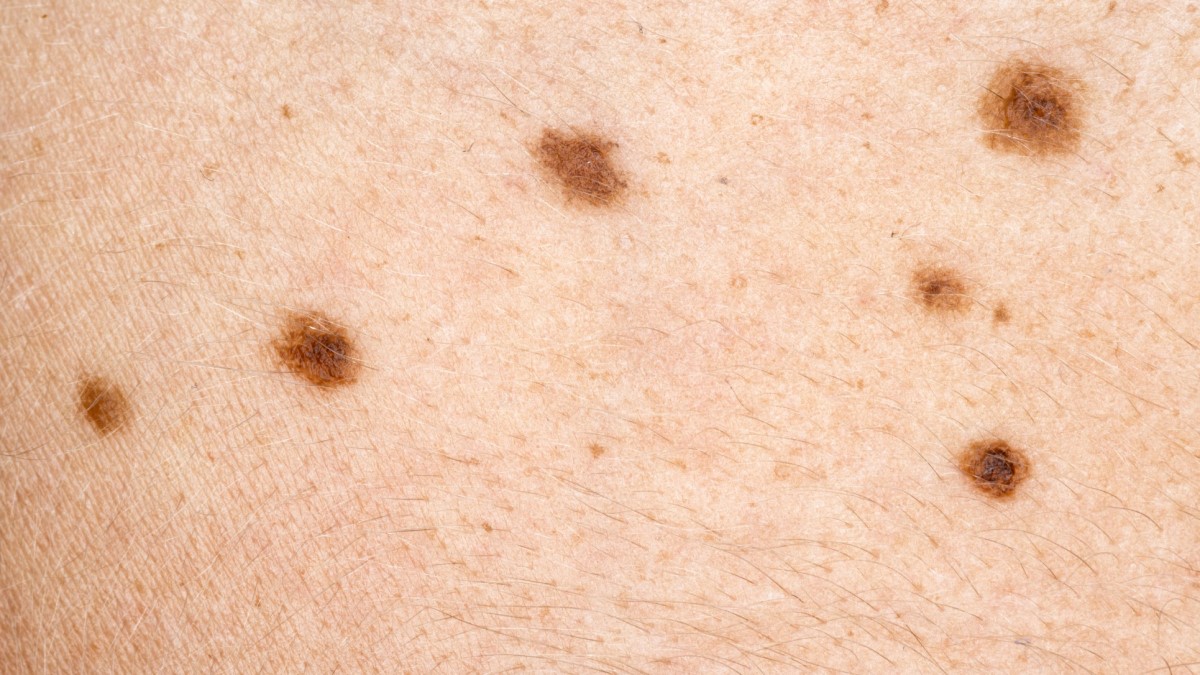
Early melanoma is typically asymptomatic but presents with visible changes that can be identified using the ABCDE criteria:
- Asymmetry: One half of the mole doesn’t match the other half
- Border: Irregular, ragged, notched, or blurred edges
- Color: Variation in color within the same mole (shades of tan, brown, black; sometimes red, white, or blue)
- Diameter: Larger than 6mm (about the size of a pencil eraser), though early melanomas can be smaller
- Evolving: Changes in size, shape, color, or elevation; new symptoms such as bleeding, itching, or crusting
Additional early signs include:
- The “ugly duckling” sign: A mole that looks different from other moles on the body
- New rapidly growing pigmented lesions
- Changes in existing moles, particularly after puberty when most moles typically stabilize
Advanced-Stage Symptoms
As melanoma progresses, symptoms may include:
Local Symptoms:
- Ulceration (breakdown of the skin over the melanoma)
- Bleeding or oozing from the lesion
- Persistent itching, tenderness, or pain
- Satellite lesions (small new growths near the primary melanoma)
- Hardening or thickening of the lesion
- Changes in sensation (numbness, tingling)
Regional Spread Symptoms:
- Enlarged, sometimes painful lymph nodes near the primary site
- Hardened or matted lymph nodes
- Visible or palpable nodules in lymphatic channels (in-transit metastases)
Distant Metastasis Symptoms:
- Unexplained weight loss
- Fatigue and weakness
- Persistent cough or shortness of breath (lung metastases)
- Headaches, seizures, or neurological changes (brain metastases)
- Abdominal pain or jaundice (liver metastases)
- Bone pain or fractures from minimal trauma (bone metastases)
- Swelling of extremities (lymphatic obstruction)
Common vs. Rare Symptoms
Common symptoms experienced by most patients include:
- Changes in appearance of a mole or pigmented area
- Development of new pigmented lesions
- Local symptoms at the site of the primary lesion (itching, tenderness)
Rarer symptoms include:
- Amelanotic Presentation: Melanomas without obvious pigmentation appearing as pink or skin-colored lesions
- Neurotropic Symptoms: Tingling or electrical sensations due to nerve involvement
- Paraneoplastic Syndromes: Rare immunological reactions affecting distant organs
- Regression Phenomenon: Spontaneous partial disappearance of the primary tumor (often with whitish areas)
- Metastasis of Unknown Primary: Presentation with metastatic disease without an identifiable primary lesion
Symptom Progression Over Time
Melanoma typically progresses through several phases:
Radial Growth Phase:
- Initial horizontal growth within the epidermis
- Minimal invasive potential
- Changes primarily in appearance (color, size, border)
- Can persist for months to years
Vertical Growth Phase:
- Invasion into deeper layers of the skin
- Development of local symptoms (elevation, firmness)
- Increasing risk of metastasis
- More rapid progression
Regional Metastasis Phase:
- Spread to regional lymph nodes
- Development of satellite or in-transit lesions
- Symptoms related to lymphatic involvement
Distant Metastasis Phase:
- Spread to distant organs
- Systemic symptoms (fatigue, weight loss)
- Organ-specific symptoms based on sites of involvement
The rate of progression varies greatly among individuals and is influenced by tumor characteristics, location, and host factors. Some melanomas may remain in situ for years, while others may rapidly progress to invasive and metastatic disease within months.
4. Causes
Biological Causes
At its core, melanoma results from the malignant transformation of melanocytes due to accumulated genetic damage. The biological mechanisms include:
- Cellular Transformation: Conversion of normal melanocytes into cancer cells through multiple genetic mutations
- Dysregulation of Cell Cycle: Uncontrolled cell division due to mutations in genes controlling the cell cycle
- Evasion of Apoptosis: Resistance to programmed cell death, allowing cancer cells to survive
- Genomic Instability: Accumulation of genetic alterations due to defects in DNA repair mechanisms
- Activation of Oncogenic Pathways: Particularly the MAPK pathway (RAS-RAF-MEK-ERK) and PI3K-AKT pathway
- Loss of Tumor Suppressor Functions: Especially the p16INK4a/CDK4/Rb pathway
- Telomerase Reactivation: Enabling unlimited replicative potential
- Angiogenesis: Formation of new blood vessels to support tumor growth
- Immune Evasion: Development of mechanisms to avoid detection and elimination by the immune system
Environmental Causes
The primary environmental factor driving melanoma development is ultraviolet (UV) radiation, which can damage DNA in melanocytes:
Ultraviolet Radiation:
- UVA: Penetrates deeply into the skin, generates reactive oxygen species
- UVB: Directly damages DNA, causing characteristic “UV signature” mutations
- UVC: Largely blocked by the atmosphere but can come from artificial sources
Sources of UV Exposure:
- Natural sunlight
- Tanning beds and sunlamps (particularly harmful due to concentrated UVA)
- Therapeutic UV treatments
- Occupational exposure to UV radiation or UV-generating equipment
Pattern of Exposure:
- Intense intermittent exposure (e.g., sunburns) appears more dangerous than cumulative exposure
- Childhood and adolescent exposure may be particularly significant
Other Environmental Factors:
- Ionizing radiation (rare)
- Chemical carcinogens (certain industrial chemicals may increase risk)
- Arsenic exposure (associated with increased skin cancer risk)
Genetic and Hereditary Factors
Approximately 10% of melanomas occur in families with inherited predisposition:
High-Penetrance Genes:
- CDKN2A (p16): Most common, accounting for 20-40% of hereditary melanoma cases
- CDK4: Rare mutations affecting cell cycle regulation
- BAP1: Associated with uveal melanoma and mesothelioma risk
- MITF: E318K variant increases melanoma risk
- POT1, ACD, TERF2IP: Telomere maintenance genes associated with familial melanoma
Moderate-Risk Genes:
- MC1R variants: Associated with red hair, fair skin, and increased melanoma risk
- MITF variants: Influence pigmentation and melanocyte function
- Pigmentation genes: SLC45A2, TYR, TYRP1, ASIP
Polygenic Inheritance: Many individuals have multiple lower-risk genetic variants that collectively increase risk
Genetically Determined Risk Factors:
- Fair skin, light hair, and eye color
- Multiple or atypical moles
- Freckle pattern and sun sensitivity
Known Triggers and Exposure Risks
Specific triggers that may initiate or promote melanoma development include:
- Severe Sunburns: Especially during childhood and adolescence
- Tanning Bed Use: Particularly before age 30, increases risk by 75%
- Immunosuppression: Organ transplant recipients, HIV/AIDS patients, and those on immunosuppressive medications
- Pre-existing Moles: Large congenital nevi and numerous acquired nevi increase risk
- Previous Melanoma: Having had one melanoma significantly increases risk for subsequent primary melanomas
- Rare Syndromes:
- Xeroderma pigmentosum (defective DNA repair)
- Familial atypical multiple mole melanoma (FAMMM) syndrome
- Li-Fraumeni syndrome
- BRCA2 mutations (associated with increased melanoma risk)
The development of melanoma typically requires a combination of genetic susceptibility and environmental exposures, with each factor contributing to the accumulation of genetic damage that ultimately leads to malignant transformation.
5. Risk Factors
Demographic Risk Factors
Certain demographic characteristics are associated with increased melanoma risk:
Age:
- Risk increases with age, with median age at diagnosis around 65
- However, melanoma is one of the most common cancers in young adults, especially women under 30
Sex:
- Men have higher overall incidence and mortality, particularly after age 50
- Women have higher rates before age 50, especially on the legs
- Men tend to develop thicker melanomas with worse prognosis
Race/Ethnicity:
- Highest rates in non-Hispanic whites
- Lower rates in Hispanic, Asian, and Black populations
- Later diagnosis and worse outcomes in minority populations when melanoma does occur
Geographic Location:
- Highest incidence in Australia, New Zealand, North America, and Northern Europe
- Proximity to the equator increases risk in fair-skinned populations
- Altitude increases UV exposure (approximately 4% increase per 300m elevation)
Environmental and Occupational Risk Factors
Various environmental and occupational exposures influence melanoma risk:
Sun Exposure Patterns:
- Intense intermittent exposure (associated with recreational activities)
- Childhood and adolescent sun exposure
- History of sunburns, especially blistering sunburns
- Living in high UV-index regions or at high altitudes
Artificial UV Exposure:
- Indoor tanning bed use (particularly harmful for young users)
- Phototherapy for skin conditions without proper protection
High-Risk Occupations:
- Outdoor workers (construction, agriculture, landscaping)
- Airline crew (increased cosmic radiation at high altitudes)
- Military personnel deployed to high-UV regions
- Petroleum and chemical workers (exposure to polycyclic aromatic hydrocarbons)
- Miners (exposure to various carcinogens)
- Firefighters (exposure to carcinogenic compounds)
Other Environmental Factors:
- Living in areas with depleted ozone layer
- Exposure to certain pesticides and industrial chemicals
- Radiation exposure (therapeutic, diagnostic, or occupational)
Genetic and Physical Risk Factors
Individual genetic and physical characteristics significantly affect melanoma risk:
Skin Phenotype:
- Fair skin that burns easily and tans poorly (Fitzpatrick skin types I and II)
- Blue, green, or gray eyes
- Blonde or red hair
- Freckles and solar lentigines (sun spots)
Nevus Phenotype:
- Multiple common nevi (>50 moles)
- Presence of atypical/dysplastic nevi
- Large congenital melanocytic nevi (present at birth)
Family History:
- First-degree relatives with melanoma (2-3 fold increased risk)
- Multiple family members affected (suggests genetic syndrome)
- Family history of pancreatic cancer or astrocytoma (may indicate CDKN2A mutations)
Personal History:
- Previous melanoma (9-fold increased risk of developing another)
- Non-melanoma skin cancers (basal cell or squamous cell carcinoma)
- History of severe sunburns
Impact of Pre-existing Conditions
Certain medical conditions affect melanoma risk and outcomes:
Immunosuppression:
- Organ transplant recipients (2-3 times increased risk)
- HIV/AIDS
- Chronic lymphocytic leukemia
- Use of immunosuppressive medications
Genetic Syndromes:
- Xeroderma pigmentosum (>1000-fold increased risk)
- Familial atypical multiple mole melanoma syndrome
- Werner syndrome
- Retinoblastoma (RB1 mutations)
Other Medical Conditions:
- Previous radiotherapy
- Parkinson’s disease (1.5-2 fold increased risk)
- Hormonal factors (pregnancy may temporarily accelerate melanoma growth)
- Vitamin D deficiency (controversial, may affect progression)
Medication Effects:
- Photosensitizing medications
- BRAF inhibitor therapy (paradoxically increases risk of non-melanoma skin cancers)
- Immunosuppressive agents
- Controversial association with erectile dysfunction medications
The interplay between multiple risk factors creates individual risk profiles, with the presence of multiple factors substantially increasing melanoma risk. Risk assessment models incorporating these factors are increasingly used in clinical practice for screening recommendations and risk management strategies.
6. Complications
Direct Complications of Primary Melanoma
The primary melanoma lesion itself can cause several complications:
- Local Tissue Destruction: Advanced primary melanomas can invade deeply, destroying surrounding skin, subcutaneous tissue, and underlying structures
- Ulceration: Breakdown of the skin surface, increasing infection risk and signifying more aggressive disease
- Bleeding: Spontaneous or trauma-induced bleeding from the tumor
- Infection: Secondary bacterial infection of ulcerated tumors
- Nerve Involvement: Pain, numbness, or paresthesia due to perineural invasion
- Satellite Lesions: Development of small tumor deposits within 2cm of the primary lesion
- Functional Impairment: Particularly for melanomas on joints, hands, feet, or functionally important areas
- Cosmetic Disfigurement: Especially relevant for visible areas like the face
Metastatic Complications
As melanoma spreads, it can affect virtually any organ system:
Lymphatic System:
- Regional lymphadenopathy (enlarged lymph nodes)
- Lymphedema (swelling due to lymphatic blockage)
- In-transit metastases (tumor deposits in lymphatic channels)
Nervous System:
- Brain metastases (causing headaches, seizures, focal neurological deficits)
- Spinal cord compression (causing pain, weakness, bowel/bladder dysfunction)
- Leptomeningeal disease (affecting the coverings of the brain and spinal cord)
Pulmonary System:
- Lung metastases (causing cough, shortness of breath, chest pain)
- Pleural effusions (fluid around the lungs)
- Endobronchial obstruction
Hepatic System:
- Liver metastases (causing abdominal pain, jaundice, liver failure)
- Portal vein thrombosis
- Ascites (fluid in the abdominal cavity)
Skeletal System:
- Bone metastases (causing pain, fractures, spinal cord compression)
- Hypercalcemia (elevated calcium due to bone destruction)
Other Sites:
- Adrenal gland metastases
- Cardiac metastases (rare but potentially causing arrhythmias, heart failure)
- Gastrointestinal metastases (causing bleeding, obstruction)
- Splenic metastases
Treatment-Related Complications
Modern melanoma treatments can themselves cause significant complications:
Surgical Complications:
- Wound healing problems
- Infection
- Scarring and contractures
- Lymphedema following lymph node dissection
- Nerve injury and sensory loss
- Seroma formation (fluid collection)
Immunotherapy Complications:
- Immune-related adverse events affecting multiple organ systems:
- Colitis (inflammation of the colon)
- Hepatitis (liver inflammation)
- Pneumonitis (lung inflammation)
- Endocrinopathies (thyroid, pituitary, adrenal dysfunction)
- Dermatitis (skin inflammation)
- Nephritis (kidney inflammation)
- Neurological problems (neuropathy, encephalitis)
- Myocarditis (heart muscle inflammation)
- Many can be life-threatening if not promptly recognized and treated
- Immune-related adverse events affecting multiple organ systems:
Targeted Therapy Complications:
- BRAF/MEK inhibitor side effects:
- Skin toxicities (rash, photosensitivity, development of squamous cell carcinomas)
- Pyrexia (fever)
- Arthralgia (joint pain)
- Fatigue
- Liver function abnormalities
- Cardiac effects (QT prolongation, decreased ejection fraction)
- Ocular toxicities
- BRAF/MEK inhibitor side effects:
Radiation Therapy Complications:
- Skin reactions
- Fatigue
- Organ-specific effects depending on treatment area
Long-term Impact and Mortality
Melanoma can have profound long-term impacts:
Physical Consequences:
- Chronic pain syndromes
- Persistent lymphedema
- Functional limitations from surgery
- Endocrine abnormalities from immunotherapy
- Chronic fatigue
Psychological Impact:
- Fear of recurrence
- Depression and anxiety
- Body image concerns
- Post-traumatic stress
- Social isolation
Mortality Rates:
- Stage-specific survival:
- Stage I: >95% 5-year survival
- Stage II: 65-85% 5-year survival
- Stage III: 30-70% 5-year survival (highly variable based on substage)
- Stage IV: Historically <10% 5-year survival, though modern treatments have improved this to 20-50% in some populations
- Stage-specific survival:
Years of Life Lost: Melanoma often affects young and middle-aged adults, resulting in substantial premature mortality
Economic Impact: Lost productivity, caregiver burden, and direct healthcare costs
Survivorship Issues: Long-term surveillance requirements, chronic medication needs, and management of treatment sequelae
Despite significant advances in treatment, metastatic melanoma remains a potentially fatal disease with substantial morbidity. However, early detection and intervention can prevent many of these complications, emphasizing the critical importance of prevention, screening, and prompt treatment.
7. Diagnosis & Testing
Clinical Examination
The diagnostic process typically begins with thorough clinical assessment:
- Full-Body Skin Examination: Systematic evaluation of the entire skin surface, including scalp, genital area, between toes, and other easily missed areas
- Dermoscopy (Epiluminescence Microscopy): Non-invasive technique using a handheld device that magnifies the skin and eliminates surface reflection, allowing visualization of deeper structures
- Digital Dermoscopy: Computer-assisted dermoscopic analysis with digital storage for monitoring changes over time
- Total Body Photography: Baseline photographs of the entire skin surface to detect new or changing lesions at follow-up visits
- Clinical Assessment Tools:
- ABCDE criteria (Asymmetry, Border irregularity, Color variation, Diameter >6mm, Evolution)
- Ugly Duckling sign (moles that look different from other moles on the patient)
- Glasgow 7-point checklist (major criteria: change in size, shape, color; minor criteria: inflammation, crusting/bleeding, sensory change, diameter ≥7mm)
Biopsy Techniques
Tissue diagnosis is essential for confirming melanoma:
- Excisional Biopsy: Removal of the entire lesion with a small margin (2mm) of normal skin; preferred method for suspected melanoma
- Incisional Biopsy: Removal of a portion of the lesion when the suspicious area is large or in a cosmetically sensitive location
- Punch Biopsy: Use of a circular tool to remove a core of tissue, including deeper layers; useful for smaller lesions
- Shave Biopsy: Generally discouraged for suspected melanoma as it may transect the base of the tumor and compromise accurate depth assessment
- Saucerization: A deep shave technique that may be appropriate for certain lesions
- Fine Needle Aspiration: Used for sampling suspicious lymph nodes, not primary skin lesions
Histopathological Assessment
Microscopic examination of biopsy specimens provides critical diagnostic and prognostic information:
Diagnostic Features:
- Atypical melanocytes
- Pagetoid spread (upward migration of melanocytes)
- Asymmetry and poor circumscription
- Cytological atypia
- Mitotic figures
- Lack of maturation with depth
Prognostic Features:
- Breslow Thickness: Measured from the top of the granular layer to the deepest point of invasion
- Ulceration: Absence of intact epidermis overlying the melanoma
- Mitotic Rate: Number of mitoses per square millimeter
- Microsatellites: Discontinuous nests of metastatic cells separated from the primary tumor
- Lymphovascular Invasion: Presence of tumor cells within blood or lymphatic vessels
- Neurotropism: Invasion of nerve sheaths
- Tumor-Infiltrating Lymphocytes: Immune cells within the tumor (favorable prognostic factor)
- Regression: Areas where the tumor has been partially destroyed by immune response
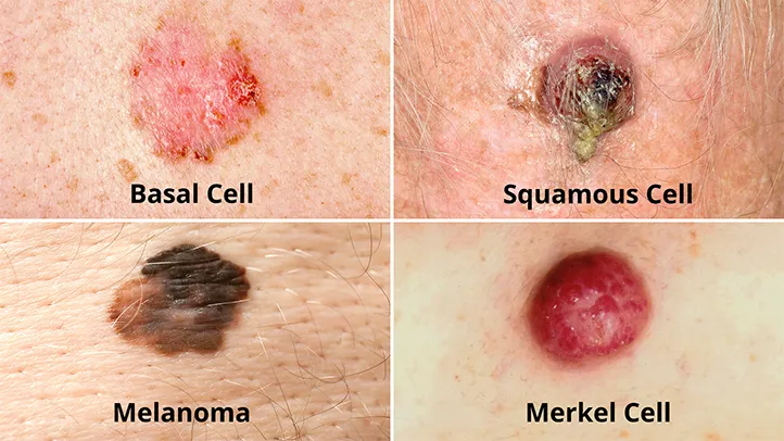
Histological Subtypes:
- Superficial spreading melanoma (most common)
- Nodular melanoma (aggressive, vertical growth phase from onset)
- Lentigo maligna melanoma (on sun-damaged skin of elderly)
- Acral lentiginous melanoma (palms, soles, nail beds)
- Desmoplastic melanoma (marked by fibrous tissue)
- Other rare variants (spitzoid, nevoid, amelanotic, etc.)
Staging Workup
After diagnosis, staging determines the extent of disease:
Sentinel Lymph Node Biopsy (SLNB):
- Identifies and examines the first lymph node(s) receiving drainage from the tumor site
- Generally recommended for melanomas ≥0.8mm thick or thinner with high-risk features
- Provides important staging and prognostic information
Imaging Studies:
- Regional Lymph Node Ultrasound: High sensitivity for detecting lymph node metastases
- CT Scan: Chest, abdomen, and pelvis to evaluate for distant metastases
- PET/CT: More sensitive for detecting metastatic disease, particularly useful for higher-stage disease
- Brain MRI: Gold standard for detecting brain metastases
- Bone Scan: If bone metastases are suspected
Laboratory Testing:
- LDH (Lactate Dehydrogenase): Elevated in advanced disease, associated with poorer prognosis
- Liver Function Tests: May indicate liver metastases
- Complete Blood Count: May show anemia or other abnormalities in advanced disease
- S100B and MIA (Melanoma Inhibitory Activity): Serum biomarkers sometimes used to monitor for recurrence
Molecular Testing
Genetic analysis provides information for treatment planning:
- BRAF Mutation Testing: Essential for determining eligibility for BRAF inhibitor therapy
- NRAS Mutation Testing: May guide clinical trial eligibility
- c-KIT Mutation Testing: Particularly relevant for acral, mucosal, and chronically sun-damaged melanomas
- PD-L1 Expression: May help predict response to immunotherapy, though not used as a strict selection criterion
- Tumor Mutational Burden: Higher mutation rates may correlate with better immunotherapy response
- Gene Expression Profiling: Tests like DecisionDx-Melanoma assess metastatic risk based on gene expression patterns
Early Detection Methods and Effectiveness
Strategies to identify melanoma at earlier, more treatable stages:
Skin Self-Examination:
- Regular (monthly) systematic self-checks
- 40% of melanomas detected by patients themselves
- Education about warning signs improves effectiveness
Professional Skin Examinations:
- Regular screening by dermatologists or trained primary care providers
- Particularly important for high-risk individuals
- Increases early detection rates
Advanced Imaging Technologies:
- Reflectance Confocal Microscopy: Non-invasive “optical biopsy” for real-time cellular resolution imaging
- Electrical Impedance Spectroscopy: Measures electrical properties of skin lesions
- Automated Image Analysis: AI-assisted evaluation of dermoscopic images
- Mobile Teledermatology: Smartphone-based applications for preliminary assessment
Effectiveness of Early Detection:
- Screening programs have shown a shift toward thinner melanomas at diagnosis
- Population-based screening may reduce melanoma mortality by 38-63%
- Targeted screening of high-risk individuals is particularly cost-effective
- Recent studies show significant survival benefit from regular skin examinations
Early detection remains the most effective strategy for improving melanoma outcomes, with the goal of diagnosing tumors before they develop invasive or metastatic potential.
8. Treatment Options
Surgical Management
Surgery remains the primary treatment for localized melanoma:
Wide Local Excision:
- Standard treatment for primary melanoma
- Margin width based on Breslow thickness:
- In situ: 0.5-1.0 cm margin
- ≤1mm thick: 1 cm margin
- 1.01-2mm thick: 1-2 cm margin
2mm thick: 2 cm margin
- May require skin grafts or flaps for larger defects
Sentinel Lymph Node Biopsy (SLNB):
- Staging procedure for intermediate and thick melanomas
- Uses radiotracer and/or blue dye to identify the first draining lymph node(s)
- Provides important prognostic information and guides adjuvant therapy decisions
Lymph Node Dissection:
- Complete removal of regional lymph node basin
- Currently recommended for clinically detected nodal metastases
- Role following positive SLNB has diminished with effective adjuvant therapies
Surgery for Metastatic Disease:
- Resection of isolated metastases can be curative in select cases
- Particularly effective for solitary lung, gastrointestinal, or brain metastases
- Often combined with systemic therapy
Mohs Micrographic Surgery:
- Specialized technique for melanomas in certain anatomic locations
- Allows tissue conservation while ensuring complete tumor removal
Adjuvant Therapy
Treatments after surgery to reduce recurrence risk:
Immunotherapy:
- Anti-PD-1 Antibodies:
- Pembrolizumab or nivolumab for stage III and resected stage IV
- Significantly improves recurrence-free and overall survival
- CTLA-4 Inhibitors:
- Ipilimumab (high-dose) approved but with significant toxicity
- Less commonly used since anti-PD-1 approval
- Anti-PD-1 Antibodies:
Targeted Therapy:
- BRAF/MEK Inhibitor Combinations:
- Dabrafenib plus trametinib for BRAF-mutant stage III melanoma
- Encorafenib plus binimetinib being studied
- Requires BRAF V600 mutation (present in approximately 50% of melanomas)
- BRAF/MEK Inhibitor Combinations:
Radiation Therapy:
- Adjuvant Radiation:
- For high-risk regional nodal basins after lymph node dissection
- May reduce regional recurrence but without overall survival benefit
- Stereotactic Radiosurgery:
- For resected brain metastases to reduce local recurrence
- Adjuvant Radiation:
Older Adjuvant Approaches (largely historical):
- Interferon alpha-2b (rarely used now)
- Biochemotherapy regimens
- Adjuvant vaccines (experimental)
Management of Metastatic Disease
Advanced melanoma treatment has been revolutionized in the past decade:
Immunotherapy:
- Anti-PD-1 Monotherapy:
- Pembrolizumab or nivolumab
- Response rates of 30-40%, many durable
- Combination Immunotherapy:
- Ipilimumab plus nivolumab
- Higher response rates (60%) but increased toxicity
- Sequential Approaches:
- Various strategies to optimize sequence and combinations
- Interleukin-2 (high-dose):
- Older approach with durable responses in small percentage
- Significant toxicity limits use
- Anti-PD-1 Monotherapy:
Targeted Therapy (for BRAF-mutant melanoma):
- BRAF/MEK Inhibitor Combinations:
- Dabrafenib plus trametinib
- Vemurafenib plus cobimetinib
- Encorafenib plus binimetinib
- Response rates of 70-80% but resistance often develops
- Median progression-free survival of 12-14 months
- BRAF/MEK Inhibitor Combinations:
Intralesional Therapy:
- Talimogene laherparepvec (T-VEC):
- Oncolytic virus therapy for accessible lesions
- Both local and systemic immune effects
- Talimogene laherparepvec (T-VEC):
Chemotherapy:
- Limited role in modern era
- Options include dacarbazine, temozolomide, carboplatin/paclitaxel
- Considered after failure of immunotherapy and targeted therapy
- Response rates of 10-15%, rarely durable
Radiation Therapy:
- Palliative radiation for symptomatic metastases
- Stereotactic radiosurgery for brain metastases
- Whole-brain radiation for multiple brain metastases
Special Situations in Melanoma Management
Leptomeningeal Disease:
- Intrathecal chemotherapy
- Whole-brain radiation
- Systemic therapy with good CNS penetration
Mucosal Melanoma:
- Often c-KIT mutations rather than BRAF
- May respond to c-KIT inhibitors (imatinib, nilotinib)
- Generally poorer prognosis than cutaneous melanoma
Ocular Melanoma:
- Liver-directed therapies for metastatic disease
- Immunotherapy less effective than in cutaneous melanoma
- Specialized approaches like liver isolation perfusion
Desmoplastic Melanoma:
- Often requires wider surgical margins
- Higher response rates to anti-PD-1 therapy
Emerging Treatments and Clinical Trials
The melanoma treatment landscape continues to evolve rapidly:
Novel Immunotherapy Approaches:
- LAG-3 Inhibitors: Relatlimab approved in combination with nivolumab
- TIM-3, TIGIT Inhibitors: Under investigation
- Bispecific Antibodies: Targeting multiple immune checkpoints
- Tumor-Infiltrating Lymphocyte (TIL) Therapy: Adoptive cell transfer
- PVSRIPO (polio virus-rhinovirus chimera): Oncolytic virus therapy
- Toll-like Receptor (TLR) Agonists: Stimulating immune response
Next-Generation Targeted Therapies:
- SHP2 Inhibitors: Targeting resistance mechanisms
- Novel BRAF Inhibitors: Improved specificity and CNS penetration
- CDK4/6 Inhibitors: For NRAS-mutant and other subtypes
- NRAS-targeted Approaches: For the second most common mutation
- Epigenetic Modifiers: Targeting gene expression regulation
Combination Approaches:
- Triplet Regimens: BRAF/MEK inhibitors plus immunotherapy
- Sequential Strategies: Optimizing order of treatments
- Neoadjuvant Therapy: Treatment before surgery
- Immunotherapy plus Radiation: Potential synergistic effects
- Novel Drug/Device Combinations: Electrochemotherapy, photodynamic therapy
Personalized Medicine Approaches:
- Liquid Biopsies: Circulating tumor DNA for monitoring and early detection of relapse
- Tumor Mutational Signatures: Predicting response to specific therapies
- Microbiome Modulation: Gut microbiome influences on immunotherapy response
- Ex Vivo Drug Sensitivity Testing: Patient-derived xenografts and organoids
The rapidly evolving treatment landscape has transformed melanoma from a disease with few effective therapies to one with multiple options and significantly improved survival outcomes, particularly for advanced disease.
9. Prevention & Precautionary Measures
Primary Prevention Strategies
Preventing melanoma by reducing risk factors and exposure:
Sun Protection Measures:
- Sunscreen Use: Broad-spectrum, SPF 30+ sunscreen applied properly and regularly
- Protective Clothing: Wide-brimmed hats, long sleeves, UV-protective fabrics
- Seeking Shade: Especially during peak UV hours (10 AM to 4 PM)
- Sunglasses: UV-blocking eyewear to protect eyes and surrounding skin
- Window Protection: UV-filtering film for car and home windows
Avoiding Artificial UV Exposure:
- Tanning Beds: Complete avoidance is recommended
- UV Lamps: Avoiding non-medical UV exposure
- Nail Salon UV Lamps: Using sunscreen or protective gloves when necessary
Population-Level Interventions:
- Public Education Campaigns: Raising awareness of melanoma risks and prevention
- School-Based Programs: Sun-protection education starting in childhood
- Shade Structures: In public spaces, playgrounds, and sports facilities
- UV Index Reporting: Making UV forecasts widely available
- Legislation: Restricting youth access to tanning beds
- Workplace Policies: Protecting outdoor workers through regulations and practices
Secondary Prevention (Early Detection)
Identifying melanoma at its earliest, most treatable stages:
Skin Self-Examination:
- Monthly comprehensive self-checks
- Using mirrors or partners to view difficult-to-see areas
- Documentation of existing moles (photography)
- Knowledge of personal “mole map” and normal patterns
Professional Skin Examinations:
- Regular screening by dermatologists or trained primary care providers
- Frequency based on risk level:
- Average risk: Every 1-3 years
- Moderate risk: Annually
- High risk: Every 3-6 months
Risk-Based Screening Recommendations:
- Tailored screening frequency based on individual risk factors
- More intensive follow-up for those with:
- Personal history of melanoma
- Multiple atypical nevi
- Familial melanoma syndrome
- Genetic risk factors
- Immunosuppression
Screening Technologies:
- Whole-body photography for baseline documentation
- Sequential digital dermoscopy imaging for monitoring
- Algorithmic risk assessment tools
- Computer-assisted diagnosis systems
Risk Reduction for High-Risk Individuals
Specialized approaches for those at elevated risk:
Genetic Counseling and Testing:
- For families with multiple melanoma cases
- Identification of hereditary syndromes
- Personalized risk management plans
Prophylactic Measures:
- Removal of atypical nevi in very high-risk patients
- Chemoprevention strategies (under investigation)
- More intensive sun protection regimens
Surveillance Protocols:
- Specialized high-risk clinics
- Comprehensive imaging and documentation
- Consideration of prophylactic surgeries in extreme risk cases
Management of Precursor Lesions:
- Monitoring versus removal of dysplastic nevi
- Treatment of actinic damage
- Addressing persistent UV-induced immunosuppression
Lifestyle and Environmental Modifications
Long-term approaches to reduce melanoma risk:
Dietary Factors:
- Antioxidant-rich diet (fruits, vegetables)
- Omega-3 fatty acids
- Vitamin D sufficiency (through diet or supplements rather than sun exposure)
- Polyphenol-rich foods (green tea, turmeric)
- Limited alcohol consumption
Physical Activity:
- Regular exercise (with sun protection)
- Maintaining healthy weight
- Potential immune-enhancing effects
Environmental Considerations:
- Awareness of altitude effects on UV exposure
- Recognition of UV reflection from water, snow, and sand
- Environmental UV monitoring
- Seasonal variations in protection needs
Special Populations:
- Children: Extra protection during critical developmental years
- Outdoor athletes: Sport-specific protection strategies
- Occupational exposure: Workplace interventions and policies
- Post-transplant patients: Intensified surveillance and protection
Education and Awareness
Knowledge-based approaches to prevention:
Public Education Campaigns:
- “Slip, Slop, Slap, Seek, Slide” (Australia’s successful campaign)
- ABCDE awareness initiatives
- Celebrity advocacy and testimonials
- Social media campaigns targeting young adults
Healthcare Provider Education:
- Primary care training in skin examination
- Recognition of atypical presentations
- Cultural competence for assessing diverse skin types
- Integration of skin checks into routine care
Community Interventions:
- Free skin cancer screenings
- Beach and pool-based educational programs
- Workplace wellness initiatives
- School curriculum incorporation
Policy Advocacy:
- Sunscreen availability in schools and public spaces
- Insurance coverage for preventive services
- Environmental protection policies affecting UV exposure
- Warning labels on tanning devices
These prevention strategies, when implemented comprehensively, have the potential to significantly reduce melanoma incidence and mortality. The success of Australia’s public health campaigns, which have led to stabilization and even decline in melanoma rates in younger populations, demonstrates the effectiveness of coordinated prevention efforts.
10. Global & Regional Statistics
Global Incidence and Prevalence
Melanoma occurrence varies dramatically worldwide:
Global Burden:
- Approximately 324,000 new cases annually worldwide
- Over 57,000 deaths per year globally
- Cumulative lifetime risk: 0.1-1.9% depending on geographic location and ethnicity
- Rising incidence in most populations over the past several decades
Geographical Variations:
- Highest Incidence Regions:
- Australia (40-60 cases per 100,000 people)
- New Zealand (30-40 cases per 100,000)
- North America (20-30 cases per 100,000)
- Northern Europe (15-25 cases per 100,000)
- Moderate Incidence Regions:
- Southern Europe (10-15 cases per 100,000)
- Eastern Europe (5-10 cases per 100,000)
- South America (3-7 cases per 100,000)
- Lowest Incidence Regions:
- Asia (0.2-4 cases per 100,000)
- Africa (1-2 cases per 100,000)
- Highest Incidence Regions:
Temporal Trends:
- Annual increase of 3-7% in fair-skinned populations over the past decades
- Plateau or slight decrease in younger age groups in countries with strong sun protection programs
- Continued increase in older age groups representing historical sun exposure
- Faster increase among women in many regions
Regional Statistics and Patterns
North America:
- United States: approximately 100,000 new cases annually
- Higher rates in northern states despite lower UV exposure
- Reflects socioeconomic factors and indoor tanning practices
- Five-year survival of 93.3% (all stages combined)
- Regional “hot spots” in higher-altitude states
Europe:
- Significant north-south gradient with higher rates in Scandinavia
- Rising rapidly in Central and Eastern Europe
- UK: approximately 16,000 new cases annually
- Norway has one of the highest rates in Europe (29.6 per 100,000)
- Mediterranean countries show lower rates despite higher UV exposure
Australia/New Zealand:
- Highest global incidence (approximately 13,000 new cases annually in Australia)
- Success of public health campaigns showing impact on younger cohorts
- Melanoma is the 3rd most common cancer in Australia
- “Melanoma capital of the world” due to combination of geography, ozone depletion, and predominantly fair-skinned population
- Pioneering prevention and early detection programs
Asia:
- Much lower incidence but often diagnosed at more advanced stages
- Japan: 1-2 cases per 100,000
- China: 0.2-0.4 cases per 100,000
- Different distribution: acral and mucosal melanomas more common
- Rising incidence with westernization of lifestyles
Africa:
- Limited data available
- Lower incidence but higher mortality-to-incidence ratio
- Non-cutaneous melanomas represent higher proportion
- Acral lentiginous melanoma predominant in Black African populations
- Healthcare access challenges affecting outcomes
Latin America:
- Brazil: 2.8-3.4 cases per 100,000
- Significant variation by region and ethnicity
- Mixed patterns reflecting diverse ancestry
- Limited specialized care outside major urban centers
Mortality and Survival Statistics
Global Mortality:
- Approximately 57,000 deaths annually worldwide
- Mortality rates generally correlate with incidence but with important exceptions
Mortality Trends:
- Stabilizing or decreasing in high-income countries despite rising incidence
- Reflects earlier detection and improved treatments
- Continuing to rise in many developing regions
- Gender differences: generally higher mortality in men
Survival Rates:
- Stage-Specific Survival (5-year):
- Stage I: >95%
- Stage II: 65-85%
- Stage III: 30-70%
- Stage IV: Historically <10%, now 20-50% with modern therapies
- Country-Specific Survival:
- Australia: 92% 5-year survival (all stages)
- United States: 93.3% 5-year survival
- Eastern Europe: 74-84% 5-year survival
- African countries: Often <40% 5-year survival
- Stage-Specific Survival (5-year):
Survival Disparities:
- Socioeconomic status strongly influences outcomes
- Rural vs. urban differences in access to specialized care
- Racial disparities: lower survival in non-white populations
- Insurance status impact on treatment access and outcomes
Epidemiological Patterns and Trends
Age Distribution:
- Median age at diagnosis: 65 years
- Rising incidence in younger adults, particularly women
- Different age distribution by subtype (e.g., nodular melanoma more common in older adults)
Gender Patterns:
- Higher incidence in men after age 50
- Higher incidence in women before age 50
- Anatomic differences: trunk more common in men, extremities in women
- Survival advantage for women across all stages
Anatomic Site Trends:
- Increasing trunk melanomas in men
- Increasing lower extremity melanomas in women
- Both potentially related to fashion and sun exposure patterns
- Shifting distributions with sun protection awareness
Prognostic Trends:
- Decreasing average tumor thickness in developed countries
- Improved staging through sentinel node biopsy
- Increasing proportion of in situ melanomas
- Treatment advances dramatically improving stage IV survival
These global and regional statistics highlight the complex interplay of genetics, behavior, environment, and healthcare systems in determining melanoma risk and outcomes. They also underscore the potential impact of coordinated public health initiatives in reducing the burden of this disease.
11. Recent Research & Future Prospects
Latest Research Breakthroughs
Recent scientific advances have transformed our understanding and treatment of melanoma:
Tumor Microenvironment Discoveries:
- Characterization of immune suppressive mechanisms in the tumor microenvironment
- Identification of stromal cell contributions to melanoma progression
- Recognition of metabolic interactions between melanoma and immune cells
- Understanding of extracellular matrix remodeling in invasion
Genetic and Molecular Insights:
- Comprehensive molecular classification beyond BRAF/NRAS/NF1
- Identification of rare driver mutations with therapeutic implications
- Epigenetic mechanisms controlling melanoma plasticity and drug resistance
- Non-coding RNA involvement in melanoma progression
Immunological Advances:
- Characterization of neoantigens and their role in immunotherapy response
- Understanding mechanisms of primary and acquired resistance to immunotherapy
- Identification of predictive biomarkers for treatment selection
- Microbiome influences on melanoma progression and treatment response
Technological Innovations:
- Single-cell RNA sequencing revealing unprecedented tumor heterogeneity
- Spatial transcriptomics mapping the architecture of the tumor ecosystem
- Artificial intelligence applications in diagnosis and prognosis
- Liquid biopsy technologies for minimal residual disease detection
Translational Research Advances:
- Patient-derived xenografts and organoids for personalized therapy testing
- CRISPR-based functional genomics identifying new therapeutic targets
- Novel animal models better reflecting human melanoma biology
- Validation of circulating biomarkers for early detection and monitoring
Emerging Treatment Approaches
Novel therapeutic strategies showing promise:
Next-Generation Immunotherapies:
- Novel Immune Checkpoint Inhibitors:
- Anti-LAG-3 (relatlimab) approved in combination with nivolumab
- Anti-TIGIT, anti-TIM-3, and other emerging targets
- Bi-specific and Multi-specific Antibodies:
- Targeting multiple immune checkpoints simultaneously
- Engaging T cells and targeting tumor antigens
- Engineered Cytokines:
- Modified IL-2, IL-12, and IL-15 with improved safety profiles
- Tumor-targeted cytokine delivery systems
- Adoptive Cell Therapies:
- Tumor-infiltrating lymphocyte (TIL) therapy
- TCR-engineered T cells targeting melanoma antigens
- CAR-T cell approaches for solid tumors
- Personalized Cancer Vaccines:
- mRNA vaccines targeting patient-specific neoantigens
- In situ vaccination approaches
- Novel Immune Checkpoint Inhibitors:
Targeted Therapy Innovations:
- Overcoming Resistance Mechanisms:
- SHP2 inhibitors blocking multiple resistance pathways
- Novel MAPK pathway inhibitors with improved properties
- Autophagy inhibitors to overcome metabolic adaptation
- Expanding Molecular Targets:
- CDK4/6 inhibitors for NRAS-mutant melanoma
- MDM2 antagonists for p53 wild-type tumors
- Drugs targeting other genetic subtypes (NF1, GNAQ/GNA11)
- Overcoming Resistance Mechanisms:
Novel Combination Approaches:
- Rational Triplet Therapies:
- BRAF/MEK inhibitors plus immunotherapy
- Multiple immune checkpoint blockade plus targeted therapy
- Sequencing Strategies:
- Optimal timing and order of different treatment modalities
- Intermittent dosing regimens to manage resistance
- Neoadjuvant Therapy:
- Presurgical systemic therapy to improve outcomes
- Pathological response assessment to guide adjuvant treatment
- Rational Triplet Therapies:
Specialized Approaches for Difficult-to-Treat Melanomas:
- Uveal Melanoma:
- Tebentafusp (first approved immunotherapy)
- Novel liver-directed therapies
- Mucosal Melanoma:
- Combination immunotherapy approaches
- Targeted therapies based on unique genetic profiles
- Brain Metastases:
- Blood-brain barrier penetrant drugs
- Novel radiation and drug combinations
- Uveal Melanoma:
Ongoing Clinical Trials
Key studies shaping the future of melanoma care:
Adjuvant/Neoadjuvant Setting:
- KEYNOTE-716: Pembrolizumab in stage IIB/C melanoma
- CheckMate 76K: Nivolumab in stage IIB/C melanoma
- NADINA: Neoadjuvant combination therapy in stage III
- SWOG S1801: Comparing neoadjuvant vs. adjuvant pembrolizumab
Advanced Melanoma:
- RELATIVITY-047 Follow-up: Long-term outcomes with nivolumab plus relatlimab
- COSMIC-021: Cabozantinib plus atezolizumab
- LEAP-003: Lenvatinib plus pembrolizumab
- TRIdENT: Triple combination of immunotherapy and targeted therapy
Novel Approaches:
- KEYNOTE-942: Personalized mRNA vaccine plus pembrolizumab
- IOV-COM-202: TIL therapy combined with checkpoint inhibitors
- IGNYTE: Tumor-selective oncolytic virus RP1 plus nivolumab
- INTRIGUE-1: Intratumoral TLR9 agonist CMP-001 plus pembrolizumab
- TIDAL: Tumor-infiltrating lymphocyte therapy lifileucel
Special Populations:
- GEM1402: Novel approaches for uveal melanoma
- CA045-009: Nivolumab plus relatlimab in melanoma brain metastases
- PACMEL: Paclitaxel plus pembrolizumab in mucosal melanoma
- NIPU: Ipilimumab plus nivolumab after radiation in uveal melanoma
Future Research Directions
Areas with potential for major advances:
Precision Oncology Applications:
- Comprehensive Biomarker Profiles: Beyond single gene mutations to integrate multiple data types
- Artificial Intelligence Algorithms: For treatment selection and response prediction
- Real-time Monitoring: Adapting therapy based on molecular response
- Minimal Residual Disease Detection: Ultra-sensitive technologies for recurrence prediction
Novel Therapeutic Targets:
- Metabolic Vulnerabilities: Targeting melanoma-specific metabolic adaptations
- Epigenetic Modulators: Drugs affecting chromatin structure and gene expression
- RNA-based Therapeutics: Targeting previously “undruggable” targets
- Cancer Stem Cell Strategies: Addressing the self-renewing melanoma subpopulation
Preventive and Early Intervention Approaches:
- Chemoprevention Strategies: For high-risk individuals
- Immune Surveillance Enhancement: Boosting natural anti-tumor immunity
- Field Cancerization Treatments: Addressing UV-damaged skin areas
- Risk Prediction Models: Integrating genetic, phenotypic, and environmental factors
Treatment De-escalation and Quality of Life:
- Identifying Minimal Effective Therapy: Reducing toxicity while maintaining efficacy
- Treatment Discontinuation Strategies: Identifying patients who can safely stop therapy
- Survivorship Interventions: Managing long-term effects of treatment
- Value-based Treatment Approaches: Balancing efficacy, toxicity, and cost
Global Access and Implementation:
- Cost-effective Approaches: For resource-limited settings
- Technology Transfer: Expanding access to advanced diagnostics and treatments
- Telemedicine Applications: Remote expert consultations and monitoring
- Simplified Treatment Protocols: Adapted for diverse healthcare systems
The melanoma research landscape is extraordinarily dynamic, with rapid translation from laboratory discoveries to clinical practice. Continued innovation promises to further improve outcomes for patients across the spectrum from prevention to advanced disease management.
12. Interesting Facts & Lesser-Known Insights
Historical and Cultural Perspectives
Fascinating aspects of melanoma’s place in history and culture:
- Ancient Recognition: Hippocrates (460-370 BCE) described “black cancer” in his writings, likely including what we now know as melanoma
- Presidential History: President Jimmy Carter had a melanoma that metastasized to his brain in 2015; he was successfully treated with radiation and immunotherapy, becoming a high-profile success story
- Sunshine Paradox: The relationship between sun exposure and melanoma was only recognized in the mid-20th century; before this, sunbathing was actually prescribed as a health treatment
- Military Impact: During World War II, melanoma rates increased in soldiers stationed in the Pacific theater, providing some of the first epidemiological evidence linking intense sun exposure to melanoma risk
- Art and Diagnosis: In some Renaissance paintings, subjects are depicted with suspicious moles that modern dermatologists have retrospectively identified as likely melanomas
Unusual Biological Features
Unique characteristics that set melanoma apart:
- Spontaneous Regression: Melanoma is one of the few cancers that occasionally undergoes spontaneous regression, likely due to immune recognition; this phenomenon helped inspire modern immunotherapy
- Pregnancy Effects: Melanoma is one of the few cancers that can metastasize across the placenta to affect the fetus, though this is extremely rare
- Dual Origin Theory: Recent evidence suggests melanocytes may arise from both neural crest cells and a separate population of Schwann cell precursors, potentially explaining some of melanoma’s heterogeneity
- Melanin Double-Edge: The pigment melanin can both protect against UV damage and, paradoxically, generate free radicals that damage DNA when exposed to UV radiation
- Dormancy Champion: Melanoma holds the record for the longest documented dormancy period before recurrence—more than 40 years in some cases
Surprising Risk Factors and Associations
Unexpected connections to melanoma risk:
- Red Hair Beyond UV Sensitivity: Redheads have increased melanoma risk even in non-sun-exposed areas due to pheomelanin chemistry producing DNA-damaging free radicals independently of UV exposure
- Parkinson’s Disease Connection: People with Parkinson’s disease have a significantly higher risk of melanoma (and vice versa), suggesting shared biological pathways
- Viagra Use Association: Several studies have found associations between phosphodiesterase-5 inhibitor use (e.g., Viagra) and melanoma risk, though causality remains debated
- Pilot and Flight Attendant Risk: Aviation professionals have approximately twice the melanoma risk of the general population, potentially due to increased cosmic radiation at altitude
- Artificial Light Exposure: Some evidence suggests that exposure to artificial light at night may influence melanoma risk through disruption of circadian rhythms and melatonin production
Myths and Misconceptions
Common misunderstandings about melanoma:
Myth: “Dark-skinned people don’t get melanoma” Fact: While less common, melanoma in darker skin types often occurs in non-sun-exposed areas (palms, soles, nail beds, mucous membranes) and is frequently diagnosed at later stages
Myth: “All melanomas develop from moles” Fact: Only about 20-30% of melanomas arise from pre-existing nevi; the majority develop as new lesions on previously normal skin
Myth: “Melanoma is always dark or black” Fact: Amelanotic melanomas have little to no pigment and can appear pink, red, or skin-colored, making them particularly challenging to detect
Myth: “Sunscreen completely prevents melanoma” Fact: While sunscreen helps reduce risk, no sunscreen blocks 100% of UV radiation, and many people apply it inadequately; comprehensive sun protection strategies are necessary
Myth: “Tanning beds are safer than natural sunlight” Fact: Tanning beds emit primarily UVA radiation at intensities 10-15 times greater than midday sun, significantly increasing melanoma risk
Myth: “Indoor workers have lower melanoma risk” Fact: The “indoor worker paradox” shows that office workers often have higher rates of melanoma than outdoor workers, likely due to intense intermittent sun exposure during recreation
Occupation and Lifestyle Impacts
How specific lifestyles affect melanoma risk and outcomes:
- Professional Athletes: Outdoor athletes experience significantly elevated melanoma risk, with golfers and tennis players showing rates 2-3 times higher than the general population
- Welders: Arc welding produces intense UV radiation, creating occupational risk even without sun exposure
- Transplant Recipients: Organ transplant recipients have a 2-5 fold increased melanoma risk due to immunosuppression, and their melanomas tend to be more aggressive
- Photographers and Artists: Historically, photographers and artists working with certain chemicals experienced higher rates, leading to the discovery of chemical carcinogens that can trigger melanoma independently of UV exposure
- Astronauts: Space travelers face unique melanoma risks due to cosmic radiation exposure, with NASA implementing specialized screening protocols
Emerging Science and Future Directions
Cutting-edge research with potential future impact:
- Microbial Influence: Emerging evidence suggests that the skin microbiome may influence melanoma development and response to therapy
- Chronotherapy: Timing treatments according to circadian rhythms may enhance efficacy and reduce side effects
- Extracellular Vesicles: Melanoma cells release exosomes that prepare distant sites for metastasis through “pre-metastatic niche” formation
- Electromagnetic Treatments: Tumor-treating fields (alternating electric fields) are showing promise in early trials for advanced melanoma
- Evolutionary Approaches: Adaptive therapy strategies that maintain stable disease rather than attempting complete eradication may prevent resistance development
- Photoprotection Beyond Sunscreen: Oral photoprotective supplements and DNA repair enzymes applied topically represent novel approaches to protection
- Sensory Technologies: “Electronic noses” and optical spectroscopy devices that can “smell” or “see” molecular signatures of melanoma are in development for non-invasive diagnosis
This complex and dynamic disease continues to challenge and inspire the medical and scientific communities. From its ancient recognition to today’s revolutionary treatments, melanoma research exemplifies how scientific understanding can transform a once-devastating diagnosis into a manageable and often curable condition when caught early.
This comprehensive report provides an in-depth overview of melanoma, from its biological mechanisms and historical context to cutting-edge treatments and future research directions. The information represents current medical understanding based on recent research and clinical practice guidelines. As melanoma research continues to evolve rapidly, ongoing education and awareness remain essential components of efforts to reduce the impact of this significant public health challenge.