Diffusion
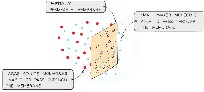
Molecules and ions in fluids (liquids and gases) move randomly and continuously whereas in solids, they vibrate on a spot. Particles change direction when they bump into each other. When they are closer together (higher concentration of a substance) they tend to collide more often & therefore get diffused till they are evenly spread within a fluid.
Diffusion is:
- the net movement of particles
- from a region of their higher concentration to a region of their lower concentration (down a concentration gradient)
- as a result of their random movement
*A concentration gradient is a concentration slope which goes from up to down, similarly to the process of diffusion.

The energy for diffusion comes from the kinetic energy of random movement of molecules and ions known as Brownian motion.
Substances move into and out of cells by diffusion through the cell membrane.
The relationship between the difference in concentration and the rate of diffusion is directly proportional. This means that the more the difference in concentration (steeper slope), the more the diffusion and visa-versa.
Difference in Concentration ∝ Diffusion Rate
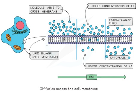
For living cells, the principle of the movement down a concentration gradient is the same, but the cell is surrounded by a cell membrane which can restrict the free movement of the molecules.
The cell membrane is a partially permeable membrane – this means it allows some molecules to cross easily, but others with difficulty or not at all. The simplest sort of selection is based on the size of the molecules.
Diffusion helps living organisms to:
- obtain many of their requirements
- get rid of many of their waste products
- carry out gas exchange for respiration
Diffusion in Life Processes
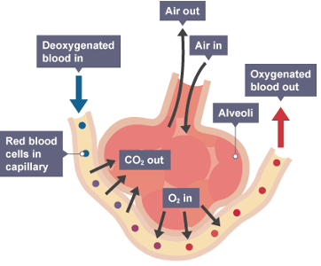
Diffusion occurs all the time in living organisms and it does not require energy from the body. One of the places in the human body in which diffusion takes place is the alveoli in the lungs. Over here the diffusion of oxygen and carbon dioxide takes place. The concentration changes all the time as we breathe as the carbon dioxide will diffuse and exit the body, whereas the oxygen will enter and diffuse inside. Equilibrium is never reached so that the concentration gradient shall be maintained. Diffusion rates get lower when one holds their breath, thus making the heart pump faster.
Similarly, in plant tissues, they take in carbon dioxide for photosynthesis and produce oxygen. Some of the oxygen is used up for respiration and the rest is diffused out. Only the excess can be and will be diffused.
There are several other places in which diffusion takes place in living organisms. The diffusion of fluids is thus considered to be a very important process. Diffusion additionally doesn’t require any energy to take place, meaning that only a substances presence with a concentration gradient available will allow diffusion to take place. Some more of these life processes are shown below.
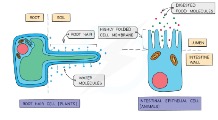
- The absorption of water in root hair cells
- Gas exchange in the stomata
- Diffusion of oxygen from inside to outside in a leaf
- Saprophytes absorbing nutrients (extracellular digestion – enzymes secreted)
- Gas exchange in the alveoli
- Absorption of glucose in the ileum
- Exchange of glucose at organs
| SITE | MOLECULES MOVING | FROM | TO |
| SMALL INTESTINE | DIGESTED FOOD PRODUCTS – GLUCOSE, AMINO ACIDS, FATTY ACIDS AND GLYCEROL ETC. | LUMEN OF SMALL INTESTINE | BLOOD/LYMPH IN VILLI FOUND COVERING SMALL INTESTINE WALLS |
| LEAF | OXYGEN | AIR SPACES BETWEEN MESOPHYLL CELLS | MITOCHONDRIA IN ALL CELLS |
| LEAF | CARBON DIOXIDE | AIR SPACES BETWEEN MESOPHYLL CELLS | CHLOROPLASTS IN MESOPHYLL CELLS |
| LEAF | WATER VAPOUR | STOMATAL PORES | AIR OUT SIDE STOMATA |
| LUNGS | OXYGEN | AL VEOLAR AIR SPACE | BLOOD IN CAPILLARIES AROUND AL VEOLI |
| LUNGS | CARBON DIOXIDE | BLOOD IN CAPILLARIES AROUND AL VEOLI | AL VEOLAR AIR SPACE |
Adaptations to Speed up Diffusion
Diffusion may not be the fastest process and that is why organisms have several adaptations to speed up diffusion:
- Short diffusion distances: blood vessels such as capillaries are only 1 cell thick to allow quick diffusion taking place (thin walls and openings present)
- Concentration gradients are maintained: oxygen and carbon dioxide can always diffuse in and out of the body as the carbon dioxide leaves and oxygen circulates in the body (quick removal from circulation so that equilibrium is not reached – high and low in gradient shall not change as diffusion will stop in this case)
- Large diffusion surface area: root hair cells have a root hair which increases the surface area for the diffusion of water (larger surface area allows more diffusion)
- Higher temperatures: the higher the temperature, molecules have more energy and move faster which allows more collisions against a membrane allowing more molecules to diffuse
Osmosis – A special case of diffusion
Osmosis is a kind of diffusion but only of water molecules through a membrane which will only allow the diffusion of small water molecules known as a partially permeable membrane.
Osmosis is:
- the net movement of water molecules
- from a region of higher water potential (dilute solution) to a region of lower water potential (concentrated solution) [down a water potential gradient]
- through a partially permeable membrane
This partially permeable membrane is the cell membrane of a cell. A water potential gradient is a concentration gradient of water potential, thus going from high to low.
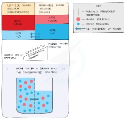
Water diffuses through partially permeable membranes by osmosis and water moves in and out of cells by osmosis through the cell membrane.
Osmosis in Living Organisms
All living organisms have a partially permeable cell membrane. Plants have a freely permeable cell wall (will have bigger openings to allow larger molecules in).
Animal Cells:
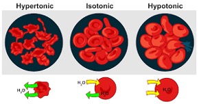
Hypertonic Solution: lower water potential compared to the cell; exosmosis (water exits from the cell) takes place; cell becomes flaccid (loose or droopy) and becomes shrivelled or crenated (in the case of RBC’s) when too much water exits the cell. This will carry lesser oxygen. |
Isotonic Solution: equal water potential compared to the cell; osmosis is equal (entry and exit of water – no net movement of water); cell remains in their regular shape. |
Hypotonic Solution: higher water potential compared to the cell; endosmosis (water enters the cell) takes place; cell becomes turgid (bloated) and will swell and then burst / lyse (haemolysis occurs) when too much water enters the cell (in animal cells as there is no cell wall to keep the cell rigid ) and only the ‘ghost’ (the membrane) of the RBC is left behind. |
As such damaging cases may occur, the kidney in the human body or an animal body plays a vital part in a process known as osmoregulation (regulates the water content in the blood).
Plant Cells:
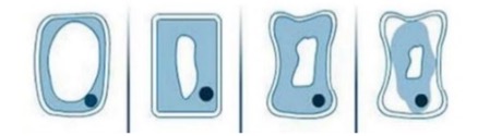
- The first cell is present in a hypotonic solution – the cytoplasm pushes hard against the cell wall (turgor pressure) and the cell becomes turgid.
- The second cell stays the same – the cytoplasm just presses against the cell wall.
- The third cell is present in a hypertonic solution – the cytoplasm starts to push away from the cell wall, thus making it flaccid (floppy).
- The last cell is in a hypertonic solution with a big water potential difference and the cytoplasm the cell membrane pushes away from the cell wall making the cell plasmolysed (through the process of plasmolysis – shrinkage of cell contents, including the cell membrane away from the cell wall).
When plasmolysis occurs for too long the cell will end up dying. Thus, when one has a throat infection, one is recommended to have hot water with salt (low water potential) so that exosmosis occurs from the bacteria making it go under plasmolysis until it gets plasmolysed and dies.
The empty space in a plasmolysed cell is filled up of the solute that concentrates or reduces the water potential of the water outside the cell through the process of diffusion that couldn’t occur when the cell membrane (partially permeable) was sticking to the cell wall (salt in the bacteria example).
Plants are supported by the pressure of water inside the cells pressing outwards on the cell wall.
When the turgor pressure is high it will not allow any more water to enter the cell even if there is a higher water potential outside the cell. When there is enough water applying pressure on an inelastic cell wall, it supports the cell by the turgor pressure within the cell.
Plant roots are surrounded by soil water and the cytoplasm of root cells has a lower water potential than the soil water. This means water will move across the cell membrane of root hair cells into the root by osmosis. The water moves across the root from cell to cell by osmosis until it reaches the xylem. Once they enter the xylem, they are transported away from the root by the transpiration stream, helping to maintain a concentration gradient between the root cells and the xylem vessels.
For a cell to not be affected by haemolysis or plasmolysis, it needs to be in an isotonic solution.
If turgor pressure exceeds wall pressure (pressure to ensure cell doesn’t burst), the cell will burst in animal cells. In plant cells, the cell won’t burst as it has a cell wall and the wall pressure will never be less than the turgor pressure. It may be equal though.
Homeostasis
Homeostasis is the maintenance of a constant internal environment. Osmoregulation is one of such processes.
Active Transport
Root hair cells contain a high concentration of mineral ions, so that there is a lower water potential in the cell to ensure a sufficient amount of water to enter through endosmosis. The water doesn’t stay in the cell, but will pass on to the other cells through osmosis. This is known as cell-to-cell osmosis.
These cells have a high mineral ions concentration, so it is important to take in more ions against the concentration gradient. Thus, there is a 3rd mode of transport.
Diffusion and osmosis don’t require energy to occur (passive transport), but to take in mineral ions or other particles the cells use energy to take in particles by a method known as active transport.
Active transport is:
- the movement of particles through a cell membrane
- from a region of a lower concentration to a region of higher concentration (against a concentration gradient)
- using energy from respiration
- and protein carriers in the cell membrane
Some examples of active transport are:
- Uptake of ions by plant root hairs
- Uptake of glucose by epithelial cells of the villi and kidney tubes
These cells always have a higher number of mitochondria to provide maximum energy from respiration.
Carrier proteins play an important role in this process.
Steps of active transport are:
- The carrier protein (cell membrane made of lipoprotein) of the membrane recognises a particle and holds it (it has a specific shape to hold a specific particle – many different types and shapes of carriers – won’t allow any other particle except the one it is functioned to).
- With energy provided from respiration, the carrier protein is driven across the membrane.
- The particle is released into the cell.
- The protein carrier returns to collect more of the same particle.
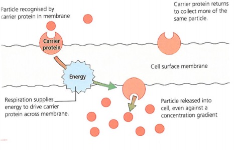
The protein carriers will not carry any foreign organisms as the shape of the carrier will not be able to carry any other shape.
Carrier proteins, like enzymes, are specific and are able to pick and choose particular molecules from a mixture. Therefore, the process of active transport is specific as the cell can chose which molecule it will absorb from its surroundings.
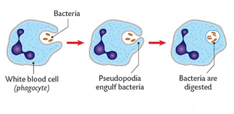
Using Phagocytosis
Some particles are too large to cross a membrane by diffusion or active transport. A few specialised cells have developed methods to take the particles. The cell surface membrane flows around the particles and engulfs them by a process known as phagocytosis.
White Blood Cells (also known as phagocytes) show this process. These cells have pseudopodia (false feet) and show amoeboid movement.
The phagocyte action involves 3 main steps:
- The white blood cell has a long, lobed shape nucleus which allows them to squeeze out of leaky capillaries. This process is known as diapedesis. They get attracted to wounds or sites of infection by chemical messages known as histamine.
- The phagocyte recognises the pathogen and flows around (engulfs) it, enclosing it in a sac.
- Digestive enzymes secreted from an organelle known as lysosomes are poured into the sac and the pathogen is destroyed or killed. They could also kill themselves in this process and hence are known as suicidal bags or they use the pathogen’s nutrients for its own benefits.
Movement In and Out of Cells FAQs
What controls movement in and out of the cell? Which organelle is responsible?
The primary structure that controls the movement of substances into and out of the cell is the **Cell Membrane (also called the Plasma Membrane)**. While not strictly an "organelle" in the sense of membrane-bound internal structures like mitochondria, it is a crucial component of the cell responsible for regulating transport.
How does the cell membrane control what enters and leaves the cell?
The cell membrane is **selectively permeable** (or semipermeable). This means it allows certain substances to pass through while preventing others. Its structure, a phospholipid bilayer embedded with various proteins, facilitates this control:
- Small, nonpolar molecules (like oxygen and carbon dioxide) can often pass directly through the lipid bilayer.
- Larger, polar, or charged molecules (like glucose, ions, and amino acids) usually require the help of transport proteins embedded in the membrane to move across.
What is the movement of substances in and out of cells called?
The overall process of substances moving across the cell membrane is called **Cell Transport** or **Membrane Transport**. This includes various mechanisms depending on the substance and whether energy is required.
What are the main types of cell transport?
Cell transport mechanisms are broadly categorized into:
- **Passive Transport:** Does not require the cell to expend energy. Substances move down their concentration gradient (from an area of high concentration to low concentration). Examples include simple diffusion, osmosis, and facilitated diffusion (which uses transport proteins but no energy).
- **Active Transport:** Requires the cell to expend energy (usually ATP) to move substances, often against their concentration gradient (from low to high concentration). This process uses specific transport proteins ("pumps") or vesicles (like endocytosis and exocytosis).
Why is the movement of substances in and out of cells important?
This movement is fundamental for cell survival and function:
- **Nutrient Uptake:** Cells need to import essential nutrients like glucose, amino acids, and ions for energy and building blocks.
- **Waste Removal:** Metabolic waste products need to be exported to prevent toxicity.
- **Gas Exchange:** Cells exchange gases like oxygen (needed for respiration) and carbon dioxide (a waste product).
- **Communication:** Cells exchange signaling molecules to communicate with each other.
- **Maintaining Homeostasis:** Transport processes help regulate internal conditions like pH, ion concentrations, and water balance.