
Nephrocalcinosis — A Comprehensive, Source-Backed Report (2025)
1) Overview
What is it? (Concise definition)
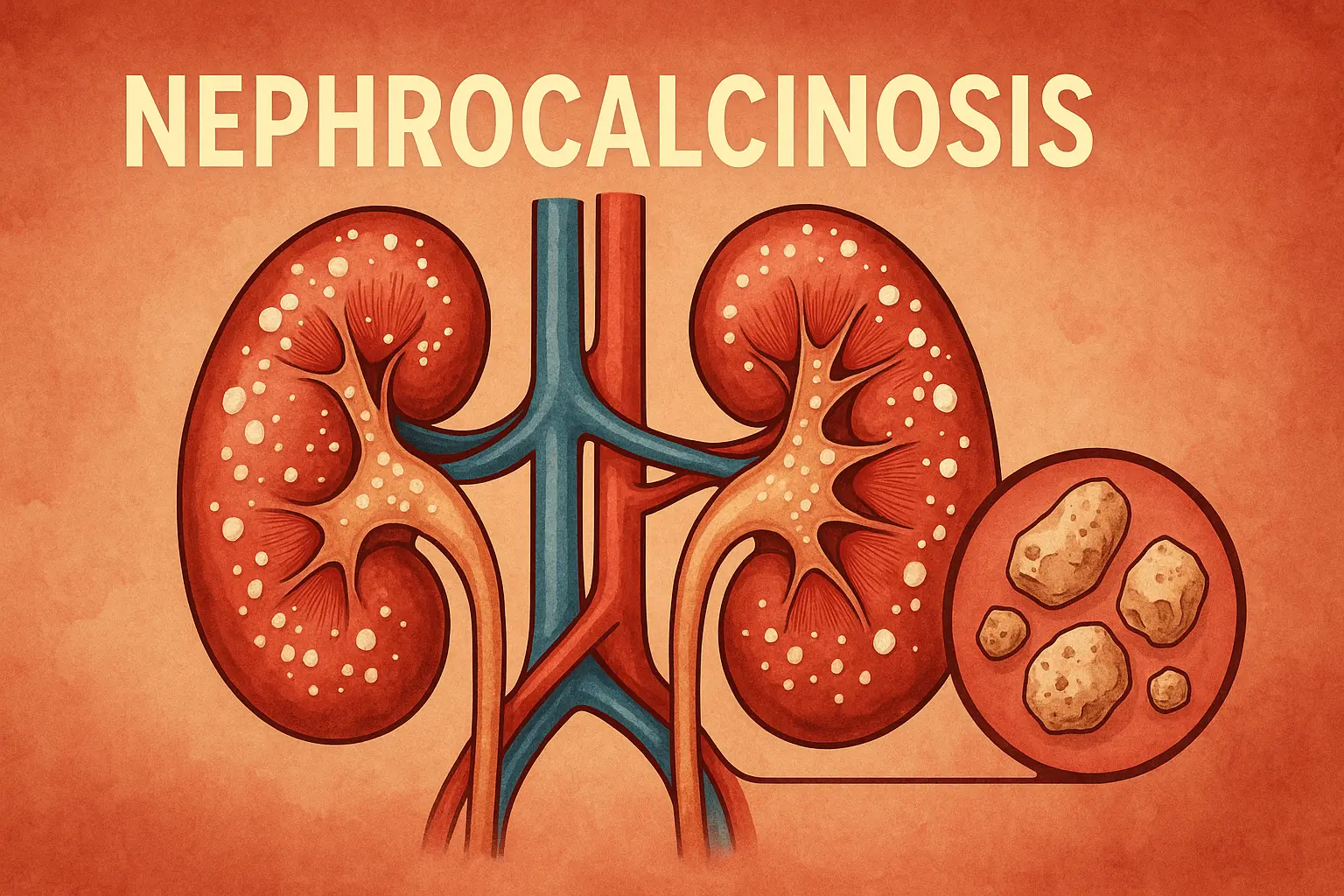
Nephrocalcinosis is the pathologic deposition of calcium salts within the renal parenchyma (cortex and/or medulla), most often calcium phosphate and/or calcium oxalate. It is a radiologic and histologic finding that signals an underlying disorder of mineral handling—typically hypercalciuria, hyperphosphaturia, and/or hyperoxaluria—rather than a standalone disease. The term was coined by Fuller Albright in 1934.
Organs affected
The kidneys are affected—usually the medulla (medullary nephrocalcinosis), less commonly the cortex (cortical nephrocalcinosis). Medullary involvement is most frequent and shows characteristic echogenic pyramids on ultrasound and punctate calcifications on CT.
Prevalence & significance
Population-level prevalence is not well defined because nephrocalcinosis is often asymptomatic and detected incidentally. Notable high-risk groups include preterm neonates (reported in up to ~40% in some cohorts) and patients with metabolic/tubular disorders (e.g., distal renal tubular acidosis, medullary sponge kidney, primary hyperoxaluria). The finding warrants evaluation because it is associated with nephrolithiasis and, in certain etiologies, progression to CKD.
2) History & Discoveries
Origins of the concept. The term nephrocalcinosis was introduced by Fuller Albright (1934) in the context of hyperparathyroidism; radiologic/pathologic characterization expanded through work by Anderson (1940s) and Reginald Carr (1950s), giving rise to the eponym “Anderson–Carr kidney.”
From definition to mechanisms. Modern reviews emphasize biochemical drivers (urine supersaturation, pH, citrate) and link nephrocalcinosis to stone pathogenesis (e.g., Randall’s plaque), reframing it as a signpost to underlying disorders.
Neonatal era. With widespread neonatal intensive care and loop diuretic use, prematurity-associated NC was recognized; later analyses nuanced the furosemide link and highlighted multifactorial contributors (immaturity, supplements, chronic illness).
Genetic breakthroughs. Identification of CYP24A1 (vitamin D catabolism) in idiopathic infantile hypercalcemia and Dent disease genes (CLCN5/OCRL) established clear molecular routes to hypercalciuria/NC.
3) Symptoms
Early/common
Often asymptomatic; findings arise on ultrasound/CT. When present, symptoms usually reflect the underlying cause or stones: flank pain/renal colic, hematuria, dysuria.
Advanced/uncommon
Polyuria/polydipsia from impaired concentrating ability; manifestations of distal renal tubular acidosis (dRTA); features of hypercalcemia (nausea, constipation), or, rarely, acute kidney injury in specific causes.
Progression over time
Trajectory depends on etiology. Without addressing the driver (e.g., dRTA, primary hyperoxaluria, hyperparathyroidism), recurrent stone events and chronic interstitial injury may lead to CKD; effective etiologic therapy can stabilize or improve outcomes.
4) Causes
Biological (metabolic/tubular) causes—most to least common in practice
Hypercalciuria states: idiopathic hypercalciuria; primary hyperparathyroidism; sarcoidosis; vitamin D intoxication; milk-alkali syndrome.
Tubular disorders: distal renal tubular acidosis (dRTA); Bartter syndrome; Dent disease (CLCN5/OCRL); medullary sponge kidney (urinary stasis + tubular defects).
Hyperoxaluric states: primary hyperoxaluria (PH1–3), enteric hyperoxaluria; these favor calcium oxalatedeposition and stones with frequent NC.
Neonatal/prematurity environment: prematurity, mineral supplements, chronic lung disease therapies; the role of furosemide is controversial and appears not uniformly causal in modern cohorts.
Genetic/hereditary
CYP24A1 (vitamin D 24-hydroxylase) variants → hypercalcemia/hypercalciuria/NC from impaired vitamin D inactivation; can present from infancy to adulthood.
Dent disease (CLCN5/OCRL, X-linked) → LMW proteinuria + hypercalciuria ± NC/stones/CKD.
Triggers/exposures
Excess vitamin D or calcium supplements; prolonged immobilization; high urine pH with hypocitraturia; infections/tuberculosis (rare, cortical patterns).
5) Risk Factors
Age: peaks in preterm neonates; also children/adolescents with metabolic/tubular disorders; adults with endocrine disease (e.g., hyperparathyroidism) or stone diatheses.
Genetic background: CYP24A1, CLCN5/OCRL; family history of stones/tubulopathies.
Comorbidities/contexts: dRTA, medullary sponge kidney (MSK), primary hyperoxaluria, malabsorptive GI disease; high-dose supplements; chronic immobilization.
Procedural/iatrogenic: long courses of loop diuretics in neonates (contextual, multifactorial).
6) Complications
Nephrolithiasis (stones): common co-traveler; contributes to pain, obstruction, infections.
Chronic kidney disease (CKD): risk varies by etiology—high in primary hyperoxaluria and some tubulopathies; in CKD/ESRD cohorts, microscopic NC prevalence rises with CKD stage (>50% in ESRD).
Tubular dysfunction: impaired concentrating ability; association with dRTA and secondary nephrogenic diabetes insipidus in some settings.
Mortality/disability: Nephrocalcinosis itself is rarely directly fatal; risk is mediated through stone complications, infections, and progressive CKD in high-risk disorders.
7) Diagnosis & Testing
Imaging
Ultrasound: first-line; shows increased medullary echogenicity and can grade NC.
Non-contrast CT: highly sensitive for calcifications; helps distinguish parenchymal (NC) from collecting systemcalcifications (stones).
Laboratory evaluation (identify the cause)
Serum: Ca, P, bicarbonate, creatinine, PTH, 25-OH/1,25-OH₂ vitamin D (hypercalcemia work-up).
Urine: 24-h urine for Ca, citrate, oxalate, phosphate, creatinine (or spot Ca/Cr in children); urine pH (dRTA).
Genetics (when indicated)
CYP24A1 testing for hypercalcemia/NC with vitamin D sensitivity; CLCN5/OCRL testing for suspected Dent disease.
Early detection & effectiveness
In at-risk neonates and children with stone/NC on imaging, systematic metabolic testing detects a cause in the majority (~80%)—improving targeted management.
8) Treatment Options
Core principle: Treat the underlying disorder and reduce urinary supersaturation.
General measures (most patients)
High fluid intake to maintain dilute urine; sodium restriction; balanced calcium intake (avoid excess); manage hypocitraturia with potassium citrate (except in very alkaline urine).
Etiology-specific
Idiopathic hypercalciuria: diet (lower Na/animal protein), thiazide diuretics if needed.
dRTA: alkali therapy (potassium citrate/bicarbonate) to normalize bicarbonate and reduce calcium phosphate precipitation.
Primary hyperparathyroidism: parathyroidectomy when indicated.
Medullary sponge kidney: stone prevention—hydration, citrate, address hypercalciuria; monitor for infections and CKD.
Primary hyperoxaluria (PH1):
RNAi therapies that lower oxalate production: lumasiran (Oxlumo®, FDA 2020); nedosiran (Rivfloza®, FDA 2023)—both reduce urinary oxalate and are transforming outcomes; transplant remains for advanced disease.
CYP24A1-related hypercalcemia/NC: avoid vitamin D excess; case-based regimens (e.g., azoles/inducers) under specialist care.
Stones & procedures
Manage urolithiasis per standard algorithms (analgesia, expulsive therapy, endourology as needed). Persistent obstruction/infection requires urgent urologic care.
9) Prevention & Precautionary Measures
Primary prevention depends on genetic counseling/testing in heritable causes (e.g., Dent disease, CYP24A1), prudent supplement use (avoid unnecessary vitamin D/calcium megadoses), and hydration in stone formers.
Secondary prevention targets recurrence: dietary sodium reduction, adequate dietary calcium (with meals), citrate repletion, and etiologic therapies noted above.
Neonatal care: judicious mineral/diuretic use, routine renal ultrasound surveillance in very preterm infants at risk.
Vaccines/screening: No disease-specific vaccines; follow standard immunizations. Screening is targeted to high-risk groups (prematurity; children with stones; families with tubulopathies).
10) Global & Regional Statistics
General population: exact global prevalence and incidence are unknown due to asymptomatic cases and heterogeneous reporting; nephrocalcinosis should be viewed as a marker of underlying conditions.
Neonates: imaging-detected NC in up to ~40% of preterm neonates in some series.
Comorbidity settings: microscopic NC is common in advanced CKD (>50% in ESRD biopsy cohorts), reflecting disordered mineral metabolism.
Condition-specific: MSK is uncommon overall (≈1 in 5,000) but enriched among calcium stone formers (≈12–20%); NC is frequent in MSK due to collecting duct changes.
11) Recent Research & Future Prospects
Genetic & molecular diagnostics: increasing yield for monogenic causes (Dent disease spectrum; CYP24A1; others), enabling precision therapy and family counseling.
Transformative therapies for oxalate disorders: Lumasiran and Nedosiran (RNA interference) markedly reduce urinary oxalate and are being tracked in long-term registries; access/earlier use may prevent NC-driven renal decline in PH1.
Neonatal outcomes: contemporary pharmacoepidemiology suggests furosemide may not independently raise NC risk when modern care is optimized, refocusing attention on multifactorial contributors in NICUs.
Imaging & quantification: advances in ultrasound grading and CT characterization may standardize monitoring and prognostication.
12) Interesting Facts & Lesser-Known Insights
Name game: You’ll still see the historical terms “Albright’s calcinosis” and “Anderson–Carr kidney.” The modern usage is broader and radiology-driven.
It’s a compass, not the destination. Nephrocalcinosis is a diagnostic gateway—its presence should trigger a systematic search for the cause (often successful in children).
Women, kids, and genetics: Don’t overlook Dent disease in boys with LMW proteinuria/hypercalciuria or CYP24A1-related hypercalcemia in infants and even adults with “unexplained” NC.
Practical Diagnostic Checklist (for clinicians)
Confirm NC on US ± CT and distinguish from stones.
Serum: Ca, P, HCO₃⁻, creatinine, PTH, 25-OH/1,25-OH₂ D.
Urine: 24-h (Ca, citrate, oxalate, phosphate, creatinine) or spot Ca/Cr (children); urine pH.
Etiology tests as indicated: autoimmune sarcoid work-up, genetics (CYP24A1; CLCN5/OCRL), stone analysis.
Selected References (open-access or high-quality summaries)
StatPearls: Nephrocalcinosis — definition, causes, work-up, management.
Radiopaedia — medullary NC imaging and differentials.
Kidney International reviews — mechanisms, history, and risk of CKD/ESKD.
AAP NeoReviews (2024) — neonatal NC (preterm, contributors, monitoring).
NIDDK/StatPearls on MSK — epidemiology and link to NC and stones.
RNAi therapies for PH1 — Lumasiran and Nedosiran approvals and outcomes.